The Ubiquitin Editing Enzyme A20
Maintains Immune Homeostasis and
Prevents Autoimmunity
Rita M. Tavares
Dissertation presented to obtain the Doctorate degree (Ph.D.) in
Biology at Instituto de Tecnologia Química e Biológica da
Universidade Nova de Lisboa
ISBN: 978-989-20-2300-7
Com o apoio da FCT e do FSE no âmbito do Quadro Comunitário de
Apoio, BD n.º SFRH/BD/15222/2004
The work compiled in this thesis was undertaken in the scope of the
Gulbenkian PhD Program in Biomedicine (PGDB), Portugal.
The first year of the program (2003/2004) was spent at graduate courses
and other advanced training at Instituto Gulbenkian de Ciência, Oeiras, Portugal,
and was funded by Fundação Calouste Gulbenkian.
The research work was carried out between 2004 and 2010, at the
University of California, San Francisco, under the supervision of Dr. Averil Ma.
Fundação para a Ciência e Tecnologia (FCT) / Fundo Social Europeu
(FSE) awarded the doctorate fellowship SFRH/BD/15222/2004 from 2004 to 2008.
Work in the laboratory of Dr. Averil Ma was supported by the National Institutes of
Health, the Kenneth Rainin Foundation and the Alliance for Lupus Research.
Or
How A20 restricted most of my 20
ʼ
s
TABLE OF CONTENTS
Acknowledgments 11
Summary 17
Sumário 19
Chapter 1 – Introduction 23
From Immunity to an Immune System 25
Evolution of Immunity 26
Evolution of Theories and Models of Immunity 27
Scope of this thesis 29
Toll-like-receptors 31
TLR signaling 32
TLRs in health and disease 35
B cells 37
B cell selection 38
Germinal Centers 40
B cells in autoimmunity and cancer 42
The transcription factor NF-κB 44
Regulation of NF-kB through ubiquitylation 45
The ubiquitin editing enzyme A20 47
A20 in autoimmunity and cancer 49
Chapter 2 – Homeostatic MyD88-dependent signals cause lethal inflammation in the absence of A20
53
Summary 57
Introduction 61
Results 64
Discussion 82
Methods 89
Acknowledgments 93
References 95
Chapter 3 – The Ubiquitin Modifying Enzyme A20 restricts B-cell survival
and prevents autoimmunity 103
Summary 107
Introduction 109
Results 111
Discussion 133
Acknowledgments 138
Methods 139
References 143
Chapter 4 – Final Discussion 151
Appendix – Unpublished data and future perspectives 161
References 175
Acknowledgments
First and foremost I want to thank my parents, Nazaré e João: por me criarem e me darem o vosso amor incondicional, nunca existirão palavras suficientes para descrever o meu amor infinito por vocês. Durante estes seis anos da minha vida, mesmo com uma distância física tão grande, vocês estiveram sempre muito perto, e nos momentos em que me senti mais perdida, como que no meio do oceano que nos separava, numa noite escura, saber que vos tenho foi a minha bússola mais fiel e que me manteve sempre a navegar até bom porto.
With equal love, I am grateful to my sisters, Maria João e Joana: e quero que saibam sempre que faço qualquer coisa por elas, que dou a vida ou um braço, ou uma perna, mas que nunca saberei gostar delas menos do que isso, e que quero sempre estar perto das suas vidas, mesmo que haja esse tal oceano entre nós. Vocês continuam a ser sempre as coisas mais preciosas que tenho.
To my grandparents, and in particular to my grandfather António: a todos eles, os meus avós que já cá não estão, e em particular ao meu avô António, que ainda me viu a meio deste doutoramento. Ficam as imensas saudades e o desejo de que o Avô ainda me tivesse visto chegar ao fim desta etapa. Eu sei o quão orgulhoso estaria, e não fosse eu ateia convicta, desejaria que hoje, em qualquer lado, ele soubesse o quanto está sempre presente em mim, como um exemplo de vida e dessa beleza que tinha de ser a melhor pessoa do mundo. Este doutoramento custou-me estar longe demais quando ele disse adeus.
An additional word of gratitude goes to my extended family of uncles, aunts and cousins, who provide extra unconditional love even if they see me too seldom.
enough; and all the hallway conversations about Portugal and the dinners and patuscadas with Sílvia Vilarinho.
I am grateful for more of the wonderful friends I made in San Francisco: Géraldine Bienvenu, Cédric Louvet, Sílvia Curado, Vincent Denef, Marta Gaglia, Laurence Clement, Yann Rignault, Markus Hagele, Tatiana Hochgreb, Marine Champsaur, Alexandre Fernandes, André Guedes, Rui Cunha, Hans Dooms. A special word to Lionel Christiaen, because he was such an important part of my growing process, personal and professional, in fact. To my classmates become close friends from PGDB: Renata Brito, Tiago Carvalho, Joana Sá, Marta Vitorino. Meeting all of you guys was the first time I found so many people all at once that I could connect with and that changed my life. And a mention of childhood friends Paula Pinto, Catarina Trindade, Marta Varandas, Mafalda Ferreira; college friends: Ema Alves, Ana Caetano, Sandrina Nóbrega Pereira, Liliana Pires. And in these last few months, Rodrigo Ábreu and Nuno Almeida.
I am also in debt to Deborah Raphael and Erik Sandegard: they know who they are, and they got me closer to know who I am.
All these friends (and so many more people that I would run out of space to mention) played their roles in getting me where I am today, and were part of the meaning my life had way beyond science. This PhD would not have been worth without them. So every day I am grateful for having had so many rich personal experiences.
From my early steps in science I would like to thank Cecília Arraiano and her lab for having trained me as an undergrad student at ITQB, and in particular to José Andrade and Sandra Viegas.
I am thankful to all my lab mates without exception, because I have learnt something from all of them. A particular word of gratitude to those who also became good friends: Gianna Hammer (a precious role model as a woman scientist and a strong woman, also a warm and lovely friend), Julio Barrera (for the absolute infinite help and loyalty, I truly love him and wish so much he can be happy), Albert Lee (a wonderful and loyal friend), Bettina Lee (a very sweet friend), Nataliya Shiffrin, Tim Lu, Mike Whang, Alex Agelidis. Joseph Callahan, Shigeru Oshima, Osamu Hitotsumatsu, Rommel Advincula, Archer Smith, Sara Gozalo, Leesun Kim, Erwan Mortier, Min Wang, Bao Dong were wonderful colleagues to have around. Emre Turer was not such a good colleague to have around but I am appreciative of his contribution to my projects with some of his work. To David Boone, I am grateful for his support and teachings in my first year in the lab; Thank you for the friendship and for believing in me from the beginning.
I am very thankful to Patrizia Scapinni, that collaborated in the B cell project and provided help at a crucial step and turning point of that story. I am also grateful for additional precious collaboration from Cliff Lowell and PJ Utz labs (Chih-Long Liu) in that same project. Very appreciated technical help at UCSF came from Cliff McArthur, Shuwei Jiang, Ivy Hsieh, Jean Publicover.
To Averil Ma: thank you for taking me in your lab and always believing in me even
opportunity. I hope the papers are a good pay off! Well, that work was a pleasure for me, and that is the most important feeling I look for in my life.
To Barbara Malynn: I could not thank you enough for all your help. From the time
you taught me how to do ES cell transfections to start co-guiding the B-cell project, your help and mentorship have always propelled my progress at crucial steps and that has had a huge positive impact. Additionally, you were another one of my role models of female scientist and strong woman that have been so important in these past formative years. You have inspiring patience, sensibility and rigor in many facets of life. My profoundly felt thank you, for sharing all that with me.
To Sukalyan Chatterjee, for the huge impact he had in that first year of the
PGDB and the continued immense support he has given me until the completion of it all. Thank you for the investment you took in organizing our courses and for all the time you have taken for advising me in particular, and for sharing so much fine knowledge, from science to Bob Dylan, Woody Allen and alike. You were indeed a mentor beyond science, pushing for intellectual commitment and questioning all. You contributed enormously to the momentum I needed to start a bigger, richer and more adventurous journey in my life. I cannot thank you enough for that.
To Manuela Cordeiro, I thank the incredibly efficient administrative help
throughout the years, always available for all students, always on the studentsʼ side, a constant reference back at the IGC, a source of constancy herself.
I thank Miguel Soares, Jocelyne Demengeot and Thiago Carvalho for also being a source of academic help at the IGC.
I am very grateful to Ana Maria Portocarrero for making the PhD degree burocracy at ITQB/UNL feel like a piece of cake and not like burocracy at all, throughout every single year of PhD. Additionally, I thank Fátima Madeira for that same efficient help in these last few months of the degree completion.
Finally, I would like to thank the remaining PhD defense jury members, Paulo Vieira, Bruno Silva-Santos, Jorge Carneiro, for their prompt availability in taking part in this process, their careful reviewing of this thesis, and making the defense questioning a pleasurable experience of intellectual exchange. And I thank the president of the jury from ITQB, Luís Paulo Rebelo.
To Tony DeFranco I would like to thank his continued availability throughout the period I was at UCSF, from being a Journal Club mentor, to reading our manuscript or be willing to be part of my thesis jury. His academic commitment is inspiring and has given me its share of feeling supported along the road. For similar reasons, I would like to thank Jason Cyster, as well as say I feel grateful to the Department of Immunology at UCSF, and the institution of the University of California, San Francisco, where I met so many wonderful people, from the cafeteria workers to the senior scientists, which made for such a rich experience every day.
Very importantly, I would like to finish by thanking the main institutions that have
supported my PhD: Fundação Calouste Gulbenkian / Instituto Gulbenkian de
Ciência – being part of a program like PGDB was an immense privilege and made for an
Tecnologia (and Fundo Social Europeu), for the last 4 years of funding that provided such a competitive advantage in joining the top institutions in the world; And Instituto de
Tecnologia Química e Biológica / Universidade Nova de Lisboa, for the easiness with
Summary
The immune system is vital to ensure the surveillance of organisms against
pathogens or malignant cells. However, the negative regulation of the immune
system is equally essential, and defects in the termination of immune signals can
result in autoimmunity and other pathologies.
The functioning of the immune system results from the integration of
signals between and within cells. For some time, studies in immune signaling have
focused on the molecular events playing a role in activating such cascades.
However, little was known about how to turn off those signals. Just recently, new
research started shedding light into mechanisms that ensure the negative
regulation of immune signaling.
The enzyme A20 previously has been shown to be a fundamental
intracellular negative regulator of immune signals. Mice deficient in the gene that
codes for the A20 protein, Tnfaip3, have massive generalized inflammation and
die prematurely. A20 deficient cells fail to terminate TNF and TLR induced
responses.
In the first part of this work, we aimed to define the basal signals
responsible for triggering the spontaneous phenotype of A20-/- mice. While
genetically removing TNF signals by obtaining A20-/-TNF-/- double-deficient mice
does not rescue the disease observed in single A20-deficient mice, animals
lacking the common TLR adaptor protein MyD88 (A20-/-MyD88-/-) have a clear
amelioration of the A20-/- phenotype, indicating that there are tonic TLR signals
that need to be constantly terminated by A20. We have shown that these signals
are likely initiated in the gut, since treatment with broad-spectrum antibiotics that
reduces commensal intestinal flora also ameliorates disease. Additionally, we
In the second part of this work, we used a floxed allele of Tnfaip3 to generate mice that lack A20 specifically in B-cells. While these mice develop
normally and show no signs of the general inflammation observed in A20 globally
deficient mice, Tnfaip3fl/fl and Tnfaip3fl/+ CD19-Cre mice have disrupted lymphoid
homeostasis and develop lupus-like autoimmunity. A20 deficient B-lymphocytes
hyper-respond to signals initiated by the BCR, TLRs and CD40. We have
demonstrated that A20 is necessary to restrict cell survival as A20-deficient
B-cells are resistant to Fas-mediated cell death. This provides one mechanism by
which A20 can prevent B-cell mediated autoimmunity.
All together, this work bestows several new insights on the mechanisms by
which A20 plays a central role in maintaining the homeostasis of both the innate
and the adaptive immune system. We show that there are tonic innate immune
signals that need to be constantly terminated by A20. Furthermore, we show for
the first time in vivo that A20 also plays a role in lymphocyte function. These
findings relate to recent genetic associations between human A20, autoimmune
Sumário
O funcionamento do sistema imunitário é fundamental aos organismos
desenvolvidos para garantir a prevenção contra microorganismos patogénicos ou
células malignas. Ainda assim, a regulação negativa do sistema imunitário é
igualmente essencial, e defeitos na terminação de sinais imunitários podem
resultar em auto-imunidade e outras patologias.
O funcionamento do sistema immunitário resulta da integração de sinais
entre e dentro das células. Na sua fase inicial, a investigação em sinalização
imunitária dedicou-se principalmente aos processos moleculares responsáveis
pela activação destas cascatas de sinalização. No entanto, pouco se sabia
acerca de como terminar estes sinais de activação, de forma a que não se
prolongassem indefinidamente. Só já mais recentemente, nova investigação
começou a fornecer pistas acerca dos mecanismos que asseguram a regulação
negativa da sinalização imunitária.
Alguma desta investigação recente demonstrou que a enzima A20 é um
regulador negativo intracelular de sinais imunitários fundamental. Ratinhos
deficientes no gene que codifica a proteína A20, Tnfaip3, desenvolvem níveis
elevados de inflamação em vários órgãos e morrem prematuramente. Células que
são deficientes em A20 não possibilitam a terminação de respostas induzidas por
Tumor Necrosis Factor (TNF) ou Toll-Like-Receptors (TLRs).
Na primeira parte deste trabalho, o nosso objectivo foi definir quais os
sinais existentes naturalmente, responsáveis por iniciar o fenótipo espontâneo de
ratinhos A20-/-. Eliminámos geneticamente sinais mediados por TNF, obtendo
ratinhos A20-/-TNF-/- e observámos o mesmo nível inflamação registada em
ratinhos A20-/-. No entanto, animais deficientes na proteína adaptadora comum à
inflamação comparativamente a A20-/-. Estas observações indicam que há sinais
inerentes mediados por TLRs que devem ser constantemente terminados através
da acção de A20. Demonstrámos ainda que a origem mais provável destes sinais
se encontra no intestino, já que o tratamento com antibióticos de largo espectro,
que reduz a flora intestinal comensal, resulta num efeito semelhante.
Demonstrámos ainda que A20 pode também regular sinais de TLRs que não
dependem de MyD88.
Na segunda parte deste trabalho, usámos um alelo “floxed” de Tnfaip3 e
gerámos ratinhos sem A20 única e especificamente em células B. Estes ratinhos
desenvolvem-se normalmente e não mostram sinais de inflamação generalizada
como observado em ratinhos globalmente deficientes em A20. Ainda assim,
ratinhos Tnfaip3fl/fl e Tnfaip3fl/+ CD19-Cre apresentam alterações evidentes na
homeostase dos seus linfócitos e desenvolvem uma doença com características
semelhantes a lúpus. Linfócitos B deficientes em A20 respondem
exageradamente a sinais iniciados pelo B-Cell-Receptor (BCR), TLRs e CD40.
Demonstrámos que A20 é necessária para restringir a sobrevivência de células B,
já que células B deficientes em A20 são resistentes à morte celular mediada por
Fas. Esta descoberta sugere um mecanismo através do qual A20 pode prevenir a
auto-imunidade mediada por células B.
Este trabalho fornece novos esclarecimentos sobre os mecanismos
através dos quais a proteína A20 representa um papel central na manutenção da
homeostase tanto do sistema imunitário inato como do adaptativo. Demonstrámos
que há sinais imunitários inatos inerentes que devem ser constantemente
descontinuados através da acção de A20. Mais ainda, demonstrámos pela
primeira vez, in vivo, que A20 também têm um papel em linfócitos, o qual está
relacionado com recentes associações genéticas entre A20 humano, doenças
CHAPTER 1
From Immunity to an Immune System
Throughout evolution, systems of defense against opportunistic pathogens
arose in nearly all organisms. Such systems have been crucial for the fitness of
progressively more complex species. Even though vertebrates continued to evolve
more sophisticated immune processes, immunity can be found in invertebrates
and plants, and remarkably, several of the same molecular pathways are shared
among such different organisms (Howard and Jack, 2007; Litman and Cooper,
2007).
Jawed vertebrates as mammals evolved some of the most complex
immune systems, and humans and mice have been at the center of much of the
research in the field of Immunology. From the early days of vaccine discovery by
Edward Jenner and Louis Pasteur, to the recent development of vaccinations and
treatments against cancer, this research has had wide implications in medicine
(Janeway, 2005; K. Abbas et al., 2007).
Vertebrate immune systems clear pathogenic infections as well as
recognize and eliminate malignant cells, and as a result immunodeficiency is
highly deleterious. Nonetheless, exaggerated immune responses can also harm
the organismʼs own cells and cause equally severe pathologies. Therefore it is
now extensively recognized that proper functioning of the immune system is
fundamental and needs to be finely tuned (J. Kindt et al., 2007; Janeway, 2005).
Plants and invertebrates have several forms of immune defense but are
jawed vertebrates that have evolved the most complex immune systems.
Vertebrates like humans and mice have specialized immune cells, and have
dedicated immune organs that allow the appropriate development of these cells as
well as their efficient migration through the organism. Immune cells circulate
surveillance and specialized lymphoid organs where they either develop (primary
lymphoid tissues – bone marrow and thymus) or mount an immune response
(secondary lymphoid tissues – spleen and lymph nodes). Immunity in vertebrates
has been classified in two broad types: innate immunity and adaptive immunity
(Janeway, 2005; K. Abbas et al., 2007).
Evolution of immunity: Innate Immunity and adaptive immunity
Innate immunity is common to vertebrates, invertebrates and plants. This
kind of immunity has been characterized by being non-specific, and comprises
anatomic barriers, physiological barriers, phagocytic and endocytic activities,
production of anti-microbial proteins, and inflammatory barriers to foreign
microbes (J. Kindt et al., 2007). Vertebrates like mice and humans also have
professional innate immune cells (macrophages, neutrophils, dendritic cells (DCs))
that develop in the bone marrow and then circulate through the body or reside at
particular locations. These cells are often the first set of cells to be recruited to a
site of infection and mediate several of the aforementioned activities (K. Abbas et
al., 2007).
The adaptive immune system is found only in jawed vertebrates and is
characterized by being specific, enabling the development of memory for
particular pathogens. Lymphocytes serve as the adaptive immune arm, and are
comprised mainly of T and B cells. Conventional B and T lymphocytes can have
their DNA somatically rearranged during their development, which allows each cell
to express a different but specific receptor of its own. Each clone then goes
through several steps of selection that guarantee its receptor can recognize an
antigen – a small piece of a certain molecule - that, all working correctly, will
recognize foreign components (non-self) but not self constituents. When a given
clone is activated by the presence of its antigen, it rapidly proliferates and
will be maintained as a pool of memory cells, eventually granting a faster and
stronger secondary response (Janeway, 2005).
T-lymphocytes recognize antigens through the T cell receptor (TCR). To be
activated, the TCR needs to bind antigens processed and presented by MHC
(Major Histocompatibility Complex) molecules in Antigen Presenting cells (APCs)
as DCs, illustrating the crosstalk between the innate and adaptive immune
systems. Activated T-cells produce cytokines that aid the clearance of the
infection (T helper cells) or directly kill infected or transformed cells (Cytotoxic
T-cells), in what has been designated as the Cell-mediated Response (K. Abbas et
al., 2007).
B-lymphocytes recognize antigens through the B cell Receptor (BCR) and
are the cells responsible for producing antibodies (immunoglobulin), mediating the
so-called Humoral Response. Binding of antibodies to antigenic particles activates
molecular mechanisms that assist phagocytosis or killing of pathogens by other
immune cells (Janeway, 2005).
Evolution of theories and models of immunity: from self-non-self recognition to the danger model
Once the existence of an immune system was recognized the central
question in the field of immunology was (and still is to a certain extent) how does
the system decide what it reacts to or not? How does it not attack the own
organism and develop what has later been termed tolerance toward self? Or is it
ignorance?
The immunity model that has prevailed the longest, and is still the
theoretical framework for much of the research, is self-non-self recognition. In
Acquired Immunity (Burnet, 1959), in which he proposed that each lymphocyte
expresses a single surface receptor for a foreign particle and that self-reactive
lymphocytes are deleted early in development. In 1970, this model was further
developed by Bretscher and Cohn (Bretscher and Cohn, 1970); after the discovery
of hypermutation in activated B lymphocytes and thus the renewed chance for
creation of autoreactive cells, these scientists proposed that there had to be a
second signal for a cell to initiate a response, and that the second signal was
coming from an helper cell (later found to be a T-cell). Soon after, Lafferty and
Cunningham proposed a similar model for T-cell activation, which instead of “help”
required “costimulation” (Lafferty and Cunningham, 1975).
For a while it was thought that lymphocytes were responsible for initiating
the immune response. However, the need for a second signal posed a problem: if
a second signal (like costimulation) was often not coming from a lymphocyte, and
thus the adaptive immune harm of the response was not solely responsible for its
initiation, how was self-reactivity avoided?
It is clear that the innate immune system recognizes foreign invaders
(non-self) in a very different manner than cells of the adaptive immune system.
However, the idea that innate immunity provides “non-specific” protection at
mucosal barriers is an overly simplistic definition. In 1989, Charles Janeway Jr.
proposed the revolutionary Pattern Recognition Theory (Janeway, 1989). He
predicted that innate immunity encompassed the expression of Pattern
Recognition Receptors (PRRs), which could recognize Pathogen Associated
Molecular Patterns (PAMPs). Importantly, these PAMPs would consist of patterns
not found in the host, and thus would be specific to the invading microbe or foreign
body. PRRs would be responsible for directly sensing pathogens before
intervention of the adaptive immune system. He called it discrimination between
infectious non-self and noninfectious self, and evolutionarily this process of
discriminating foreign bodies in an organism pre-dates the straight self / non-self
proved true and provided much of the framework that propelled a large amount of
the Immunology research in the following decades. The discovery of
Toll-Like-Receptors (TLRs) as the proposed PRRs was the first big step (Medzhitov, 2009).
Nevertheless, the infectious non-self model still has its limitations. Namely,
if one thinks about the numerous instances in which there is the need to recognize
self that might be harmful, like tumours, or the need to be tolerant of non-self that
might be harmless, like commensals. Matzinger (from discussions with Fuchs) has
proposed a Danger model (Matzinger, 1994). She adds another layer at the apex
of the immune response, to include cells and signals from all tissues. Matzingerʼs
viewpoint is that the immune system does not care as much about non-self
discrimination as it does need to protect against danger, and that the initiation and
polarization of an immune response depend on danger signals from the tissue
affected by any sort of threat (Matzinger, 2002).
A number of Danger Associated Molecular Patterns (DAMPs) released by
injured tissues have now been described. Remarkably, besides identifying new
receptors, it was found that some PRRs also recognize DAMPs (Matzinger, 2002;
Rubartelli and Lotze, 2007; Zhang et al., 2010). Furthermore, it has also been
shown that several cell types in a plethora of tissues can respond to and produce
cytokines, express PRRs, or have specialized, resident immune cells, contributing
not only for the initiation but also the polarization of an immune response
(Janeway, 2005; Matzinger, 2002). Hence, it seems clear that this dialog between
tissues and the immune system is really happening, and playing a prominent role
in the overall immune outcome.
Scope of this thesis: what breaks tolerance and do we really need a unifying model?
Nevertheless, is there really a need for a unifying model and de we have to
layers of immune recognition and alert have to be taken into account, with
absolutely the same level of significance. If at times there have been trends to
favor this or that aspect of immunity (sometimes just a single cell type), it might as
well be about time to beware of such biased approaches. The barriers of what the
immune system is have always been loose, after all. Notwithstanding a few
defined immune organs, all the same immune cells can travel through numerous
tissues (with a few exceptions, as the privileged brain) and in those instances it
becomes harder to define what is the system itself versus what is an immune
defense process that might as well not immediately involve an immune cell.
The scope of this work shines some light into just that. Perturbations in a
single protein, in distinct aspects of the immune system (Toll-like-receptors in
innate immune cells in chapter 2, B cells in chapter 3) depict how different cells
and tissues play equally important yet distinct roles in maintaining the
homeostasis of the immune system. Even if some outcomes do result more
dramatic, altered intracellular regulation of a number of signals, in distinct cell
types, appears each time to be sufficient to break immune tolerance at those
various levels. In fact, conditional gene targeting will probably still be teaching us a
great amount of new principles about the workings of these complex networks in
many years to come. And just as selective pressures are diverse, there might as
well not be a single model of immunity that fits it all. Or at least our knowledge will
still have to evolve further to be able to create a broad enough theory. Ultimately,
while interpretations might always be based on the wrong preconceptions, robust
Toll-like-receptors
TLRs are the broadest class of PRRs identified to date, first found in flies,
and soon established to have an essential role in the immune systems of
vertebrates like those of humans and mice. In mice and humans, TLRs can be
expressed in both innate and adaptive immune cells, as well as in non-immune
cells. However, their highest levels of expression are found in dedicated innate
immune cells like macrophages, dendritic cells (DCs) or neutrophils (Beutler,
2009; Kawai and Akira, 2010; Medzhitov, 2009).
Demonstrating that innate immunity is more specific than previously
thought, each TLR binds only a few specific ligands of its own. Confirming
Janewayʼs prediction that PRRs would recognize patterns associated specifically
with the invading pathogen and not the host itself, TLRs recognize motifs such as
those of lypopolisacharyde (LPS), found in the wall of Gram negative bacteria
(TLR4), double stranded RNA, which can be found in viruses (TLR3), or
methylated DNA, from bacteria (TLR9). There have been 12 TLRs described in
mice, and 10 in humans. TLRs 1-9 are conserved in both species (Kawai and
Akira, 2010). Table 1.1 summarizes some basic features of TLRs.
Table 1.1 Mouse TLRs, their principal ligands and downstream adaptor proteins. Adapted from Beutler, 2009.
TLR Ligands Adaptors
TLR1/2 Glycolipids, Triacyl lipoproteins MyD88,Tirap
TLR2/6 Lipoteichoic acid, Zymosan, Diacyl lipoproteins MyD88, Tirap
polyI:C, dsRNA
TLR3 TRIF
Flagellin
TLR5 MyD88
MyD88 ssRNA, imiquimod, loxoribine, other
TLR7
TLR9 CpG, methylated DNA MyD88
Profilin TLR11 MyD88 ? TLR12 ? ? TLR13 ?
TLR activation triggers signaling cascades that result in the expression of
effector genes. Effector genes mediate direct functions in the activated cells, but
also the expression of surface co-stimulatory molecules and a large number of
cytokines and chemokines. Many of these are pro-inflammatory cytokines like IL-6
(Interleukin-6), IL-1β, TNF-α (Tumor Necrosis Factor α) (Beutler, 2009).
Production of cytokines mediates the amplification and specification of the
immune response and appropriate clearing of a particular infection. There has
been ample evidence that innate immune system activation through TLRs is an
essential component of effective immunity, and is critical for the efficient activation
of the adaptive immune response (Beutler, 2009; Hou et al., 2008; Kawai and
Akira, 2010).
Toll-like-receptor signaling
All TLRs except TLR3 bind the adaptor protein MyD88 (Myeloid
differentiation primary response gene (88)). Upon TLR engagement, kinases and
adaptor proteins are recruited by MyD88 and lead to the activation of downstream
transcription factors. The main transcription factor responsible for the expression
of pro-inflammatory cytokines downstream of MyD88 is NF-κB (Nuclear factor
kappa-light-chain-enhancer of activated B cells) (Hayden and Ghosh, 2008).
Figure 1.1 summarizes some of the main players in the MyD88-dependent
Figure 1.1 – TLR signaling through MyD88.
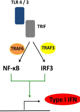
TRIF (TIR-domain-containing adapter-inducing interferon-β) is an alternate
TLR adaptor. TLR3 signals exclusively through TRIF, and TLR4 signals through
both MyD88 and TRIF. The TRIF-dependent pathway also activates NF-κB (and
thus expression of pro-inflammatory cytokines), but it is best known for activating
the transcription factor IRF3 (Interferon Regulatory Factor 3). IRF3 is the main
factor responsible for the expression of type I interferons (IFN) alpha (IFN-α) and
beta (IFN-β) (Kawai and Akira, 2010). IFNs are critical anti-viral cytokines, and
thus the TRIF-dependent response is characterized by its anti-viral action. Even
though IRF3 is the transcription factor leading to the most robust type I IFN
transcription, IFNs have NF-κB binding sites in their promoters, such that both
transcription factors can contribute to optimal cytokine production (Sun and Ley,
2008). There are two distinct pathway branches that activate either NF-κB or
IRF3. These pathways were defined through the use of knockout cells from
different TRAFs (TNF receptor associated factors), adaptor proteins downstream
of TLRs (Häcker et al., 2006; Oganesyan et al., 2006). The pathway branch
!"#$
01233$
!#456$
7#48%$
788%$
79:;$
leading to the activation of NF-κB depends on TRAF6, whereas the one that
activates IRF3 depends on TRAF3. Figure 1.2 illustrates these findings.
Figure 1.2 – TLR signaling through TRIF.
Nevertheless, this is only a simplified description of the prominent TLR
signaling pathways, as they occur in innate immune cells (such as macrophages)
and in response to most foreign agents. Thus, there are a number of variations in
these signaling cascades, depending on cell types, ligands, or even cellular
location of the receptors (Hayden and Ghosh, 2008; Kawai and Akira, 2010). The
case of the specialized innate immune cells known as plasmacytoid DCs (pDCS,
as opposed to conventional DCs, cDCs) illustrates this point well. These cells are
known for being able to produce large amounts of type I interferons in response to
TLR7 and TLR9 stimulation (Liu and Nussenzweig, 2010; Marshak-Rothstein,
2006). These are intracellular TLRs that respond to ssRNA and CpG DNA motifs,
and thus detect viral and bacterial nucleic acids, respectively (Blasius and Beutler,
2010). Even though these TLRs signal through MyD88, and in other cell types
!"#$%$&$'$
!#()$
*)+,-$
(#)'$
!./0$($()*$
!#1)'$mediate only the production of pro-inflammatory cytokines through NF-κB, in
pDCs the MyD88-dependent pathway can lead to the production of type I IFNs. It
is thought this is due to high basal expression of another transcription factor in
these cells, IRF7, that also transcribes IFNs α and β. IRF7 can be activated by the
MyD88-dependent pathway. (Häcker et al., 2006; Kawai and Akira, 2010;
Oganesyan et al., 2006).
Additionally, the activation state of the cells can also contribute to the
differences in their response. For example, it is known that NF-κB can mediate the
expression of other transcription factors, as IRFs (Hayden and Ghosh, 2008; Sun
and Ley, 2008). Consequently, a secondary response, after the initial NF-kB
activation, can vary from a primary one.
Toll-like-receptors in health and disease
Generation of knockout mice of different TLRs or their common adaptors
results in impaired responses to pathogens and confirmed their essential role in
immunity (Beutler, 2009). More recently it has become increasingly evident that
appropriate TLR mediated responses are also critical in additional facets of the
health of mice and humans (Kawai and Akira, 2010; Marshak-Rothstein, 2006). It
was shown that TLR stimulation by commensal flora is essential for gut
homeostasis (Rakoffnahoum et al., 2004). Furthermore, colonization by particular
types of commensals can determine the kind of global response initiated by the
immune system, and whether this is beneficial or harmful to the host (Ivanov et al.,
2009). Finally, failure to regulate TLR responses can lead to chronic inflammation
and autoimmune disease. In fact, besides the need for modulating the typical
responses of TLRs to microbes, there has been increasing evidence that some
TLRs, like intracellular TLRs 7 and 9, can recognize endogenous components,
such as those of dying cells, and thus contribute to the triggering of a number of
In conclusion, TLRs have emerged as fundamental components of the
innate immune system. It is also recognized that they contribute to the
orchestration of the whole immune response along with the adaptive immune
system. Inappropriate TLR responses appear to be related to a number of
pathologies. Hence, it seems imperative to understand the regulation of TLR
B cells
B cells first develop in the bone marrow, from which they emigrate as
immature B cells. They conclude most of their differentiation to mature B cells in
the spleen, after which they can circulate in the blood stream or through other
peripheral lymphoid organs (Allman and Pillai, 2008).
Figure 1.3 schematizes the life of a B cell.
Figure 1.3 – Development and differentiation of B cells from the bone marrow to the periphery. Adapted from Dörner, 2009.
The antigen recognition component of the BCR is membrane-bound
immunoglobulin (Ig). Two designated heavy chains and two light chains constitute
immunoglobulin protein. Once a B-cell is activated, its ultimate goal is to generate
and secrete large amounts of antigen-specific immunoglobulin. There are several
isotypes of Ig, and the primary humoral response is initially characterized by IgM
undergo a process called class-switch recombination: the Ig chains expressed are
changed in order to produce other antibody isotypes, such as IgGs, IgA or IgD,
each with particular characteristics appropriate for the type of response required
(Goodnow et al., 2010; Janeway, 2005).
Immunoglobulin, including the BCR, binds unprocessed antigen, meaning
that on the contrary of T lymphocytes, B cells do not recognize processed
peptides presented by MHC molecules. Instead, they bind intact antigen, which
they can access directly or extract from innate immune cells (Cyster, 2010; Qi et
al., 2006). B cells can function as APCs themselves by expression of MHC class II
molecules (K. Abbas et al., 2007).
Besides producing antibodies, B cells also produce cytokines and interact
with other immune cells, contributing to the intricate conduction of the immune
response. In fact, B lymphocytes express a diverse repertoire of different
receptors besides the BCR, such as TLRs and TNFR family receptors, like CD40
and BAFFR (B-call Activating Factor Receptor). B cells integrate all these different
stimuli in an effort to mount the most appropriate immunological outcome for the
host (Dörner and Lipsky, 2006; Sen, 2006).
B cell selection
Like T cells, B cells go through steps of selection during their development
that ensure their receptor binds a foreign element (positive selection) but not a
self-antigen (negative selection). B cells are selected at several stages of
development, allowing the diversification of checkpoints and decreasing the
likelihood of failure (Goodnow et al., 2010; Shlomchik, 2008). The two main
checkpoints of negative selection happen in the bone marrow and in structures
called germinal centers (GCs), present in the spleen and lymph nodes. Both in the
selection of autoreactive B cells can happen via three main mechanisms: clonal
deletion (death of the autoreactive cell), anergy (the cell is turned unresponsive) or
a process called receptor editing, in which the light chain of the BCR (at first κ) is
replaced by an alternative one (type λ) by DNA rearrangement (Shlomchik, 2008;
von Boehmer and Melchers, 2010; Yurasov and Nussenzweig, 2007).
It is accepted that selection takes in at least one basic principle: one signal
alone is not enough to base an appropriate fate decision. The simplest version of
this theory was first described as a two signal model, but today we understand
that it might be more than a binary mechanism (Bretscher and Cohn, 1970;
Goodnow et al., 2010; von Boehmer and Melchers, 2010). Regardless, itʼs
probably safe to say that there should always be at least 2 signals, if not more, for
a B cell to survive and be “given permission” to proliferate when encountering its
antigen. Accordingly, much of this selection will involve growth versus death
decisions (Shlomchik, 2009).
The first selection signal is usually from the BCR, and binding of antigen.
To ensure that the B cell generates a productive BCR that recognizes an antigen
(positive selection), weak BCR stimulation or no stimulation at all lead to B cell
anergy or death, respectively. In fact, B cells die easily without any stimulation, in
what can be designated as “death by neglect” (Sen, 2006). Nevertheless,
excessively strong BCR stimulation without any other signal also leads to B cell
death, likely ensuring the avoidance of what can be the signal of an abundant
self-antigen (Dörner and Lipsky, 2006). Hence, strength of signal is also decisive. The
second stimulus is usually co-stimulatory, and promotes B cell survival and even
proliferation; it can be TLR stimulation, from antigenic moieties that most likely
belongs to a pathogen, or TNFR family stimulation, like BAFFR in the bone
Autoreactive B cells can also be killed through direct stimulation of their
death receptors. The most notable case is that of Fas (also known as CD95),
which is expressed in B and T cells (Shlomchik, 2008). Fas deficient mice, and
more strikingly, mice that lack Fas specifically on GC B cells, have profound
autoimmunity, characterized by expansion of B an T cells and production of
self-reactive antibodies (Hao et al., 2008; Rathmell et al., 1995). Activated T cells can
also express FasL, and just as they give a survival and growth signal to B cells
through CD40L, they will provide a death signal instead. Curiously, still, it was also
observed that an activated B cell expressing B7.2 (also known as CD86) can bias
a T cell to actually give a weak survival signal through FasL (Rathmell et al.,
1998).
Germinal Centers
There is an extensive cross talk between B and T cells that modulates both
sides of a response, i.e., the humoral and cell mediated arms of adaptive immunity
(Dörner and Lipsky, 2006). The privileged meeting point for lymphocytes is
generally in GCs, where B cells and T cells, along with a network of DCs (follicular
DCs) are in tight communication. These structures enhance the likelihood of
exposure to antigens (Goodnow et al., 2010).
After leaving the bone marrow (and thus having already passed central
tolerance selection), naïve B cells will traffic until they are exposed to antigen and
first get activated. An activated B cell can differentiate into either a plasma cell, a
GC B cell or a memory B cell. Plasma cells are characterized by making large
amounts of antibodies and memory B cells for being long lived. GC B cells divide
rapidly and can themselves become memory cells or plasma cells. Even though
we understand that these decisions also depend on the integration of several
signals, the outcome cannot be easily predicted (Goodnow et al., 2010;
In GCs, B cells divide rapidly, and this rate of division leads to expression
of enzymes that allow both Class Switch Recombination and Somatic
Hypermutation. Somatic hypermutation results in changes of about one nucleotide
per cell division in the Ig complimentary determining regions of the B cell DNA.
This nucleotide change results in either a silent mutation, where the same amino
acid is encoded, or a replacement mutation, where a different amino acid is
encoded. Ultimately the cell expresses a different amino acid and an altered BCR
in the regions that determine antigen recognition. This can result in higher, lower
or even inexistent affinity for the antigen that was bound by the BCR in the first
place. High affinity clones are likely selected to survive and enriched in the
resulting clonal population, whereas low affinity ones most likely die through an
inability to compete for antigen and survival signals. This process is called Affinity
Maturation (Yurasov et al., 2005).
During somatic hyper-mutation, however, some of these matured receptors
can bind self-antigen creating another chance of escape of autoreactive cells.
Therefore, there is the renewed need for negative selection. Once more, the
outcome depends on the integration of several signals, to which the B cells are
exposed during their stay at the GC. BCR strength is integrated with any TLR
signals and the extensive crosstalk with the T cells. Besides expressing MHCII,
activated B cells may also often express B7.1 (CD80), B7.2 (CD86), and ICOS-L
(Inducible T cell Costimulator – Ligand), which are ligands for CD28, CTLA4
(Cytotoxic T-Lymphocyte Antigen 4), or ICOS on T cells, and act as
co-stimulators. In turn, activated T cells will express CD40-L, Fas-L, and secrete IL-4.
It is recognized that all of these factors play critical roles in the selection process.
Still, mathematical modeling would probably be needed to try to predict the result
of the integration of all the different signal combinations and their different
intensities (Dörner and Lipsky, 2006; Goodnow et al., 2010; Rathmell et al., 1996;
Figure 1.4 depicts some of these interactions.
Figure 1.4 – B – T – myeloid cell interactions at Germinal Centers
B cells in autoimmunity and cancer
Failure to appropriately regulate each one of the processes described
above can result in impaired selection, escape of autoreactive cells and ultimately
systemic autoimmune disease. In fact, it is well documented that conditions of B
cell mediated autoimmune disease as Systemic Lupus Erythematosus (SLE) are
the consequence of defects in B cell selection, which result in a profound “break of
tolerance” to self (Cappione et al., 2005; Fairhurst et al., 2006; Yurasov et al.,
2005).
!"#$ !%&'(($
)*#$ "+,-$
)*#$
./'(012$&'(($
.3"$44$
)%&'(($
)"#$ .3"$44$
!5678!5698 4":;*$
"+9<8")*=,8 4":;$
"+,-*$ >?@*$ >?@$
4*%,$
Besides the intricate control network required for maintaining tolerance,
regulation of B cell function is also necessary to avoid cancer. B cell lymphomas
are the result of uncontrolled expansion of a certain B cell population, and often
show deregulated B cell activity (Küppers, 2009).
BCR stimulation leads to MAPK (Mitogen-activated protein kinases) and
NF-kB activation. Similarly, TLRs, CD40 and the BAFFR also activate NF-κB. We
could then hypothesize that much of the signal integration that mediates selection
decisions and proper functioning of B cells depends on NF-kB signals strength
The transcription factor NF-κB
NF-κB was first indentified as a regulator of the kappa light chain of B cells.
Soon it was discovered it functioned as the central transcription factor downstream
of a diverse number of immune signaling networks. Over the past two decades,
increasingly more functions of NF-kB have been described, many in immune
related functions but some also in additional settings. Furthermore, the activation
of NF-kB has turned into an excellence model of inducible gene expression, which
principles of functioning go beyond the immune system (Hayden and Ghosh,
2008).
There are 5 different forms of NF-κB: p65 (RelA), p50 (NF-κB1), p52
(NF-κB2), cRel and RelB. All of these forms have in common a Rel homology domain
(RHD) that accounts for DNA binding and homo- and heterodimerization,
necessary for transcriptional activity. Inducible gene expression is achieved given
that NF-κB is found in inactive states prior to a stimulating signal (Hayden and
Ghosh, 2008).
In the predominant canonical pathway, IκBα (Inhibitor of NF-κB) binds
p50/RelA dimers in the cytoplasm, keeping them from travelling to the nucleus and
initiating transcription. Active IKKs (IκB kinases) mediate the phosphorylation and
consequent degradation of IκBα, which allows initiation of transcription by NF-κB
dimers. In the non-canonical pathways, instead of IκB proteins, unprocessed
forms of NF-κB keep dimers inactive. In B cells, downstream of CD40, the
BAFFR, or TNFRII, IKKα phosphorylates p100, resulting in its proteolysis into the
active form p52 and release of p52/RelB dimers (Hayden and Ghosh, 2008; Sun
and Ley, 2008).
The families of receptors discussed so far – TLRs, BCR, TNFR family
activates some additional transcription factors or other downstream responses of
their own. Every one of these NF-κB activation pathways has its particular version
of signaling intermediates, and in a given cell type results in the expression of a
specific transcriptional program by NF-κB. Nonetheless, such routes seem to work
by combining some of the same modules that in the end results in IKK activation
(Kawai and Akira, 2010; Sen, 2006; Sun and Ley, 2008).
Regulation of NF-κB through ubiquitylation
While the first decade of research after the identification of NF-κB quickly
provided a lot of knowledge about its activation mechanisms, less was understood
about how to turn off these signals. Thus there remained a gap in our
understanding in the regulation of inflammatory activity. Still today not much is
known about what happens to stop NF-κB at the transcription level. But, in this
past decade there has been remarkable progress on understanding the regulation
of upstream cascades that signal to activate IKKs (Hayden and Ghosh, 2008; Sun
and Ley, 2008).
Ubiquitylation is the post-translation addition of one or more (chains)
ubiquitin molecules to proteins. Ubiquitin is a 76 amino acid protein that can be
covalently affixed to other proteins. It requires E1 (activating), E2 (conjugating)
and E3 (ligase) enzymes, as illustrated in Figure 1.5. Ubiquitylation was first
identified as the addition of chains of ubiquitin with covalent bounds between
lysines at position 48. These so-called K48 chains normally target proteins for
degradation. However, it was found that mono-ubiquitylation and other types of
ubiquitin chains can mediate various non-degrative functions. The most notable
non-degrative ubiquitin chain type is K63 ubiquitin, which is necessary for diverse
Figure 1.5 – Protein ubiquitylation. Sun, 2008.
More recently, ubiquitylation events have been identified in a number of
signaling intermediates that lead to NF-kB activation. Notably, IKKγ is K63
ubiquitylated, as well as a number of TRAF molecules, namely TRAF6. Alongside
with these discoveries, several proteins were described as negative regulators of
immune signals. Some of the most significant NF-kB signaling regulators turned
out to be ubiquitin-editing-enzymes, that can either remove ubiquitin chains
(deubiquitylating enzymes, or DUBs) or mediate ubiquitin addition as E3 ligases.
Importantly, a remarkable enzyme, A20, can perform both functions (Coornaert et
al., 2009; Sun and Ley, 2008).
E1 E2 E2
Ub Ub
Ub Ub Ub Ub Ub Ub Ub
Ub
Ub Ub E3
E3 E3 Substrate protein Ubiquitin precursor ADP ATP K48-linked polyubiquitin chain
Ub Ub Ub Ub
Ub K63-linked polyubiquitin chain Ub Monoubiquitin Non-degradative functions
t Protein trafficking
t Protein–protein interaction
t Functional activation
t Receptor endocytosis
t DNA repair and replication
The ubiquitin editing enzyme A20
The A20 protein is encoded by the Tnfaip3 gene. It was identified 20 years
ago as an NF-κB inducible gene downstream of TNFα and initial observations
suggested it could be part of a regulatory negative feedback mechanism (Krikos et
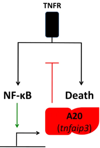
al., 1992; Opipari et al., 1990). The generation of A20 knockout mice confirmed its
fundamental role in terminating NF-κB signals. A20-/- mice present with
multi-organ spontaneous inflammation and cachexia and die prematurely by 3-4 weeks
of age. A20 deficient mouse fibroblasts (MEFs) and thymocytes are
hyperresponsive to TNFα, showing prolonged NF-κB activation and TNF-induced
apoptosis (Lee et al., 2000).
Figure 1.6 – Failure to terminate TNF-induced NF-κB and cell death responses in A20 deficient mice.
Analysis of the A20 sequence identified an N-terminal OTU (Ovarian
Tumor) cysteíne protease domain and seven C-terminal Zinc Fingers. Further
+.!7(
.!/23(
:;<(
/
!"#$%&'
0#
research demonstrated that A20 is a dual function ubiquitin-modifying enzyme: It
can remove K63 ubiquitin chains from RIP-1 (Receptor Interacting Protein 1),
which needs them to operate as an essential mediator of TNFR induced NF-κB
activity; Additionally, A20 can also use its zinc fingers to act as an E3 ligase and
add K48 ubiquitin chains to RIP-1, targeting it for degradation (Wertz et al., 2004).
The latter observations uncovered a novel mechanism by which signals can
be terminated through regulation of different ubiquitylation events. Nonetheless,
there is still a lot to be understood about the many potential functions of this
enzyme. Namely, it is remains unclear if there is ubiquitin type specificity for each
one of A20ʼs domains, whether that depends on each particular target, and finally,
whether additional targets exist but remain unidentified. Furthermore, very
recently, an additional mechanism for A20ʼs action has been proposed, with
evidence showing that A20 could antagonize TRAF E3 ligase activities by
impeding them from binding E2 ligases (Shembade et al., 2010).
In spite of A20 being a potent inhibitor of TNF induced signals, A20-/-TNF
-/-mice also displayed spontaneous multi-organ inflammation and early lethality as
A20-/- mice (Boone et al., 2004). This indicates signals other than TNF are
responsible for initiating the spontaneous inflammation in the absence of A20.
RAG (Recombination Activation Genes) deficiency (results in the absence of T an
B cells) also does not rescue the A20-/- mice phenotype, demonstrating that B and
T lymphocytes also are not responsible for initiating the generalized inflammation
in these animals. On the other hand, A20 was shown to be required to terminate
TLR and NOD (Nucleotide-binding oligomerization domain-containing) protein
signals, and promote deubiquitylation of TRAF6 and RIP2, respectively (Boone et
al., 2004; Hitotsumatsu et al., 2008).
More recently, some hints into the regulation of A20 itself have been
with A20 on signaling regulation have been described (Iha et al., 2008; Oshima et
al., 2009; Papoutsopoulou et al., 2006; Shembade et al., 2007; Shembade et al.,
2009; Wullaert et al., 2006). Secondly, post-translational modifications of A20
were also shown to have a role in signaling regulation. Downstream of TNF and
LPS, IKKβ phosphorylates A20, which increases its inhibitory potential by and
unknown mechanism (Hutti et al., 2007); In T an B cells, MALT1 can cleave A20
and disrupt its inhibitory activity (Coornaert et al., 2008).
A20 in autoimmunity and cancer
In the past 2 years there has been a striking number of reports of genetic
associations between TNFAIP3 polymorphisms and human autoimmune
diseases: Inflammatory Bowel Disease (IBD), Systemic Lupus Erythematosus
(SLE), Rheumatoid Arthritis (RA), Type I Diabetes (T1D), Coeliac Disease and
Atherosclerosis (Bates et al., 2009; Duan et al., 2009; Graham et al., 2008;
Kawasaki et al., 2010; Musone et al., 2008; Orozco et al., 2009; Plenge et al.,
2007; Prahalad et al., 2009; Thomson et al., 2007; Trynka et al., 2009).
Additionally, somatic mutations and deletions of A20 were found in various B cell
lymphomas at remarkably high frequencies (Compagno et al., 2009; Honma et al.,
2007; Honma et al., 2009; Kato et al., 2009; Malynn and Ma, 2009; Novak et al.,
2009; Schmitz et al., 2009).
In conclusion, A20 has emerged as a fundamental regulator of immune
signals. Its magnitude and versatility, suggest there is still a lot to be learned about
its further roles in different cell types and pathways, and the variations of
mechanism it uses to accomplish its tasks. Additionally, the recent associations
THESIS AIMS AND RATIONALE
In the first part of this thesis work, described in chapter 2, we aimed to
define which basal in vivo signals are responsible for initiating the systemic
inflammation in the absence of A20. While TNF signals were excluded from
accounting for triggering the multi-organ disease, it was found that A20 terminates
TLR responses. Given that TLRs are essential and proximal regulators early
during immune responses, and thus could trigger a widespread activation of the
immune system, we asked whether TLR signals had to be constantly regulated by
A20 in homeostatic conditions (i.e. without further external stimulation). Moreover,
given that there is permanent TLR stimulation in the gut, we hypothesized this
could be the site of initiation of such signals.
In chapter 3, our goal was to identify additional roles for A20 in the adaptive
immune system. Precisely, we asked whether A20 regulates B cell function.
Whereas in most cell types A20 expression is low before stimulation dependent
induction, in B and T cells the baseline expression of A20 is higher, suggesting it
could have a role in lymphocytes. Moreover, B cells are regulated by signals that
lead to activation of NF-κB and which share some of the same intermediate
molecules participating in A20 targeted pathways. Finally, the GWAS (Genome
Wide Association Studies) that linked A20 with B cell mediated autoimmune
disease (SLE) and the frequent somatic deletions and mutations of A20 in B cell
CHAPTER 2
Homeostatic MyD88-dependent signals cause lethal
inflammation in the absence of A20
Emre E. Turer, 1,* Rita M. Tavares, 1,2,* Erwan Mortier, 1 Osamu Hitotsumatsu
, 1 Rommel Advincula , 1 Bettina Lee, 1 Nataliya Shifrin , 1 Barbara A. Malynn ,
1
and Averil Ma 1
The Journal of Experimental Medicine, Vol. 205, No. 2, February 18, 2008, 451 -
464
1
Gastrointestinal Division, Department of Medicine, Biomedical Sciences Program,
University of California, San Francisco,
San Francisco, CA 94143
2
Ph.D. Programme in Biomedicine, Instituto Gulbenkian de Ciência, 2781-901
Oeiras, Portugal
*
These authors contributed equally to this work
SUMMARY
Toll-like receptors (TLRs) on host cells are chronically engaged by
microbial ligands during homeostatic conditions. These signals do not cause
inflammatory immune responses in unperturbed mice, even though they drive
innate and adaptive immune responses when combating microbial infections. A20
is a ubiquitin-modifying enzyme that restricts exogenous TLR-induced signals. We
show that MyD88-dependent TLR signals drive the spontaneous T cell and
myeloid cell activation, cachexia, and premature lethality seen in A20-deficient
mice. We have used broad spectrum antibiotics to demonstrate that these
constitutive TLR signals are driven by commensal intestinal flora. A20 restricts
TLR signals by restricting ubiquitylation of the E3 ligase tumor necrosis factor
receptor – associated factor 6. These results reveal both the severe
proinflammatory pathophysiology that can arise from homeostatic TLR signals as
well as the critical role of A20 in restricting these signals in vivo. In addition, A20
restricts MyD88-independent TLR signals by inhibiting Toll/ interleukin 1 receptor
domain – containing adaptor inducing interferon (IFN) β – dependent nuclear
factor KB signals but not IFN response factor 3 signaling. These findings provide
Abbreviations used: BMDM, bone marrow – derived macrophage; HSC,
hematopoietic stem cell; IRAK-M, IL-1R – associated kinase M; IRF, IFN response
factor; MCP, monocyte chemoattractant protein; mRNA, messenger RNA; PAMP,
pathogen-associated molecular pattern; poly (I:C), poly-inosine:cytosine; RIP,
receptor-interacting protein; R.U., relative units; SIGIRR, single Ig and TIR
domain; SOCS, suppressor of cytokine signaling; TIR, Toll/ IL-1 receptor; TLR,
Toll-like receptor; TRAF, TNF receptor – associated factor; TRIF, TIR domain –