Ana Sofia Martins Roda
BSc in BiochemistryNanoparticles for recognition and delivery in metastatic
colorectal cancer cells
Dissertation for the Master degree in Biochemistry for Health
Supervisor: Arturo Álvarez-Bautista, PhD, iBET
Co-supervisor: Catarina Duarte, PhD, iBET
Setembro, 2016
I
Ana Sofia Martins Roda
BSc in BiochemistryNanoparticles for recognition and delivery in metastatic
colorectal cancer cells
Dissertation for the Master degree in Biochemistry for Health
Supervisor: Arturo Álvarez-Bautista, PhD, iBET
Co-supervisor: Catarina Duarte, PhD, iBET
Instituto Tecnológico de Química e Biologia (ITQB NOVA)
III
[
Nanoparticles for recognition and delivery in metastatic colorectal cancer cells
]Copyright © [Ana Sofia Martins Roda], Instituto de Tecnologia Química e Biológica António Xavier; Faculdade de Ciências e Tecnologia, Universidade Nova de Lisboa.
O Instituto de Tecnologia Química e Biológica António Xavier e a Universidade Nova de Lisboa têm o direito, perpétuo e sem limites geográficos, de arquivar e publicar esta dissertação através de exemplares impressos reproduzidos em papel ou de forma digital, ou por qualquer outro meio conhecido ou que venha a ser inventado, e de a divulgar através de repositórios científicos e de admitir a sua cópia e distribuição com objetivos educacionais ou de investigação, não comerciais, desde que seja dado crédito ao autor e editor.
V
‘A vida está em constante mudança’ ‘A vida está em constante evolução’ Não saiba eu outra coisa… Mas quero saber mais, Quero evoluir mais! Quero viver a vida e que ela viva comigo Estou adsorvida à sua superfície e vou avançar, E absorver a mim tudo o que ela tiver para me dar!
VII AGRADECIMENTOS
Em primeiro lugar quero agradecer ao meu orientador, Dr. Arturo Álvarez Bautista pela oportunidade de integrar este plano de dissertação de mestrado e por todo o apoio e formação prestada no decorrer da tese, bem como a sua humildade e constante preocupação para comigo. Teve sem dúvida uma contribuição muito valiosa, tanto a nível profissional como pessoal. Obrigada.
À minha co-orientadora, Drª Catarina Duarte, quero agradecer pela sua importante contribuição na escolha desta dissertação, pelo conhecimento que partilhou e pela sua vocação para a ciência, inspiradora e contagiante, instruindo sempre no sentido da evolução. Um especial apreço pela sua humildade e pela sua coragem. Palavras para quê?. Seja sempre a pessoa fantástica que é.
A todos os membros que integram ou integraram o grupo dos Nutracêuticos e Libertação Controlada, agradeço pela receção e integração no grupo e pelo apoio prestado a todos os níveis. Um especial obrigado à Maria João, Luís Martins, Daniel Deodato, Agostinho e Liliana Rodrigues, que despenderam do seu tempo para me auxiliar em diversas ocasiões. Quero também agradecer à Drª Teresa Serra e Drª Ana Matias cuja postura e envolvimento no grupo contribuiu para o decorrer do projeto, nas melhores condições possíveis. E como não podia deixar de ser, às minhas companheiras e colegas de mestrado e do grupo, Joana Guerreiro, Carolina Pereira e Lucília Pereira. Em especial à minha colega e agora amiga, Anyse Pereira, que me acompanhou, lado a lado, em todos os momentos. Obrigada por teres entrado na minha vida e por contribuíres para o meu bem-estar e para minha evolução pessoal.
Ainda no contexto do mestrado, não posso deixar de agradecer a todos os grupos e pessoas que direta ou indiretamente, tornaram possível a concretização da parte experimental da tese. Em especial, às colaborações do grupo ‘Homogeneous catalysis’ (Drª Beatriz Royo, ITQB), à Drª Isabel Nogueira (IST), ao grupo do professor João Paulo Crespo (FCT/UNL) e por fim, ao ‘Colon Pathology Study Group’ (Cristina Albuquerque, IPO), envolvido em parceria no projeto e cuja contribuição foi crucial. Para além disso, também a professora Teresa Catarino e o professor Pedro Matias merecem um especial agradecimento, pelo seu profissionalismo enquanto coordenadores de mestrado, pelo seu interesse e constante preocupação por manter-nos sempre informados e pela vontade de acompanhar e contribuir para o nosso percurso.
O agradecimento mais importante dirige-se aos meus pais, que com muito esforço tornaram possível esta oportunidade. Muito obrigado. A toda a minha família, obrigada por contribuírem para a pessoa que sou, em especial aos meus tios, Luís Martins e Carla Firmo, que desde cedo me motivaram e me instigaram a evoluir e sempre se mostraram interessados e disponíveis. São sem dúvida, um modelo de inspiração.
A todos os meus amigos, obrigada por serem o meu pilar nesta jornada. Inês Brito, Paulo Oliveira, Khrystyna Kucheryava, não tenho palavras para descrever a vossa contribuição. Obrigada pela vossa amizade, pelo vosso apoio incondicional, adoro-vos. Diana Silva, obrigada pela tua energia, sempre positiva e contagiante.
Às minhas quatro e para sempre amigas, Ana Raquel Maia, Ana Sofia Narciso, Ana Paulino e Maria Inês Marreiros. Obrigada pela longa e fiel amizade.
Daniel Vilar Jorge, a ti um especialíssimo obrigado, pela tua amizade, pelo teu conhecimento, pela tua disponibilidade e pelo teu apoio incondicional.
Catarina Silva. És tu, somos nós! Não preciso de dizer mais nada. Adoro-te.
Rúben Santos, mereces um grande obrigado, pela tua amizade e por me incentivares e te disponibilizares a ajudar-me nesta fase tão importante.
IX Abstract
Colorectal cancer is a global health concern. The high incidence of colorectal metastasis, mainly in the liver, triggers an increase of the mortality rate and greatly reduces the effective cure chances. For this reason, the investigation in the area is now focused on efficient detection and elimination of metastasis. Nanotechnology has become a fundamental research field since it provides promising perspectives regarding specific, oriented and sustained delivery of loaded nanoparticles for nanoteragnostic approaches. The current project aims to develop a nanocarrier, strategically constructed for specific administration, recognition and therapy of colorectal liver metastasis. Concerning this goal, two different approaches were concurrently developed: a green-therapeutic technology and a novel nanoparticulate system, by nanoprecipitation and inverse microemulsion, respectively. The green approach focused on the encapsulation of a natural anticancer agent, phenetyl isothiocyanate (PEITC) in methoxy polyethylene glycol-co-poly ε-caprolactone (mPEG-co-PCL) nanoparticles. The development of this system did not advance significantly since more promising perspectives in a shorter period of time were obtained for the simultaneous developments of chitosan-collagenase nanosystems. Chitosan-chitosan-collagenase NPs were designed and developed for the first time due to their innovative properties regarding specificity in drug delivery and release. These nanosystems presented spherical shape and sizes in the range of 100 to 500 nm. The studies of pH and crosslinking influence in network swelling suggested higher swellings for more acidic pH and lower crosslinker contents. It was also postulated that the crosslinking degree influences differently the loading capacity and release efficiency of nanoparticles. As nanocarrier, chitosan-collagenase NPs demonstrated acceptable loads of the chemotherapeutic agent 5-fluorouracil for in vitro tests in HT29 cell lines but low release efficiencies.
Keywords: metastatic colorectal cancer; polymeric nanoparticles; drug delivery; mPEG-co-PCL; chitosan; collagenase
XI Resumo
O cancro coloretal é a terceira causa mundial de morte por cancro. A alta incidência de metástases, sobretudo no fígado, impulsiona a sua taxa de mortalidade e reduz substancialmente as hipóteses de cura. Por esse motivo, a investigação na área está focada na deteção e eliminação eficiente de metástases. A nanotecnologia tem demonstrado perspetivas promissoras no desenvolvimento de nanosistemas para libertação dirigida, controlada e continuada de agentes encapsulados para nanoteragnóstico. O presente trabalho tem como objetivo alargado o desenvolvimento estratégico de nanopartículas para administração, reconhecimento e terapia especializada do cancro coloretal metastático. Nesse intuito, foram desenvolvidos simultaneamente dois sistemas nanoparticulados. Uma abordagem ecológica focou a encapsulação de um agente anticancerígeno natural, PEITC, em mPEG-co-PCL, por nanoprecipitação. Até ao momento, o desenvolvimento deste nanosistema não avançou significativamente. Paralelamente, nanopartículas de quitosano-collagenase foram projetadas e desenvolvidas pela primeira vez por microemulsão inversa, devido às suas propriedades potenciais na otimização de terapêuticas. Estes nanosistemas revelaram morfologia esférica, com tamanhos na ordem dos 100 aos 500 nm. Os estudos da influência do pH e do grau de entrecruzamento no inchamento das redes nanoparticuladas sugeriram maiores inchamentos com o aumento de acidez e diminuição de entrecruzante. Postulou-se também que o aumento de entrecruzante confere à rede nanoparticulada maior capacidade de encapsulação e menor eficiência de libertação. A encapsulação de 5-fluorouracil demonstrou ser aceitável para testar em linhas celulares HT29, apesar da obtenção de baixas eficiências de libertação.
Palavras-chave: cancro colorectal metastático; nanopartículas poliméricas; libertação de fármacos; mPEG-co-PCL; quitosano; colagenase
XIII CONTENTS
1. OBJECTIVES AND FRAMEWORK ... 1
2. INTRODUCTION ... 3
2.1. Colorectal cancer epidemiology ... 3
2.1.2. Conventional therapies ... 3
2.2. Nanotechnology... 4
2.2.2. Nanotechnology scale ... 5
2.3. Anticancer reduced scale approved therapies ... 5
2.3.2. Nanotherapies for colorectal cancer ... 7
2.4. The role of polymeric nanoparticles in nanotheragnostic development ... 12
2.5. mPEG-co-PCL nanoparticles by nanoprecipiation... 12
2.6. Chitosan ... 13
2.6.1. Chitosan-based nanosystems for colorectal cancer ... 14
2.7. Collagenase application in nanotechnology ... 16
2.8. Chitosan-collagenase nanoparticles by inverse microemulsion ... 16
3. EXPERIMENTAL SECTION... 19
3.1. Materials ... 19
3.2. Synthesis and characterization of block copolymer mPEG-co-PCL ... 19
3.3. Synthesis of mPEG-co-PCL nanoparticles ... 19
3.4. Characterization of mPEG-co-PCL nanoparticles ... 20
3.5. Synthesis of chitosan and chitosan-collagenase nanoparticles ... 20
3.5.1. Isolation and drying of chitosan and chitosan-collagenase nanoparticles ... 21
3.5.2. Characterization of chitosan and chitosan-collagenase NPs ... 21
3.5.3. 5-Fluorouracil loading into chitosan and chitosan-collagenase nanoparticles ... 21
3.5.4. 5-Fu in vitro release ... 22
4. RESULTS AND DISCUSSION ... 23
4.1. Block copolymer mPEG-co-PCL ... 23
4.2. mPEG-co-PCL nanoparticles ... 25
4.3. Chitosan and chitosan-collagenase crosslinked nanoparticles ... 33
4.3.1. Load and release of 5- fluorouracil ... 38
5. CONCLUSIONS ... 43
6. FUTURE WORK ... 45
7. REFERENCES ... 47
XV FIGURE INDEX
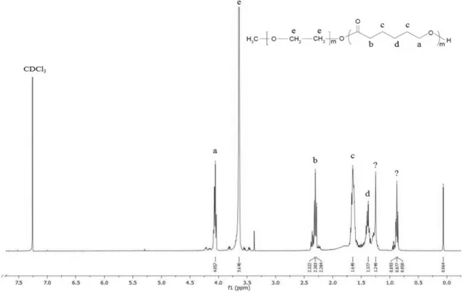
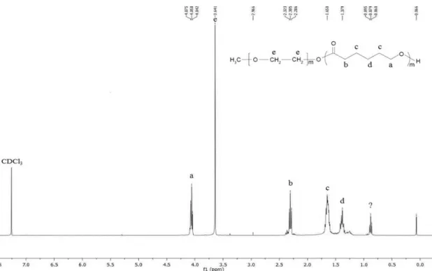
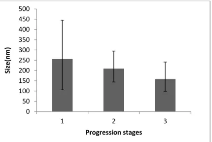
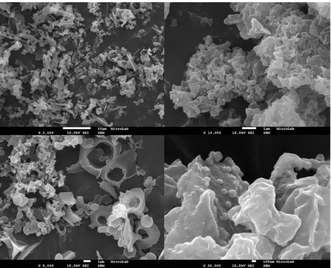
Figure 1.1 – Schematic representation of the thesis major goal and the designed approaches and its advantages for the purpose. ... 1 Figure 2.1 - Schematic representation of methoxy polyethylene glycol (mPEG) and ε-caprolactone (CL) copolymerization, to form copolymer mPEG-co-PCL, adapted from Xiong et al., 2015. ... 12 Figure 2.2 - Schematic representation of crosslink between chitosan (left) and genipin (right), from Lins et al., 2014. ... 17 Figure 4.1 – 1H-NMR spectrum obtained for chemical characterization of mPEG-co-PCL, obtained by a preliminary synthesis, similar to procedure described (data supported by literature published spectra - Figure 8.1, in appendix.) ... 23 Figure 4.2 - 1H-NMR spectrum obtained for chemical characterization of mPEG-co-CPL, synthesized by the described procedure (data supported by literature published spectra – Figure 8.1, in appendix.) ... 24 Figure 4.3 - Representation of size ranges obtained for the principal stages of optimization, ordered by progression (ascending from 1 to 3). ... 25 Figure 4.4 - Schematic representation of nanoparticles agglomeration, adapted from the online page of Product and Process Engineering - Delft University of Technology. ... 26 Figure 4.5 – FEG-SEM images obtained from samples obtained during optimization stages of mPEG-co-PCL nanoprecipitation. Left – Impurities (non spherical structures); Right – resin effect. (Scale bar: left top - 10 µm; left bottom - 1 µm; right top - 1 µm; right bottom - 100nm). . ... 26 Figure 4.6 – FEG-SEM images of mPEG-co-PCL nanoparticles, synthesized in the latest stage of optimization (Scale bar: left - 1 µm; right - 100nm). ... 28 Figure 4.7 - Graphical representation of the intensity of each size range (population), obtained for empty and loaded mPEG-co-PCL NPs, indicating its median intensity (columns) and the respective variations (error bars). The population 1corresponds to the smallest size range and population 3 identify the larger ones. ... 28 Figure 4.8 – Graphical representation of the population sizes obtained for empty and loaded mPEG-co-PCL NPs, indicating their median size (columns) and the respective variations (error bars). ... 29 Figure 4.9 – FEG-SEM images obtained for coumarin-6-loaded mPEG-co-PCL nanoparticles (Scale bar: 1 µm). ... 30 Figure 4.10 – Summary of the median sizes measured by DLS and its variations, before and after submitting a sample of mPEG-co-PCL loaded with PEITC to periods of agitation, dilution and subsequent rest. ... 31 Figure 4.11 – FEG-SEM image of structures that may correspond to nanoparticles obtained by nanoprecipitation of mPEG-co-PCL and PEITC (Scale bar: 100nm). ... 32
XVI
Figure 4.12 - FEG-SEM image of non-defined structures, obtained by nanoprecipitation of mPEG-co-PCL and PEITC (Scale bar: left top - 10 µm; left bottom - 10 µm; right top - 1 µm; right bottom – 10 µm). ... 33 Figure 4.13 – FEG-SEM images of nanoparticles dried by lyophilisation. Left – chitosan nanoparticles; right – chitosan-collagenase nanoparticles (Scale bar: 100nm). ... 34 Figure 4.14 - FEG-SEM images of nanoparticles dried using rotary evaporation. Left – chitosan nanoparticles; right – chitosan-collagenase nanoparticles (Scale bar: top - 100 nm; bottom - 10 µm). 35 Figure 4.15 - FEG-SEM images of nanoparticles dried using NADIR UP-10 membrane. Left – chitosan nanoparticles; right – chitosan-collagenase nanoparticles (Scale bar: top – 100 nm; bottom - 10 µm). ... 36 Figure 4.16 - FEG-SEM images of nanoparticles washed and dried using ethanol and diethylether. Left – chitosan nanoparticles; right – chitosan-collagenase nanoparticles (Scale bar: top - 100 nm; bottom - 1 µm). ... 37 Figure 4.17 –Graphical representation of 5-Fu loading outcomes for chitosan-collagenase nanoparticles synthesized with a genipin:polymer ratio of 1:10, 1:5, 2:5 and 10:1. ... 40 Figure 4.18 - Graphical representation of 5-Fu release outcomes from previously loaded chitosan-collagenase nanoparticles synthesized with a genipin:polymer ratio of 1:10, 1:5, 2:5 and 10:1. ... 41 Figure 4.19 - Graphical representation of 5-Fu release outcomes from chitosan-collagenase nanoparticles submitted to equal load pH (top) and equal release pH (bottom). ... 42 Figure 8.1 - 1
H-NMR spectrum obtained for mPEG-co-PCL and its corresponding chemical structure (represented by letters) by a) Xiong et al., 2015 and b) Baimark, 2009. ... 52 Figure 8.2 – a) Schematic representation of ε-caprolactone ring opening polymerization; b) NMR spectrum of poly(ε-caprolactone) and corresponding chemical groups. Adapted from Long et al., 2010. ... 52
XVII TABLE INDEX
Table 2.1 - List of approved and marketed nanoparticulate systems for cancer therapy (updated to August 2016). ... 7 Table 2.2 - List of nanoparticulate systems for cancer therapy in current clinical trials (updated to August 2016). ... 9 Table 4.1 - DLS results obtained for empty mPEG-co-PCL nanoparticles, in different stages of optimization, ordered by progression (ascending from 1 to 3). ... 25 Table 4.2 – Summarized information regarding size range and polydisperisty of mPEG-co-PCL nanoparticles, according to the results published by the cited authors. ... 27 Table 4.3 - DLS results obtained for the loaded mPEG-co-PCL nanoparticles, in the first stages of synthesis in comparison the optimized results obtained for empty nanoparticles. ... 29 Table 4.4 . DLS results obtained for the PEITC loaded mPEG-co-PCL nanoparticles, before and after submitting the sample to periods of agitation, dilution and subsequent rest. ... 31 Table 4.5 – Estimated parameters after 5-fluorouracil encapsulation in chitosan and chitosan-collagenase nanoparticles, at a pH of 5.5. ... 38 Table 4.6 - Estimated parameters obtained by 5-fluorouracil encapsulation at pH 5.5 and 3, in chitosan-collagenase nanoparticles, synthesized using genipin:polymer ratios of 1:10, 1:5 and 2:5. ... 39 Table 4.7 - Estimated parameters obtained by 5-fluorouracil release at pH 5.5 and 3, from the previously loaded chitosan-collagenase nanoparticles, synthesized using genipin:polymer ratios of 1:10, 1:5 and 2:5. ... 40
1
1. OBJECTIVES AND FRAMEWORK
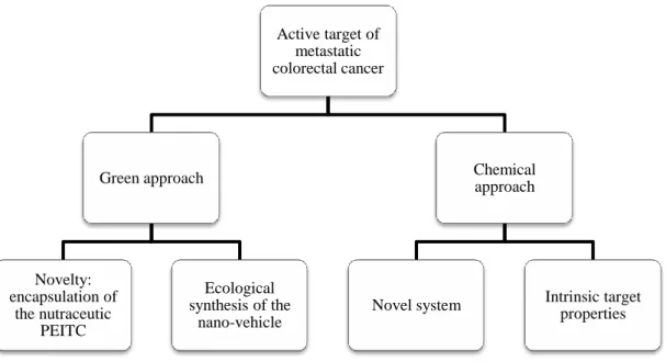
Figure 1.1 – Schematic representation of the thesis major goal and the designed approaches and its advantages for the purpose.
The work developed during this master thesis is part of a project with higher scope, evaluated, approved and financed by iNOVA4health, as a promising translational program for advanced precision medicine. With a three year initial timetable, it has the final goal of obtaining specific nanoparticulate therapeutic systems for differential recognition of the several forms of metastatic colorectal cancer.
The master thesis focused on liver metastatic colorectal cancer, the most common metastasized site. The predicted tasks included the synthesis, characterization and functionalization of two parallel systems, with polymeric composition but different designs. One pathway was directed to a green approach, by using a clean and sustainable methodology for the encapsulation of two, independent agents, the coumarin – 6 (C6), a fluorescent marker used for imaging and phenetyl isothiocyanate (PEITC), a natural therapeutic drug. PEITC was chosen mainly due to its nutraceutical nature, contributing to a chemopreventive/chemotherapeutic green approach. In turn, since C6 is an agent with published results regarding its incorporation in mPEG-co-PCL system, its encapsulation was reproduced for comparative purposes with literature. Besides, its fluorescent properties can be potentially used for monitoring of nanoparticles during in vitro delivery assays.
As further explained below, methoxy poly (ethylene glycol) nanocarriers were chosen mainly because of their ‘green’, fast and simple synthesis procedure, well described by several authors in the literature and considered reliable, reproducible and adequate for pharmaceutical application. The
Active target of metastatic colorectal cancer Green approach Novelty: encapsulation of the nutraceutic PEITC Ecological synthesis of the nano-vehicle Chemical approach
Novel system Intrinsic target properties
2
reproduction of the published results would allow us to obtain monodisperse ecologic nanosystems whose composition would be immediately ready to perform functionalization studies without additional surface modifications. Besides, the quick synthesis would enable to initiate the functionalization assays in early stages, allowing the test and optimization of several biomarkers and its specificity for colorectal metastatic cancer.
Concurrently, a potentially innovative chemical approach to obtain a novel system was designed based on the nano-combined advantages of chitosan and collagenase compounds. It is expected to form a promising drug vehicle mainly due to its intrinsic biomarker properties, deeper penetration ability and pH response, which translates into selective accumulation in metastatic cancer cells, allows to reach the tumour inner core, and the drug delivery occurs preferentially in acidic tumoral pH. Moreover, since it was expected to functionalize nanoparticles surface to target metastatic colorectal cancer, the load capacity and release efficiency of this system was planned to be tested using a commonly chemotherapeutic agent applied in the treatment of this type of cancer, 5-fluorouracil, for further in vitro assays in colorectal cancer cell lines.
Following this framework, the thesis structure will be divided into five main sections, Introduction, Experimental Section, Results and Discussion, Conclusions and Future work. The introductory section will first focus on the state of the art of colorectal cancer and nanotechnology, which are the bases of the project, then funnelling to the thematic contextualization of the principal materials involved in the project. The experimental section will contain a full description of the materials and procedures used. The section of results and discussion contains the outcomes of the experimental work developed as well and its evaluation, founded on the basis of literature available knowledge and expected results by comparative analysis. The Conclusions section will summarize the main outcomes of the experimental work and the future predicted tasks and further perspectives of the project will be summarized in the section of Future work.
PROJECT MAIN GOALS:
The main goal of this project was the synthesis and characterization of polymeric nanoparticles (PNPs) with nanometric size for parental administration of therapeutic drugs and further functionalization with specific markers for hepatic metastatic colorectal cancer.
3 2. INTRODUCTION
2.1. Colorectal cancer epidemiology
Colorectal cancer is a global health concern, with an incidence rate rising over the years. In 2012, presented an incidence of 1,36 million new cases, being the third cause of cancer death worldwide (Cancer Research UK, 2012). Regarding Europe, colorectal cancer represents the second position in terms of cancer occurrence and mortality, with register of about 447,000 cases and 215,000 deaths. In Portugal, it is also the second most common and lethal cancer, with an estimated prevalence of about 7130 cases and 3800 deaths (Ferlay et al., 2013). The late diagnosis, which often occurs in advanced stages of the disease, mainly metastatic, severely contributes to the current mortality rates. The hepatic colorectal cancer metastasis is the most frequent (Masi et al., 2011).
2.1.2. Conventional therapies
… colorectal metastasis as a ‘stone’ on the path to healing.
The available therapies are used for cancer treatment in general, presenting low specificity and efficiency. The most common therapy applied to treat colorectal cancer is surgery which comprises surgical removal of the tumour and part of the contiguous healthy tissue. However, it is not efficient in advanced stages of the disease and not applicable in cases where the removal can compromise the organ function. There are other therapies aiming at the reduction or eradication of the tumour and/or metastasis but all of the available treatments have the disadvantage of affecting healthy cells.
Chemotherapy consists on the intravenous administration of anticancer agents, having a systemic effect, which causes several collateral effects. Besides, the cells can become resistant to the drugs, compromising the efficiency of the treatment.
Radiotherapy is mainly applied as adjuvant and despite being based on local administration of high energy radiation, has similar drawbacks regarding secondary effects and resistance.
On the other hand, ablation is based on local destruction by injection of a needle/probe into the tumour as source for direct treatment, but it is only efficient for small tumours or metastasis. Embolization can be applied for larger tumours/metastasis in the liver and consists on blocking the branches of the hepatic artery, since it is the main blood source of liver cancer cells. Despite the fact that normal cells remain supplied by the portal vein, the blood supply of liver tissue is reduced, which can affect its normal function.
The invasive effect of most of these treatments translates into a debilitated health for life. However, the cure of a primary tumour is achievable, normally by resection (Colorectal Cancer, American Cancer Society). The incurability of the disease is mainly related to the difficulty of
4
metastasis eradication. Normally, it is possible to control its progression for a certain period, but considering the present and reachable developments, prolonging patient’s life is for now, the only achievable goal. Scientific efforts are focused on countering the inability of curing this disease, aiming to achieve efficient, long-term survival and non-pejorative treatments (Masi et al., 2011; Raval et al., 2014; van Hazel et al., 2016).
2.2. Nanotechnology
‘There's plenty of room at the bottom’ (Richard Feynman, 1959)
In the past decades, nanotechnology has arisen as a potential alternative to overcome the issues faced by the conventional medicine, regarding both diagnosis and therapy. The combination of nanoscale properties and cancer characteristics, allows by itself a preferential accumulation of nanosystems in the tumour environment (passive targeting), mainly due to the Enhanced Permeability and Retention (EPR) effect. The enhanced permeability in comparison with normal capillary systems occurs due to the leaky and defective vasculature of the tumours as a result of the extra and accelerated vascularisation in response to the nutritional needs required for the proliferative ability of cancer tissues. This phenomenon translates into gaps in the surrounding vessels, leading to extravasation of nanoparticles into cancer cells interstitium, whose retention is ensured by the characteristic absence of lymphatic drainage (Sinha et al., 2006).
In addition to the intrinsic EPR effect, there are other passive targeting strategies that can be applied to further specify the nanoparticles delivery, either by localized administration or by manipulating the nano formulation according to intrinsic characteristics of the delivery pathway. Regarding colorectal passive targeting, the main influences relate to gastrointestinal physiology, including pH, temperature, ionic strength, enzymes, mucoadherence and tumour microenvironment (Patel Parul, Satwara Rohan, & Pandya, 2012). Besides, there are active targeting approaches, based on delivery driven by functionalization with biomarkers, which restricts the distribution according to the specificity. The advanced state of investigation and exponential growth in molecular and genetic biology allows the identification of potential sensitive biomarkers and increasingly confined probes. This knowledge is very useful regarding the development of highly specific systems in terms of differential recognition, conferring strong specificity and efficiency to diagnosis and therapy approaches.
5 2.2.2. Nanotechnology scale
The size range defined for nanoparticles is commonly 1 to 100 nm, according to the ISO (International Organization for Standardization) and ASTM International (American Society for Testing and Materials) standards. However, it is not possible to define unequivocal size limits since the physicochemical properties vary with the materials and surrounding conditions as well as its size dependence. Whereas the characteristics are acceptable under the predicted safety and risk parameters, and as long as the size is adequate and favourable for the application, the nano size range can be adjusted. Currently, according to the SCENIHR (Scientific Committee on Emerging and Newly Identified Health Risks), the standards remain ambiguous, being established differently, according to the area, material and application.
Regarding formulations for health purposes, it is important to ensure a sufficient residence time in the system to enable effective action. In this sense, the lower size limit for nanoparticles should be higher than 6 nm to prevent rapid renal clearance and above 15 nm to allow accumulation in liver and spleen, if required (Choi et al., 2007).
Another application with limited size required is internalization of nanoparticles in cells. Despite depending on many other factors as charge, shape and composition, the nanoparticles size greatly influences the occurrence and effectiveness of the cell uptake. For most cases, cellular internalization is described to occur up to 100 nm, with a maximum uptake varying from 50 to 100 nm (Shang, Nienhaus, & Nienhaus, 2014). However, there are some exceptions, as in the case of polymeric nanoparticles, for which there are published results that refer cell internalization of larger nanoparticles (until 500 nm) (Rejman et al., 2004; Shang, Nienhaus, & Nienhaus, 2014).
2.3. Anticancer reduced scale approved therapies
…if the wings of a butterfly can cause a hurricane, imagine what nanoscale can achieve. In terms of current or ‘in test’ applications, the main approach is to use nanotechnology has bypass vehicle for the available or innovative therapies, using the nanosystems properties has an advantage. The most common application is the use of these systems for drug delivery, by active or passive targeting. The first approved nano-formulation by FDA (Food and Drug Administration) for this purpose, was Doxil, in 1995, a liposomal vehicle with a size range of 80 to 90 nm for the delivery of doxorubicin to treat metastatic ovarian cancer and AIDS (Acquired Immune Deficiency Syndrome)-related Kaposi’s sarcoma. All the approved and marketed anticancer nanoformulations to date are summarized in Table 2.1, and the current clinical trials for the same purpose are shown in Table 2.2. Most of the developments were based on lipossomal formulations as the optisomal technology, a 100 nm sphingomyelin/cholesterol liposome vehicle, with is first FDA approval in 2012 (Marqibo) for
6
vincristine delivery against Philadelphia chromosome-negative acute lymphoblastic leukemia. Two parallel systems using this technology, Alocrest and Brakiva, are currently in phase I clinical trials, for chemotherapeutic applications through vinorelbine and topotecan agents, respectively. Alocrest is being tested for breast and non-small cell lung cancer and Brakiva in non-small cell lung cancer, myelodysplastic Syndromes, ovarian cancer and acute myeloid leukemia.
The most recent anticancer FDA approved nanoformulation (2015) wasOnivyde, also known as MM-398 or PEP02. It has been revised and marketed in 2015 from its first U.S approval in 1996. It is indicated for metastatic pancreatic cancer therapy, in combination with fluorouracil and leucovorin. Besides, is also involved in phase II clinical trials for colorectal cancer, gastric cancer and glioma and in phase I clinical trial for solid tumours.
In addition to the targeting ability, the systems composition can be modulated to be sensitive or responsive to a certain detection or therapy application, by external stimuli. In these cases, besides the vehicle ability, they can be involved in the diagnostic or therapeutic procedure. A therapy example is the SIRT (Selective Internal Radiation Therapy), which uses modulated systems with metallic specific characteristics to direct the treatment into an unique area, where those systems are preferentially accumulated. An approved SIRT approach is the administration via hepatic artery of microbeads coated with isotope yttrium-90 (Y-90) for pancreatic and hepatic liver cancer therapy, commercially available as TheraSpheres (20–30 μm spheres from BTG International Canada Inc) and SIR-spheres with 20-60 μm, prepared by Sirtex Medical Inc.
The most recent commercialized therapy with responsive characteristics is NanoTherm, with regulatory approval by European Union since 2010, recently (2016) marketed in Germany to treat brain tumours by thermal ablation. These 15 nm aminosilane-coated superparamagnetic iron oxide nanoparticles are in active expansion, with near perspectives of commercialization in European Union. Additionally, the treatment has been adapted to prostate cancer and submitted to FDA as an Investigational Device Exemption. The modulated Nanotherm devices for treating both prostate and brain cancer are now undergoing preclinical studies in the USA.
A nanosystem can have both active and passive contributions in a treatment, when combining the encapsulating ability with sensitive therapeutic properties to external stimuli, being a therapy vehicle and part of the treatment itself. A good example is ThermoDOX, a lysolipid thermally sensitive liposome encapsulating doxorubicin, which combined with thermal therapies, enhances its efficiency, both thermic and chemotherapeutic by localizing the heat (tumoral deeper penetration) and enhancing the drug liberation (improved release by lipossomal heating)
In terms of diagnosis approaches, the only nanoparticulate system with FDA approval and clinical application (2001, Europe; 2002, Japan) was Resovist (Ferucarbotran), superparamagnetic iron oxide nanoparticles, with 120 to 180 nm, administrated as contrast/imaging agents for magnetic
7
resonance imaging (MRI). However, the product has been discontinued in Europe by the Bayer Pharma AG distributer, being marketed only by I’rom Pharmaceutical Co Ltd, Japan (Baetke, Lammers, & Kiessling, 2015; Haegele et al., 2014).
2.3.2. Nanotherapies for colorectal cancer …personalized therapies are the future.
Concerning the major therapeutic target of this thesis, hepatic metastatic colorectal cancer, there is a FDA-approved nanotherapy regarding the adjuvant application of both Y-90 SIRT (Selective Internal Radiation Therapy using yttrium-90 - SIR-spheres) and chemotherapy (Raval et al., 2014) and published and ongoing phase trials of variants of the same goal (van Hazel et al., 2016). Additionally, there are nanoformulations in clinical phase trials. The ThermoDOX, a heat-activated liposome technology, is currently in phase II clinical trials for combined administration with HIFU ( High-Intensity Focused Ultrasound) in liver metastasis (and breast cancer) (Celsion Corporation). Other liposome-based formulation, containing a cisplatin analogue, is Aroplatin, in phase II development for intravenous chemotherapy of metastatic colorectal cancer. In turn, NK012 is a nanopolymeric micelle of PEG-polyglutamate copolymer incorporating SN-38, the active metabolite of irinotecan, currently in phase II studies for several cancers, including colorectal cancer (Ganji et al., 2015; Jin, Jin, & Hong, 2014; Raval et al., 2014).
Considering the epidemiology of metastatic colorectal cancer, much effort is concentrated on finding alternative therapies to reduce mortality. In terms of nanotherapies regarding this application, there are many other formulations in preclinical trials or in current development but not yet ready for clinical evaluation.
Table 2.1 - List of approved and marketed nanoparticulate systems for cancer therapy (updated to August 2016). Nanoparticulate system Commercial name Drug/Active ingredient Size range (nm)
Application First year of approval Pegylated liposomal doxorubicin Doxil (Johnson & Johnson, USA) Caelyx (Janssen-Cilag, Europe) Evacet (Liposome company INC.,) Lipodox (Sun Pharma) Doxorubicin (Adriamycin) 80-90 Metastatic ovarian cancer HIV-related Kaposi sarcoma FDA, 1995 Liposomal
Daunorubicin Daunoxome Daunorubicin ≈ 45
HIV-related Kaposi
sarcoma FDA, 1996 Cytarabine
liposome DepoCyt Cytarabine
3000-30000 Lymphomatous meningitis FDA, 1999/2007* (continued)
8 Table 2.1 (continued) Nanoparticulate system Commercial name Drug/Active ingredient Size range/ nm
Application First year of approval Non-pegylated liposomal doxorubicin citrate Myocet Doxorubicin hydrochloride ≈180 Metastatic breast cancer, plus cyclophosphamide. FDA, 2001 (Europe & Canada) Microspheres Radionuclide/ yttrium-90 glass microspheres TheraSpheres Yttrium-90 20000- 30000 Pancreatic and
hepatic liver cancer FDA, 1999 SIR-Spheres Yttrium-90 20000- 60000 Liver metastatic colorectal cancer plus floxuridine FDA, 2002 Tripartite viral particle Rexin-G Vector dnG1/C-REX ≈180
Pancreatic cancer FDA orphan status, 2003 Chemotherapy-resistant solid malignancies Accelerated FDA Philippines approval, 2007 Soft tissue sarcoma
and osteosarcoma
FDA orphan status, 2008
Albumin-bound
Paclitaxel Abraxane Paclitaxel ≈ 130
Metastatic breast
cancer FDA, 2005 Advanced NSCLC FDA, 2012
Late-stage
pancreatic cancer FDA, 2013 PEG-asparaginase
(Pegaspargase) Oncaspar L-asparaginase 50-200 ALL
Initial US approval:1994 FDA (revised), 2006 Micellar diblock copolymeric paclitaxel Genexol-PM (Samyang, Korean) Cynviloq (Sorrento Therapeutics, Inc. and IGDRASOL, Europe) Paclitaxel 20-50 Metastatic breast cancer, NSCLC and ovarian cancer Approved and marketed in Korean (2007) Commercialized in Europe Liposomal mifamurtide muramyl tripeptide phosphatidyl ethanolamine Mepact Mifamurtide 1000-5000 Osteosarcoma Orphan medicinal product, 2004 Marketing authorization by EMA and European Commission (EU), 2009 Aminosilane-coated superparamagnetic iron oxide nanoparticles
NanoTherm - 15 Thermal ablation
in brain tumours Regulatory approval from EU, 2010 Commerciallized in Germany, 2016 Liposomal vincristine sulphate/ Optisomal Vincristine Marqibo (Onco TCS) Vincristine ~100 Philadelphia chromosome-negative ALL FDA, 2012
9 Table 2.1 (continued) Nanoparticulate system Commercial name Drug/Active ingredient Size range (nm)
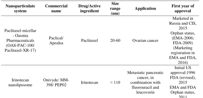
Application First year of approval Paclitaxel micellar - Oasmia Pharmaceuticals (OAS-PAC-100/ Paclitaxel-XR-17) Paclical/
Apealea Paclitaxel 20-60 Ovarian cancer
Marketed in Russia and CIS,
2015 Orphan status, (EMA-2006; FDA-2009) (Marketing registration in EMA and FDA,
2016) Irinotecan nanoliposome Onivyde/ MM-398/ PEP02 Irinotecan ≈ 110 Metastatic pancreatic cancer, in combination with fluorouracil and leucovorin Initial US approval:1996 FDA (revised), 2015 EMA and FDA
Orphan status, 2011 Note:*Year of accelerated approval and full approval, respectively.
Abbreviations/Acronyms: FDA, Food and Drug Administration; EMA, European Medicines Agency; HIV, human immunodeficiency virus; PEG, polyethylene glycol; NSCLC, non-small cell lung cancer; ALL, acute lymphoblastic leukaemia; TNF, tumour necrosis factor; USA/US, United States of America; EU, European Union; CIS, Commonwealth of Independent States
References: (Baetke et al., 2015; Haegele et al., 2014; Hafner et al., 2014; Hang, Cooper, & Ziora, 2016; Jin et al., 2014; Pillai, 2014; Raju et al., 2015; Raval et al., 2014; Sanna, Pala, & Sechi, 2014; van Hazel et al., 2016; Ventola, 2012; Weissig, Pettinger, & Murdock, 2014) and the corresponding and supplementing sources (FDA, EMA and pharmaceutical companies’ websites)
Table 2.2 - List of nanoparticulate systems for cancer therapy in current clinical trials (updated to August 2016). Nanoparticulate system Drug/Active
ingredient Potential application Phase Trial
ThermoDOX Doxorubicin
(Adriamycin)
Primary Liver Cancer (mutual radiofrequency
ablation)
Clinical Phase III (FDA Orphan Drug
Status, 2009; Fast Track designation) RCW breast cancer (plus
local hyperthermia) Clinical Phase II Liver metastasis and
breast cancer (plus HIFU)
Clinical Phase II
Paclitaxel Polyglumex/
Opaxio/ Xyotex/CT 2103 Paclitaxel
Ovarian cancer Clinical Phase III Advanced NSCLC
Clinical Phase III (Fast Track designation, FDA) Malignant brain cancer
(mutual Temozolomide and Radiotherapy)
Clinical Phase II (FDA Orphan Drug
Status, 2012) Advanced esophageal
10 Table 2.2(continued)
Nanoparticulate system Drug/Active
ingredient Potential application Phase Trial
Lipoplatin (Lipossomal cisplatin, Nanoplatin for
NSCLC) Cisplatin
NSCLC adenocarcionomas
Clinical Phase III
Pancreatic cancer
Clinical Phase II/III (EMA Orphan Drug
Status, 2007) Breast cancer; Gastric
cancer Clinical Phase II Malignant pleural
effusion Clinical Phase I
Micelplatin/Nanoplatin/NC-6004 (Cisplatin polymeric micellar nanoparticles)
Cisplatin
Pancreatic cancer Clinical Phase III Bile duct cancer;
Bladder cancer; NSCLC Clinical Phase II Head and neck cancer Clinical Phase I/II
CPX351/ Vyxeos (Lipossomal cytarabine:daunorubicin) Cytarabine: Daunorubicin (5:1) AML
Clinical Phase III (Orphan Drug Status –
FDA, 2008; EMA-2012) Breakthrough Therapy
and Fast Track status, FDA) NK105 (Paclitaxel micelle) Paclitaxel
Breast cancer, Japan Clinical Phase III Gastric cancer, Japan Clinical Phase II Solid tumours, Japan Clinical Phase I SP1049C (Micellar
doxorubicin – pluronic F127:L61 (1:8)
Doxorubicin Gastric cancer; Oesophageal cancer
Clinical Phase II/III (FDA orphan drug
status - 2007)
EndoTAG-I/lipopack Paclitaxel
Triple negative breast cancer and pancreactic
cancer (mutual with gemcitabine)
Clinical Phase II (FDA Orphan drug
status, 2006) Atragen (Liposomal all trans-
retinoic acid) Tretinoin APM Clinical Phase II
Aurimmune/CYT-6091 (colloidal gold-bound tumor
necrosis factor)
TNF NSCLC Clinical Phase II
BIND-014 Docetaxel
Squamous histology NSCLC and cervical, head and neck cancers
Clinical Phase II MBP-426 (Lipossomal oxaliplatin) Oxaliplatin Metastatic gastric, gastro-esophageal junction or esophageal adenocarcinoma Clinical Phase II MM-302 (HER2- targeted pegylated liposomal doxorubicin)
Doxorubicin Metastatic breast cancer Clinical Phase II Aroplatin (NDDP liposome) NDDP, cisplatin analog Metastatic CRC Clinical Phase II
CRLX101 (Camptothecin- cyclodextrin-polyethylene
glycol copolymer)
Camptothecin
Relapsed ovarian cancer (plus placitaxel or
bevacizumab)
Clinical Phase I/II (FDA Orphan drug
status, 2015) Metastatic renal cell
carcinoma (combined with bevacizumab)
Clinical Phase II (FDA Fast Track
11 Table 2.2(continued)
Nanoparticulate system Drug/Active
ingredient Potential application Phase Trial
LEP-ETU/NeoLipid
(Paclitaxel liposome) Paclitaxel
Metastatic breast cancer Clinical Phase II Ovarian cancer
Clinical Phase I (FDA Orphan Drug
Status, 2015) NK012/SN-38 nanopolymeric micelle Irinotecan SN-38 (Irinotecan ative metabolite) Breast cancer; CRC; Multiple myeloma; Small cell lung cancer
Clinical Phase II Solid tumours Clinical Phase I CRLX301 Docetaxel Advanced solid tumors Clinical Phase II SGT53 (Transferrin
receptor-Liposome-p53 Complex) p53-gene
Glioblastoma; Pancreatic
cancer Clinical Phase II Solid tumours Clinical Phase I
Liposomal annamycin Annamycin ALL Clinical Phase I/II
ProLindac(DACH-Pt polymer) DACH-Pt Ovarian Cancer Clinical Phase I/II Atu027 (Liposomal RNA
interference) siRNA
Pancreatic cancer Clinical Phase I/II Solid tumours Clinical Phase I Alocrest (Optisomal
Vinorelbine tartrate) Vinorelbine
Breast cancer and
NSCLC Clinical Phase I Brakiva (Optissomal topotecan
hydrochloride) Topotecan
NSCLC, Myelodysplastic Syndromes, Ovarian
Cancer and AML
Clinical Phase I
CALAA-01 (cyclodextrin
polymer-based nanoparticles) siRNA
Relapsed or refractory
cancer Clinical Phase I Docetaxel- polymeric
nanoparticle Taxane docetaxel Advanced Solid Tumors Clinical Phase I NK911 (Micellar
doxorubicin) Doxorubicin Solid Tumors
Clinical Phase I (Japan) NC-4016 (1,2- DACH-Pt-
polymeric micelles) DACH-Pt Solid Tumors Clinical Phase I S-CKD602 (Pegylated
liposomal CKD-602) Belotecan (CKD 602) Refractory solid tumors Clinical Phase I SGT 94 (Transferrin receptor -
Lipossomal RB94 plasmid) RB94 plasmid DNA Solid Tumors Clinical Phase I Lipovaxin MM
(Multi-component liposomal nanoparticle-based)
Melanoma antigens and
IFNγ Malignant melanoma
Clinical Phase I (Australia) C-VISA-BikDD liposome
(DOTAP:cholesterol)
Plasmid C-VISA
BiKDD Pancreatic cancer Clinical Phase I C225-ILS-DOX (Doxorubicin
anti-EGFR immunoliposomes) Doxorubicin Solid tumours Clinical Phase I Abbreviations/Acronyms: FDA, Food and Drug Administration; EMA, European Medicines Agency; RCW, Recurrent chest wall; HIFU, high intensity focused ultrasound; HER, Receptor tyrosine-protein kinase erbB-2; NDDP, Cis-bisneodecanoato-trans-R,R-1,2-diaminocyclohexane platinum II; DACH-Pt, diaminocyclohexane platinum; NSCLC, non-small cell lung cancer; CRC, colorectal cancer; ALM, acute myeloid leukemia; ALL, acute lymphocytic leukemia; APM, Acute promyelocytic leukemia; IFNγ, interferon gamma
References: Hafner et al., 2014; Hang et al., 2016; Jin et al., 2014; Pillai, 2014; Raju et al., 2015; Sanna et al., 2014; Ventola, 2012 and the corresponding and supplementing sources (FDA, EMA and pharmaceutical companies’ websites)
12
2.4. The role of polymeric nanoparticles in nanotheragnostic development …size and composition as favourable contribution.
Regarding biological applications, it is important to manipulate nanotechnology focusing on the primary needs of biosystems: biocompatibility, bioavailability and biodegradability. The combination of these properties allows acceptance by the biological system, delivery and effective action, active elimination of the systems and aims to prevent toxicity and immunogenic responses. Towards this purpose, polymers are a potential option, being widely applied as biomaterials in engineering, pharmacological and clinical developments (Marin et al., 2013). Polymeric chains consist on repeated simple and small units, whose arrangement and composition defines its physicochemical properties. This versatility allow to design and modulate systems to specific and diverse applications. The final properties required for a certain purpose, type of action and administration pathway, are possible to be optimized according to the type of formulation and by combination with other compounds.
Despite the interest and potential of polymer-based nanoparticles, currently there are no approved therapies based on these systems for clinical application. However, there is a great focus on its development, as its shown by the ongoing clinical trials (Table 2.2).
2.5. mPEG-co-PCL nanoparticles by nanoprecipiation …simple is better.
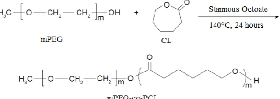
Methoxy polyethylene glycol-co-poly ε-caprolactone (mPEG-co-PCL) is an amphiphilic diblock copolymer resulting from ring-opening polymerization synthesis (Figure 2.1). In the field of nanotechnology, is described as a biocompatible and biodegradable drug delivery system with advantageous properties regarding sustained and controlled release.
Figure 2.1 - Schematic representation of methoxy polyethylene glycol (mPEG) and ε-caprolactone (CL) copolymerization, to form copolymer mPEG-co-PCL, adapted from Xiong et al., 2015.
The mPEG-co-PCL nanoparticles present spherical shape, small sizes (from 100 to 140 nm) and uniform monodispersity, which contributes to a prolonged blood circulation time, enhances the
13
accumulation in tumours and endocytic uptake and prevents rapid elimination by the reticu-loendothelial system (RES) (Baimark & Srisuwan, 2012; Baimark, 2009; Danafar & Schumacher, 2016; Xiong et al., 2015). This nanosystems design is based on ‘synergetic’ combination of both poly ε-caprolactone (PCL) and methoxy polyethylene glycol (mPEG) in order to integrate the favourable properties of the two polymers in a unique system, bypassing their individual disadvantages. PCL is commonly designed as drug carrier due to its biocompatibility, non-cytotoxicity, high solubility, low glass transition temperature (Tg) and high drug permeability, conferred by its hydrophobicity. However this hydrophobic character and its semi-crystalline structure enhance its susceptibility to RES clearance and confer low degradation rates, respectively. The copolymerization with mPEG allows to achieve a positive equilibrium in terms of biodegradability, RES clearance reduction and higher blood circulation periods, because of its hydrophilicity and flexibility (Danafar & Schumacher, 2016; Xiong et al., 2015). Besides, mPEG allows to functionalize nanoparticles surface without additional surface modifications and prevents nanoparticles agglomeration (Baimark & Srisuwan, 2012), avoiding the use of surfactants which enable to maintain the ‘green’ character of the synthesis procedure – nanoprecipitation.
Nanoprecipitation, also called solvent displacement method, is a one step procedure with easy and rapid reproduction, based on the polymers relative solubility in two miscible solvents. The standard procedure normally takes advantage of polymer solvation in a non-toxic organic solvent, where the drug and surfactants (optional) are also soluble, forming a homogenous solution. When dropped into an aqueous solution, in which the polymer is less soluble (non-solvent), the fast diffusion of the solvent causes polymer precipitation. This phenomenon is responsible for nanoparticles formation with simultaneous drug entrapment (Minost et al., 2012). Since it is a mild process, requiring low energy (Xiong et al., 2015), it can be considered a green approach, when applied without surfactant.
2.6. Chitosan
…discovered 200 years ago (Braconnot).
Chitosan is a natural cationic polysaccharide that results from partial chitin deacetylation originating random linear combinations of β-1,4-linked glucosamine and N-acetyl-D-glucosamine units. Its intrinsic properties are greatly influenced by the molecular weight, varying from 10 to 1,000 kDa, and deacetylation degree, normally in the range of 70-95% (Hamman, 2010). Given its high availability, low production cost and valuable properties regarding biocompatibility, biodegradability and low metabolic and immunogenic toxicity, it is repeatedly described in the literature as suitable delivery system in several different fields and applications. It is considered very promising regarding cancer because of its mucoadhesivity, tending to selective accumulate in mucous, preferentially in
14
anionic cancer cell surface due to its cationic nature. Other published characteristics are related with tumour growth suppression, immune system adjuvant and anti-inflammatory activity, which confer to this compound an anti-tumoral contribution (Pujana et al., 2013; Aruna et al., 2013; Park et al., 2010; Pujana et al., 2012). Besides, there are many benefits regarding other biomedical purposes – antifungal, antioxidant, anticholesterolemic, antimicrobial (Je & Kim, 2012)
Two major drawbacks in chitosan bio applications are its low solubility at physiologic pH (≈7.4) and its fast dissolution in the stomach (Park et al., 2010; Pujana et al., 2012), both explained by chitosan pKa (≈ 6.3-7) (Pujana et al., 2012). At pH above pKa, the non-protonated amino groups form strong hydrogen interactions within the chain, limiting external interactions and its solubility state. At acidic pH (below pKa), the weakening of inter-chain interactions due to amino protonation leads to chitosan dissolution. This behaviour can be controlled by using derivatives or combined systems. Moreover, this chitosan pH sensibility can be an advantage in terms of loading and strategic delivery,, for instance, to prevent drug release at physiologic pH, promoting its preferential release in tumour acidic environment or lisossomes or endossomes, in the case of internalization (Arteche Pujana et al., 2014) .
2.6.1. Chitosan-based nanosystems for colorectal cancer
The use of chitosan-based nanocarriers for pharmaceutical purposes leads to bioavailability enhancement and greater drug absorption and retention time (Aruna et al., 2013; Cerchiara et al., 2015). Concerning cancer delivery, there are a lot of published applications to generic tumour cells (Lee et al., 2014; Mehrotra, Nagarwal, & Pandit, 2011; K. Park et al., 2007; Yoon et al., 2014) and specific cancers as lung (Maya et al., 2014), liver (Guan et al., 2012; Qi, Xu, & Chen, 2007; Zhu et al., 2013), breast (Deng et al., 2014), brain (Veiseh et al., 2010), colon (Feng et al., 2015) and others. Several studies in this field have been done for colorectal cancer tissues aiming at diagnosis and therapeutics. In 2008, Yang, Chen, & Shieh reportedin vivo promising results for a chitosan-folic acid conjugated system, carrying a contrast dye (indigo carmine) for endoscopic detection of colorectal cancer cells. Similar approaches referred chitosan nanoparticles and chitosan-folic acid nano-conjugates as adequate carriers for oral administration of 5-aminolaevulinic acid (5-ALA), as fluorescent compound for colorectal cancer diagnosis. Those systems were developed with a perspective of further improvement, in order to reduce chitosan dissolution in the stomach (S. J. Yang et al., 2010; S.-J. Yang et al., 2009). Venkatesan et al presented, in 2011, promising outcomes from mouse human xenograph models for the use of a hydroxyapatite-chitosan nanosystem as transporter and delivery-agent for celecoxib and other drugs, aiming colon cancer treatment. In 2012, Xu et al published potential results for a chitosan-based nanosystem, modified with tripolyphosphate (TPP) to deliver interleukin-12 (IL-12). Focusing on the prevention of colorectal cancer liver metastasis, the
15
tests were made in vivo in mouse models, revealing an effective promotion of liver antitumor immunity. Another study, presented by Khatik et al (2013), refers the advantages of using Eudragit S 100-coated chitosan nanoparticles (ES-CS-NPs) loaded with curcumin in comparison to chitosan nanoparticles, for colon cancer treatment. The ES-coating showed benefits regarding targeted release and the results suggest that ES-CS-NPs are more biocompatible. In turn, Malhotra et al (2013) studied pegylated chitosan nanoparticles tagged with CP15 peptide to target colon cancer cells, for therapeutic gene delivery of PLK1-siRNA. The in vivo experiments using this formulation in mouse xenograft model of colorectal cancer, showed promising results as non-invasive application for gene therapy. An in vivo investigation of the combined effect of 5-fluorouracil (5-Fu) and curcumin for the treatment of colon cancer, demonstrated that the individual encapsulation in thiolated chitosan nanocarriers improves drugs bioavailability and enhances their anticancer effect, when compared to non-encapsulated combined drugs (Anitha et al., 2014). Feng et al (2015) produced a promising alternative for colorectal cancer therapy based on mucoadhesion improvement by using chitosan-carboxymethyl-chitosan-CaCl2 (CS-CMCS-Ca2+) nanoparticles loaded with doxorubicin hydrochloride (DOX). A recent investigation (Ravikumar et al., 2016) resulted in the preliminary development and characterization of chitosan-hydroxy ethyl cellulose-poly vinyl alcohol, as suitable and useful nanofibers for 5-Fu-controlled release against colorectal cancer.
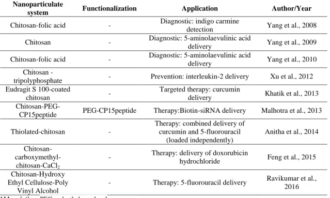
These developments are summarized in Table 2.3.
Table 2.3 – List of chitosan-based nanoparticles, in development for colorectal cancer. Nanoparticulate
system Functionalization Application Author/Year Chitosan-folic acid - Diagnostic: indigo carmine
detection Yang et al., 2008 Chitosan - Diagnostic: 5-aminolaevulinic acid
delivery Yang et al., 2009 Chitosan-folic acid - Diagnostic: 5-aminolaevulinic acid
delivery Yang et al., 2010 Chitosan -
tripolyphosphate - Prevention: interleukin-2 delivery Xu et al., 2012 Eudragit S 100-coated
chitosan -
Targeted therapy: curcumin
delivery Khatik et al., 2013
Chitosan-PEG-CP15peptide PEG-CP15peptide Therapy:Biotin-siRNA delivery Malhotra et al., 2013 Thiolated-chitosan -
Therapy: combined delivery of curcumin and 5-fluorouracil
(loaded independently)
Anitha et al., 2014
Chitosan- carboxymethyl-chitosan-CaCl2
- Therapy: delivery of doxorubicin
hydrochloride Feng et al., 2015 Chitosan-Hydroxy
Ethyl Cellulose-Poly Vinyl Alcohol
- Therapy: 5-fluorouracil delivery Ravikumar et al., 2016 Abbreviation: PEG, polyethylene glycol
16 2.7. Collagenase application in nanotechnology
Collagenase is a proteolitic enzyme responsible for the digestion of native collagen. The degradation of collagen, an important component of the extracellular matrix, helps to bypass the penetration limitations imposed by that conjunctive barrier (Goodman, Olive, & Pun, 2007; Murty et al., 2014). Therefore, collagenase has been studied for anticancer nanomedicine or nanopharmaceutical applications, as active contributor in terms of achieving a deeper tumoral penetration and consequently, higher efficiency in nanoparticles accumulation and action.
Goodman, Olive, & Pun published in 2007 the first study corroborating collagenase advantages for this purpose. The results obtained revealed a four-fold enhancement of spheroids core penetration by collagenase-coated polystyrene nanoparticles in comparison to the control albumin-coated nanoparticles. Another group showed that the pre-intravenous administration of collagenase in mice bearing mouse Lewis lung carcinoma, for further lipoplex delivery, increased tumour accumulation 1.5-fold and amplified the gene expression 2-fold (Kato et al., 2012). Moreover, in 2014, Murty et al., described nanoparticles functionalization with collagenase as a promising pathway to increase accumulation within tumours, based on tests of collagenase-pegylated gold nanoparticles in mice tumour xenografts, demonstrating an improvement of 35% in core accumulation, comparing to non-functionalized nanoparticles.
Recently, Villegas, Baeza, & Vallet-Regí (2015) presented an alternative and more complex model, for surface-transport of collagenase, aiming to avoid its exposure to blood circulating agents, as proteases. In this model, the authors attached pH-responsive polymeric-nanocapsules containing collagenase to the surface of mesoporous silica nanoparticles, to trigger collagenase release only in acidic conditions, similar to tumoral pH. Studies in three-dimensional models of human osteosarcoma cells showed considerable enhancement of tumours inner penetration.
The investigations did not report toxic effects of collagenase in human cells, being considered a safe additive for further developments.
The present work suggests a novel form of collagenase incorporation in the nanoparticulate systems and a new application for collagenase regarding colorectal cancer, as detailed below.
2.8. Chitosan-collagenase nanoparticles by inversemicroemulsion
Moilanen et al. published results in 2015 that corroborate the title of their publication, ‘Collagen XVII expression correlates with the invasion and metastasis of colorectal cancer’. The authors revealed an unexpected collagen expression in colon epithelia and its overexpression in colorectal cancer. The study suggests an overexpression enhancement in metastatic stages.
17
Based on this, we postulated the use of collagen as a biomarker for metastatic colorectal cancer and we have designed a nano-delivery system incorporating collagenase as collagen recognizer, that would contribute for a preferential accumulation of nanoparticles in metastasis of colorectal cancer.
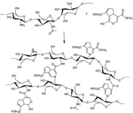
The collagenase incorporation was projected to occur through a genipin-based crosslink reaction, adapted from a chitosan-based nanospheres protocol (Pujana et al., 2013). This was possible due to the chemical groups involved in the reaction. As shown in Figure 2.2, the chitosan nanoparticles are synthesized by covalent crosslink between chitosan amine groups and the ester and dihydropyran ring of genipin. Since collagenase is also composed by amine groups, it can be crosslinked with genipin. Assuming collagenase integration as a chitosan-equivalent polymer in the synthesis procedure, simultaneous crosslink of both polymers is expected to occur, with no predefined order, forming chitosan-collagenase nanogels.
Figure 2.2 -Schematic representation of crosslink between chitosan (left) and genipin (right), from Lins et al., 2014.
The hybrid polymeric nanoparticles of chitosan and collagenase are expected to be biocompatible, bioavailable and biodegradable systems, combining the favourable properties of each polymer regarding specificity and efficiency of the delivery. Concerning nanoparticles accumulation, the use of collagen as biomarker allows to target colorectal cancer metastasis, with tumoral stronger adhesion and preferential accumulation conferred by chitosan mucoadhesivity and cationic nature. Besides, the enzymatic degradation of the extracellular tumour matrix allows the nanoparticulate
18
systems to reach the inner core, enhancing the therapeutic efficiency, also selectively directed to tumoral environment by using chitosan pH-sensitivity to trigger the drug release only in acidic environments, characteristic of tumoral surrounding.
The synthesis method reproduced, inverse microemulsion, also known as water–in-oil (W/O) microemulsion, is based on a thermodynamic equilibrium of water, oil and surfactant phases, in which the water amount is comparatively low, forming nanometric and monodisperse reverse micelles, with a polar inner core, containing polar or ionic compounds. The constant dynamic motion in solution - Brownian motion - leads to micelle collisions and fusion-fission occurrences, involving exchange and mixing of contents. The contact between reactants within micelles, forms ‘nanoreactors’, initiating the nanoparticles synthesis reaction by nucleation and controlled growth, limited and stabilized by the surfactant layer (Malik, Wani, & Hashim, 2012).
19
3. EXPERIMENTAL SECTION
3.1. Materials
Methoxy poly(ethylene glycol) (mPEG, molecular weight Mn = 5,000 Da, Aldrich, Germany), ε-caprolactone (CL) monomer (97%, Sigma-Aldrich, USA), Stannous octoate (Sn(Oct)2; 92,5-100%, Sigma, Japan), phenethyl isothiocyanate (PEITC, 99%, Aldrich, USA), coumarin-6 (C6, Santa Cruz Biotechnology, Dallas), chitosan (low molecular weight, Aldrich, USA), collagenase type I (Gibco Life Technologies, USA), genipin (98%, Wako chemicals, Germany). All solvents used were of analytical grade.
3.2. Synthesis and characterization of block copolymer mPEG-co-PCL
The methoxy polyethylene glycol-co-poly ε-caprolactone (mPEG-co-PCL) was synthesized by ring opening polymerization, adapted and optimized from Baimark, 2009; Baimark & Srisuwan, 2012; Xiong et al., 2015. The final synthesis was made as follows:
9.4 g of ε-CL (≈ 80 mmol), 2.2 g of mPEG (≈1 mmol) and 3.4 g of Sn(Oct)2 (≈ 0.1 mol% of monomer ε-CL) were added into a glass tube and submitted to a vacuum system under nitrogen refilling for about 2 hours. The tube was kept sealed and heated to ≈140°C in an oil bath for ≈24 h. The mixture was cooled to room temperature and dissolved in dichloromethane. The product was precipitated and washed in cold n-hexane or diethylether. After total evaporation of the precipitant, the final product was dried at ≈ 35°C, in an incubator. The copolymer structure was confirmed by 1
H – NMR (nuclear magnetic resonance) in a Bruker Avance III 400MHz, Fallanden equipment, using CDCl3 as solvent. This protocol was optimized and requires further optimization to obtain a purer copolymer.
3.3. Synthesis of mPEG-co-PCL nanoparticles
mPEG-co-PCL nanoparticles (NPs) were produced by nanoprecipitation, adapted and optimized from Baimark, 2009; Baimark & Srisuwan, 2012; Xiong et al., 2015.
10 mL of solvent were added to 10 mg of drug/marker and 100 mg of mPEG-co-PCL and submitted to vortex and sonification. The mixture was added dropwise to 100 mL of miliQ H2O, under stirring. The suspension was stirred overnight to remove the solvent and centrifuged to 15 000 rpm, 4°C during 60 minutes. The pellet was lyophilized during 42h.
20
Based on the cited procedures, the solvent used for both empty and C6-loaded NPs was acetone. For the loading of PEITC, a mixture of acetone:etanol (1:1) was used, taking into account the solubility properties of the copolymer and PEITC.
3.4. Characterization of mPEG-co-PCL nanoparticles
The particles size was analysed by Dynamic Light Scattering (DLS) equipment (Zetasizer Nano Series, Malvern Instruments Ltd, UK). The samples for the analysis were dissolved in filtered bidistilled water and submitted to sonication and pre-equilibration at 25°C.
Field Emission Gun Scanning Electron Microscopy (FEG-SEM) (JEOL, model JSM7001, Japan) was also used to examine particle size and morphology. The samples were prepared for observation by covering with gold/palladium (Au/Pd), in a sputter coater (Quorum Technologies, model Q150TES). Micrographs of the prepared aliquots were taken at an acceleration voltage of 15kV.
3.5. Synthesis of chitosan and chitosan-collagenase nanoparticles
The chitosan-genipin NPs were produced by inverse microemulsion, adapted from Pujana et al (2013). This synthesis was made to confirm the reproducibility of the procedure and as control, since it was applied for the development of the new system, chitosan-collagenase.
Two different microemulsions were prepared. To the first one, chitosan was prepared in 1% (v/v) aqueous acetic acid solution to a concentration of 10 mg/mL A mixture of cyclohexane:n-hexanol:chitosan was prepared to a final ratio of 2.75:1:1. Triton X-100 was added dropwise until the solution became transparent. The second microemulsion was prepared by adding genipin in excess (100 mg per mL of miliQ H2O) to a mixture of cyclohexane:n-hexanol:miliQH2O (2.75:1:1). This microemulsion was added to the first one, under stirring. After 7 days of crosslinking reaction, at room temperature, the solution was added dropwise to excess ethanol.
The chitosan-genipin-collagenase NPs were produced by reverse microemulsion, adapted and optimized from the work of Pujanaand collaborators (Arteche Pujana et al., 2013).
Two different microemulsions were prepared. To the first one, chitosan was prepared in 1% (v/v) aqueous acetic acid solution to a concentration of 10 mg/mL and mixed with collagenase dissolved in miliQ H2O, to a final ratio of 1:1. A mixture of cyclohexane:n-hexanol:chitosan-collagenase were prepared to a final ratio of 2,75:1:1. Triton X-100 was added dropwise until the solution became transparent. The second microemulsion was prepared by adding genipin to a mixture of cyclohexane:n-hexanol:miliQH2O (2.75:1:1). The amounts of genipin were adapted to final ratios of 10:1; 2:5; 1:5 and 1:10 (genipin:chitosan-collagenase). This microemulsion was added to the first one,