Ar
ti
go
*e-mail: [email protected]
ENCAPSULATION OF NAPROXEN IN NANOSTRUCTURED SYSTEM: STRUCTURAL CHARACTERIZATION
AND IN VITRO RELEASE STUDIES
Vanessa Azevedo de Mello e Eduardo Ricci-Júnior*
Departamento de Medicamentos, Faculdade de Farmácia, Centro de Ciências da Saúde, Universidade Federal do Rio de Janeiro, Av. Brigadeiro Trompowski, s/n, Cidade Universitária, 21941-970 Rio de Janeiro - RJ, Brasil
Recebido em 7/6/10; aceito em 10/1/11; publicado na web em 25/3/11
Nanoparticles were produced by solvent emulsiication evaporation method with the following characteristics: nanometric size (238 ±3 nm), narrow polydispersity index (0.11), negative zeta potential (-15.1 mV), good yield of the process (73 ± 1.5%), excellent encapsulation eficiency (81.3 ± 4.2%) and spherical shape. X-rays diffraction demonstrated the loss of drug crystallinity after encapsulation; however, the proile of the diffractograms of the poly-ε-caprolactone (PCL) nanoparticles was kept. Differential scanning calorimetry thermograms, correspondingly, exhibited the loss of drug melting peak and the increasing of the melting point of the PCL nanoparticles, evidencing an interaction drug-polymer. Naproxen release was low and sustained obeying the Higuchi´s kinetic. The results show that nanoparticles are promising sustained release system to the naproxen.
Keywords: naproxen; nanoparticles; structure characterization.
INTRODUCTION
Naproxen (NPX) is a nonsteroidal anti-inflammatory drug (NSAID) commonly used for the reduction of moderate to severe pain, fever, inlammation and stiffness caused by conditions such as osteoarthritis, rheumatoid arthritis, psoriatic arthritis, gout, injury (like fractures), tendinitis and bursitis. It works by inhibiting both the cyclooxygenase-1 (COX-1) and cyclooxygenase-2 (COX-2) enzymes. COX-2 predominates at site of inlammation and COX-1 is expres-sed normally in the gastrointestinal tract.1,2 Naproxen’s usefulness is limited by a short duration of action (8 h) when administrated orally. Repeated administrations are required for maintenance of the pharmacological action. Patients with chronic inlammatory diseases require long treatment with NSAIDs. However, complications may result from long therapies and repeated administrations such as gas-trointestinal disorder, gastritis, ulcer and bleeding.1,3 An implantable system of sustained release based on poly-ε-caprolactone (PCL) nanoparticles could sustain the release extending the pharmacological action of the NPX and reducing the frequency of administration and adverse effects.
Biocompatible and biodegradable polymers have been used to produce nanoparticles as sustained release system.4,5 Nanoparticles have several advantages of sustaining the drug release, reducing the administration frequency, decreasing adverse effects and prolonging the pharmacological action.6,7 Much attention has been focused on the biodegradable polymers such as PCL that is used to development of sustained release implants such as nanoparticles.8-12 It is biodegra-dable and has reasonable cost when compared with similar polymers utilized for encapsulation of drugs in nanoparticles. PCL suffers in vivo hydrolysis releasing monomers that are absorbed, metabolized and eliminated by organism.13 PCL nanoparticles have been used as drugs delivery systems for amphotericin B,14 indometacin,15 griseo-fulvin,16 tamoxifen,17 vaccines,18 and cyclosporine A.19 In this work, PCL nanoparticles were produced by solvent emulsion evaporation
method suitable for encapsulation of hydrophobic drugs like as NPX. The drug release from polymeric nanoparticles depends upon the rate of diffusion of the drug from delivery system, erosion of the polymeric matrix and biodegradation of the polymer. If erosion and biodegradation are slow process, the release rate is strongly inluen-ced by drug diffusion from nanoparticles. Several factors such as the interaction between drug-polymer, the state of incorporation of the drug in the delivery system, the pKa of the drug and the amount of drug loaded in nanoparticles can inluence the diffusion rate of the drug from nanostructured system.20 The state of incorporation of the drug in the system and interaction between drug-polymer are important factors that affect the release proile.6,21 The amorphous or crystalline states of the drug loaded in polymeric nanoparticles can be determined using X-rays diffraction (XRD) and interaction drug-polymer can be investigated for differential scanning calorimetry (DSC). The DSC and XRD data are often combined together to give useful information on the structural characteristics of the polymeric matrix, drug and possible interactions between drug and polymer. In this paper, we investigated as the semi-crystalline structure of the PCL nanoparticles and the drug-polymer interaction can inluence the naproxen release proile.
The aims of this work were development and characterize naproxen-loaded in poly-ε-caprolactone nanoparticles. The morpho-logy, size distribution, zeta potential, process yielding, encapsulation eficiency, study of drug-polymer interaction and in vitro release assay were evaluated.
EXPERIMENTAL Materials
Methods
Preparation of nanoparticles and process yield (%)
Nanoparticles containing naproxen were prepared by the solvent emulsiication-evaporation method (SEEM). NPX (50 mg) and PCL (150 mg) were briely dissolved in dichloromethane (DCM). This organic solution was emulsiied in 50 mL of 0.2 M phosphate buffer 1.5% PVA (w/w), pH 3.0, for 5 min using an ultrasonicator (UP100H, 100 W, 30 kHz, Hielscher, Germany) in ice bath. The solvent (DCM) was evaporated under reduced pressure (rotavapor, Heidolph, Germany) at room temperature (28 oC). Nanoparticles suspensions were puriied thrice by centrifugation at 20,000 × g (centrifuge J-25, Beckman, USA) for 20 min followed by resus-pension in water. The nanoparticles susresus-pension was transferred into a glass vial containing cryoprotector (trehalose) and frozen in a liquid nitrogen bath. Drying was carried out in a lyophilizer, yielding powder of nanoparticles. The samples were stored at room temperature (28 oC) before analysis. The lyophilized samples were resuspended with water and observed by optical microscopy to detect the presence of aggregates.
The process yield was calculated by Equation 1:
Y(%)=(MNP/MT)x100 (1)
where: Y(%)-process yield, MNP-mass of powder recovered after freeze-drying, and MT-mass of polymer, naproxen and trehalose in formulation. The encapsulation method was accomplished in triplicate (n=3).
Nanoparticles characterization
Nanoparticles were characterized in terms of size, polydispersity index (PI), zeta potential, transmission electron microscopy (TEM), encapsulation eficiency (EE), differential scanning calorimetry (DSC) and X-rays diffraction (XRD). The size and PI were deter-mined by photon correlation spectroscopy using a Zetasizer® 5000 (Malvern instruments, UK). The zeta potential was measured in a 10−3 M NaCl using the electrophoretic mode with the Zetasizer® 5000. Morphology was evaluated by transmission electron mi-croscopy (TEM), (Morgani, FEI-268). Lyophilized powder was dispersed in water and 5 µL were transferred to a grid. Uranyrl acetate solution (3%) was added for ixing and contrast. After 30 s, excess liquid was removed with ilter paper (Whatman®). Grids were placed in vacuum desiccators (Pyrex®) before analysis. The encapsulation eficiency (EE) was determined by a spectrophoto-metric method. Naproxen was dissolved in 0.2 M phosphate saline buffer (PBS), pH 7.4, and the absorbance was measured at 254 nm (spectrophotometer, V-630, Jasco). The calibration curve was linear (r = 0.9977) for naproxen of 2-20 µg mL-1. Intra and inter-days pre-cision and accuracy of the method were not greater than 3 and 2%, respectively. The naproxen concentrations were measured from the standard curve. Ten mg of freeze-dried nanoparticles were dissolved in 5 mL of hot acetone to disrupt the nanoparticles; 0.1 M PBS, pH 7.4, was added to precipitate the polymer and dissolve the drug. The suspension was iltered through a membrane ilter (pore size 0.45 µm) to remove the insoluble polymer and diluted with 0.1 M PBS (pH 7.4) to be analyzed by spectrophotometric method as described above. Encapsulation eficiency was calculated from Equation 2:
EE=(M1/Mt)x100 (2)
where: EE-encapsulation eficiency, M1-mass of naproxen in nanopar-ticles, and Mt-mass of naproxen used in formulation. The experiments of EE were accomplished in triplicate (n=3 determinations).
Differential scanning calorimetry (DSC) was utilized to investiga-te possible ininvestiga-teraction between naproxen-polymer in PCL nanoparti-cles. DSC thermograms of the naproxen, empty nanoparticles, binary mixture of naproxen-empty nanoparticles (1:3) and naproxen-loaded in PCL nanoparticles were obtained utilizing a differential scanning calorimeter (Shimadzu, DSC 50) calibrated with indium standard. The thermal behavior was studied by heating of 15 mg of the sample in a covered aluminum pan under nitrogen atmosphere. The tempe-rature range used was between 30 and 200 oC with a heating rate of 10 oC min-1. X-rays diffraction (XRD) was utilized to investigate the amorphous or crystalline state of the naproxen encapsulated in the PCL nanoparticles. The XRD diagrams of the naproxen, empty nanoparticles, binary mixture of naproxen-empty nanoparticles (1:3) and NPX-loaded PCL nanoparticles were obtained utilizing a X-ray diffractometer (Philips PW 1130/60) using Cu-k α radiation (30 kV, 40 mA). The instrument was operated in the 2θ =10-50o range at a scan rate of 1o min-1.
Solubility, in vitro releasing studies and kinetic
Naproxen solubility in the acceptor solution (0.1 M PBS, pH 7.4) was measured to establish the sink conditions of the in vitro release studies. NPX was added to acceptor solutions (n=3 determinations) and maintained at 37 °C in a water bath under continuous magnetic stirring until saturation. Samples were withdrawn, centrifuged at 20,000 x g for 30 min, iltered through a Millipore ilter (pore size 0.45 µm) and measured by spectrophotometric method (described above). Drug loaded in PCL nanoparticles, corresponding to 10 mg of naproxen, was dispersed in 0.3 mL of 0.1 M PBS, pH 7.4. Na-noparticles suspension was placed in a dialysis tube with molecular weight cut-off at 12,000 g mol-1 (Sigma-Aldrich) and closed. The tube was immersed in 100 ml of 0.1 M PBS (pH 7.4) obeying the sink conditions. The acceptor solution was stirred utilizing a paddle at a constant rate of 100 rpm and kept at 37 oC (SR8 dissolutor, Hanson). At given time intervals, six samples (n=6) of 3 mL were withdrawn for analysis. The same volume of fresh medium was placed in the dissolution vessels to maintain the sink conditions. Naproxen release from the PCL nanoparticles was measured by spectrophotometric assay (described above). The intensity of absorbance was utilized to calculate the concentrations of drug. Release proile was obtained correlating time (h) versus drug release concentration (µg mL-1). Free naproxen (10 mg) was used as control in the in vitro release studies. The naproxen release data were used to study the mechanism of drug release. They were itted utilizing the mathematical models of Zero-order, First-order, Higuchi´s model, Hixon-Crowell´s model and Korsmeyer-Peppas model.22 Equations of the mathematical models are showed in Table 1. Data were itted and the regression linear of the mathematical models was evaluated using R2 (squared correlation coeficient). The linear regression was applied in the range of 6 to 744 h.
Table 1. Applied mathematical models to the data release of the naproxen
loaded in nanoparticles
Mathematical models Equation
Zero order F=k0t
First order ln (1-F)=-kft
Higuchi F=kHt
1/2
Hixon-Crowell 1-(1-F)1/3=k
1/3t
Korsmeyer-Peppas F=kK-Pt
n
Stability of the nanostructured system
Stability studies were done by storing the nanoparticles containing naproxen (lyophilized) in amber glass bottles at room temperature. At 0, 30, 60 and 120 days we analyzed the size, polydispersity index, zeta potential, encapsulation eficiency and pH. Release study of the sample in time of 120 days was done to assess possible change in the proile of drug release and kinetics.
RESULTS AND DISCUSSION Nanoparticles characterization
Table 2 shows the results of the nanoparticles characterization. Satisfactory process yields was obtained (> 69%). The encapsulation method produced nanoparticles with diameter of 238 ± 3 nm with good polydispersity index (IP) (Table 2). IP shows that size distribu-tion is narrow and near of the monomodal. Empty nanoparticles are smaller than the nanoparticles containing naproxen. The zeta potential was negative for all samples. Naproxen-loaded in PCL nanoparticles exhibited a zeta potential lower than empty nanoparticles. Naproxen is a weak acid (pKa 4.4) and can suffer ionization in water. Naproxen adsorbed in the nanoparticles surface can suffer ionization decreasing the zeta potential of the system. The pH of aqueous external phase of the encapsulation method can inluence the encapsulation eficiency of weak acids. Naproxen is not protoned in pH 3.0 exhibiting low water solubility. Other pH values were not tested in this work. High encapsulation eficiency was obtained utilizing an acid external aqueous phase (Table 2). The shape of the nanoparticles is shown on Figure 1. The naproxen-loaded in PCL nanoparticles has spherical shape (Figure 1A) similar to empty nanoparticles (Figure 1B).
XRD and DSC studies were performed to investigate the phy-sical state of the drug in the nanoparticles and possible interaction between drug-polymer, because these aspects can inluence on the release characteristics of the drug. The nanoparticles suspensions were freeze-dried without cryoprotector (these studies in particular) to avoid interference of this substance in the experiments of DSC and RDX. Different combinations of drug-polymer may coexist in the polymeric carriers, such as: amorphous drug in either an amorphous or a crystalline polymer and crystalline drug in either an amorphous or a crystalline polymer. Moreover, a drug may be present either as a solid solution or solid dispersion in an amorphous or crystalline polymeric matrix.23-25
The solubility and physical state of the drug-loaded in PCL nanoparticles were investigated to analyze possible inluence on the drug release proile. Figure 2 shows the DSC thermograms of the na-proxen, empty nanoparticles, physical mixture of naproxen and empty nanoparticles (1:3), and drug-loaded in nanoparticles. Free naproxen exhibited a single endothermic melting peak at 158.2 °C, typical of a drug in the crystalline form (Figure 2A). Empty nanoparticles showed a single endothermic melting peak at 60.2 °C (Figure 2B). Naproxen melting peak was shifted to a higher temperature (158.7 °C) in the thermograms of the physical mixture (Figure 2C). Empty nanopar-ticles in the physical mixture exhibited an endothermic melting peak at 60.9 °C. The small shift observed may be due to an interaction between drug and polymeric nanoparticles (Figure 2C). Naproxen melting peak disappeared in the DSC thermogram of drug-loaded in PCL nanoparticles, indicating the absence of the drug crystalline state in the delivery system development (Figure 2D). During na-noparticles formation, the polymer inhibited the recrystallization of the drug. Besides, the melting peak and thermogram proile of the PCL nanoparticles was shifted to a higher temperature (64.3 oC) in the naproxen-loaded in PCL nanoparticles, evidencing an interaction between the drug and polymer.
Figure 1. Photomicrographies obtained with transmission electronic micro-copy (TEM): (A) naproxen-loaded in nanoparticles, (B) empty nanoparticles
Table 2. Characterization of naproxen-loaded in PCL nanoparticles
Samples Size
(nm)a
PIb Zeta poten-tial (mV)b
E.E. (%)a
Yield (%)a
Empty Np 212±2 (0.09) -11.3 - 69±1.8
NPX-loaded Np 238±3 (0.11) -15.1 81.3±4.2 73±1.5
aMean ± S.D. n=3 determinations. bMean n=3 determinations. NPX – naproxen. Np – nanoparticles. PI - Polydispersity Index. E.E. - Encapsula-tion Eficiency
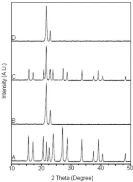
DSC thermograms showed that NPX was dispersed in the PCL nanoparticles in the amorphous form and this evidence was conir-med by X-rays diffraction (XRD) studies. The crystalline state of the naproxen was evidenced in the XRD diffractogram; the proile exhibited many diffraction bands characterizing a drug crystalline state (Figure 3A). Empty nanoparticles showed two bands in the range 20-25o (Figure 3B), conirming its semi-crystalline structure that also was observed in others works.26,27 The drug crystallinity peaks were evidenced in the binary physical mixture between na-proxen and empty nanoparticles (Figure 3C). The simple blending with empty nanoparticles did not modify the drug crystalline state. The peak of semi-crystalline structure of the PCL nanoparticles was observed in Figure 3C (20-25o). The diffraction proile of naproxen-loaded in PCL nanoparticles (Figure 3D) was similar to diffraction proile of the empty nanoparticles (Figure 3B). Drug-loaded in PCL nanoparticles did not exhibit diffraction bands associated with the drug crystalline state, conirming that the naproxen was amorphous inside the PCL nanoparticles. The evidences show that the naproxen loaded in nanoparticles is in an amorphous state of a solid molecular dispersion or a solid solution in the polymer matrix after the encapsulation.
Our results corroborate the data obtained by Javadzadeh et al..28 They entrapped naproxen in poly(
D,L-lactide-co-glicolide) acid (PLGA) (Resomer RG 502) nanoparticles using the solvent emulsiication-evaporation method. The crystallinity and interaction drug-polymer were studied by X-ray powder diffraction (XRD) and Differential scanning calorimetry (DSC), respectively. The XRD results showed consistency with the data obtained with the DSC analysis. Naproxen free exhibited a crystalline structure with dis-tinct diffraction peaks.28 PLGA displayed an amorphous structure without diffraction peaks. Naproxen-loaded in PLGA nanoparticles (naproxen-polymer, 1:5) did not show diffraction peaks, suggesting amorphization or naproxen solvation within amorphous carrier (PLGA).28 The XRD results of the physical mixture showed that the reduction in the intensity of diffraction peaks was due to dilution
effect exerted by the polymer network. 28 Concisely, DSC and XRD analysis showed decrease within crystallinity of naproxen loaded in PLGA nanoparticles. 28
It was evidenced that others drugs are able to interact with PCL after encapsulation in nanoparticles modifying the drug and polymer characteristics. The interaction between papaverine and PCL was evidenced for DSC and XRD showing that the drug was dispersed in the microparticles in an amorphous form.6 The melting peak of the papaverine was absence on the DSC thermogram of the drug-loaded PCL microparticles. However, the melting peak of the PCL micropar-ticles was shifted to a higher temperature in the papaverine-loaded in polymeric microparticles, indicating an interaction between the drug and the polymer. The diffraction proile of the papaverine-loaded PCL microparticles did not exhibit peaks associated with the drug crystalline state conirming that the papaverine was amorphous in the polymeric matrix of the PCL microparticles.6 The interaction be-tween isradipine and polymeric matrix of the PCL nanoparticles was investigates utilizing DSC and XRD.24 It was observed that isradipine was dispersed in the amorphous state in the polymeric matrix of the PCL nanoparticles. The melting peak of the drug was absence on the DSC thermogram of the drug-loaded in PCL nanoparticles. Besides, the melting peak of the empty nanoparticles and PCL nanoparticles with isradipine were similar.24 The diffraction proile obtained con-irmed the DSC data evidencing that the drug was amorphous inside polymeric nanoparticles.2
Solubility, in vitro release studies and kinetic
Naproxen is a weak acid and its solubility depends upon the pH of the dissolution medium. Drug solubility in 0.1 M PBS, pH 7.4, at 37 oC was 6.12+0.07 mg mL-1. Thus, sink conditions existed for naproxen release in this acceptor solution. Figure 4A and B shows the in vitro release studies of naproxen-loaded in nanoparticles and dissolution proile of free drug, respectively. Naproxen loaded in PCL nanoparticles produced slow and sustained release proile. Free naproxen shows a quick dissolution with 100% of the drug dissolved in the receptor medium in 30 min (Figure 4B). In the in vitro release study, naproxen-loaded in nanoparticles and free drug were placed in a dialysis tube and dropped into acceptor medium. Sink condition was obeyed to avoid saturation of the receptor medium of the in vitro release studies. Naproxen has 13% burst effect for 24 h, slow releasing during a period of 744 h and nearly 70% of drug was released from PCL nanoparticles during the study. Since the particles had been pre-pared to remove existing free drug, the initial burst could not be due to free drug. It could probably be caused by molecules in the outer layer of the PCL nanoparticles. After 24 h, naproxen had a low releasing rate into the acceptor medium (Figure 4A). The release rate of naproxen from nanoparticles was very slow due to: the low permeability of the water in the hydrophobic matrix of the PCL nanoparticles and the interaction drug-polymer that dificult the drug dissolution by water retarding the naproxen release. It was reported that others drugs can exhibit sustained release from PCL micro or nanoparticles. Jeong et al.6 found that the papaverine release proile from PCL microparticles exhibited a burst effect followed by a slow release for 1 week. They evidenced by DSC and XRD that papaverine was dispersed in the amorphous form in the PCL microparticles. The drug release rate was determined not by the slow erosion of nanostructured system but by the diffusion of the drug through the polymer matrix.6 Verger et al.24 studied the inluence of the interaction drug-polymer in the release of isradipine from PCL nanoparticles. The drug exhibited a sustained release from the semi-crystalline structure of the delivery system. Interaction drug-polymer and structure were evidenced by DSC and XRD.24 It is reported that isradipine was amorphous and molecularly dispersed in the semi-crystalline matrix of the PCL nanoparticles. Figure 3. X-rays diffraction proile obtained from naproxen (A), empty
Naproxen-loaded in PCL nanoparticles has a slow release and our data are similar to results obtained by Veronese et al.29 and Javadzadeh et al..28 Veronese et al.29 loaded naproxen in biodegradable
polyorga-nophosphazene microspheres. The encapsulation method was similar to the method used in our work (solvent emulsiication-evaporation method) obtaining naproxen-loaded in polyorganophosphazene microspheres with size distribution between 10 and 100 µm. By the appropriate choice of pH (3.0) and composition of the external phase, naproxen presented an encapsulation eficiency higher than 80%.Naproxen-loaded in polyorganophosphazene microspheres (naproxen-polymer, 1:5) has a slow release and nearly 96% of drug was released from microspheres during 824 h.29 It is known that drugs loaded in polyorganophosphazene microspheres are released by the cooperative mechanism of diffusion and bioerosion.29 Some parameters such as amount of entrapped naproxen and the type of polymer interfere with the mechanism of drug release. Thus, the lowering of the release rate when the naproxen content is decreased, seems to indicate that the diffusion through the polymeric matrix is the main mechanism of drug release.29
Javadzadeh et al.28 loaded naproxen in poly(
D,L -lactide-co-glycoli-de) acid (PLGA) (Resomer RG 502) nanoparticles. Naproxen-loaded in PLGA nanoparticles was formulates using a solvent emulsiication-evaporation method. The organic and aqueous phase were composed of dichloromethane-acetone (1:1) and polyvinyl alcohol (PVA) (0.5%, w/v), respectively. The nanoparticles were characterized by X-ray diffraction (XRD) (evaluation of crystallinity), Fourier transform infrared spectroscopy (FT-IR) and differential scanning calorimetry (DSC) (evaluation of chemical interaction drug-polymer), and in vitro release study. All the formulations resulted in nanoparticles with size distribution between 352-571 nm, spherical shape and encapsulation eficiency of 80%.28 The naproxen-loaded in PLGA nanoparticles displayed lower crystallinity and no chemical interactions between the naproxen and PLGA.28 The naproxen-loaded in nanoparticles exhibited the slower release rate in comparison with the free drug and physical mixture.28 Naproxen-loaded in PLGA nanoparticles (naproxen-PLGA, 1:5) exhibited a burst effect of close to 15% within the irst hour followed by a slow release. During the release study, nearly 80% of drug was released in 20 h. PLGA offered lesser resis-tance to naproxen diffusion than the PCL. This fact can be evidenced by the observation of the naproxen release proiles from nanoparticles. PLGA nanoparticles released 80% of naproxen in 24 h, while PCL released 11% in the same period.
Several mathematical models were used to investigate the me-chanism of the naproxen release from PCL nanoparticles.22,30 The drug release depends upon: adsorption through the nanoparticles matrix, diffusion through the nanoparticles matrix, particles erosion, a combined erosion and diffusion process and polymer degradation (chemical or enzymatic hydrolysis). The application of the correct mathematical model allows us to analyze about release rate, points of dissolution change and mechanisms of drug release. The squared correlation coeficients (R2), slope (a), linear coeficient (b), rate constant (k) obtained after regression linear utilizing mathematical models are exhibited in Table 3. Figure 4C shows the linear regression obtained utilizing Higuchi´s model31 that provided the highest value of squared correlation coeficient (R2).
Figure 4. In vitro release proile of naproxen loaded in PCL nanoparticles (A), dissolution proile of free naproxen (B) and regression linear of the release data utilizing the Higuchi’s model (C). Each point represent the mean + S.D. of n=6 determinations
Table 3. Squared correlation coeficient (R2) and coeficients obtained after linear regression of the release data utilizing ive mathematical models
Mathematical Model R2 a b k n
Zero order 0.9706 0.08 14.098 0.08 (h-1)
-First order 0.9815 0.0015 0.0911 0.0015 (h-1)
-Higuchi 0.9932 2.5619 2.9378 2.5619 (h-1/2)
-Hixon-Crowell 0.9789 0.0004 0.0371 0.0004 (h-1/3)
-Korsmeyer-Peppas 0.9931 0.4977 0.3925 2.5044 (h-n) 0.4978
Squared correlation coeficients (R2) obtained with the Zero-order model was 0.9706. This model can be used to describe the drug release from several types of delivery systems such as matrix tablets for drugs with low solubility32 and osmotic systems, where drug release would be directly proportional at time. First-order kinetics33,34 gave a R2 of 0.9815. First-order model can describe the release proile from the delivery systems containing hydrophilic drugs dispersed in porous matrices, where drugs would be release at the rates proportional to the amounts of drug remaining in the interior of the delivery system.35 Hixson-Crowell’s cube root model was applied in the data release and provided a R2 of 0.9789. This model can be applied to the delivery systems whose drug release rate is proportional to the surface area of the system such as the erosion-dependent release systems.36-38
Higuchi’s mode31 has been based on the Fick’s Law where the release occurs by the diffusion of drugs within the delivery system. In this case, the cumulative released amount of the drug is proportio-nal at square root of time. The relation can be used to describe drug dissolution from several types of modiied-release pharmaceutical dosage forms.39 If the relation squared root of time versus cumula-tive amount of drug released yields (Table 1) a straight line then the particular dosage form is considered to follow Higuchi kinetics.39 Under some experimental situation the release mechanism can deviate from Fickian diffusion, following an anomalous transport (no-Fickian release).39 In these cases a more generic equation can be used. Korsmeier-Peppas et al.39,40 developed a mathematical model relating exponentially the drug release to the elapsed time. The model is described in Table 1. Peppas39,41 used the release exponent (n) in order to characterize different release mechanism. If the n value is 0.5 or less, the release mechanism follow Fickian diffusion (Higuchi model), and higher values 0.5 < n < 1 for mass transfer follow a non-Fickian model denominates anomalous transport. The drug release follows Zero-order drug release or Case-II transport if the n value is 1. For the values of n higher than 1 (n=1), the mechanism of drug release is regarded as super Case-II transport.39,41 This model is used to analyze the release of pharmaceutical polymeric dosage forms when the release mechanism is not well known or when more than one type of release phenomena was involved.39 This model is limited to the irst 60% of the cumulative amount of drug release. The n value could be obtained from slope (a) of the straight line of log cumulative amount of drug release (%) versus log time.39 For the models of Zero-order, First-Zero-order, Higuchi and Hixon-Crowell, the rate constant (k) was obtained from the slope (a) of the straight line (Table 3). The rate constant (k) for the Korsmeyer-Peppas model was determined using the release exponent (n) and the equation described in Table 1.
As shown in Table 3, Higuchi’s model31 and Korsmeyer-Peppas model39-41 gave the highest values of squared correlation coeficient (R2). Thus, these mathematical models would be the most suitable models to describe the naproxen release from PCL nanoparticles.42 Both Higuchi and Korsmeyer-Peppas models are widely used to cha-racterize the release mechanism. In Higuchi approach, the fraction of drug released is proportional to the square root time. The rate constant (k) is a constant characteristic of the formulation.42 For Korsmeyer-Peppas, k is also the rate constant and n is the release exponent charac-terizing the diffusion mechanism. For n=0.5, the Korsmeyer-Peppas model becomes identical with Higuchi model.42,43 This fact can be observed in our studies (Table 3). The release exponent (n) obtained was nearly 0.5. Thus, the Korsmeyer-Peppas model suggests that the release of the naproxen-loaded in PCL nanoparticles is controlled by Fickian diffusion. The k values obtained with the Higuchi and Korsmeyer-Peppas models were quite similar. The results obtained with the Korsmeyer-Peppas model corroborate the data obtained with the Higuchi model.
The polymer hydrolysis and polymeric matrix erosion could
in-luence in the drug release from nanoparticles, however, these process are very slow. The result suggests that the release of naproxen from PCL nanoparticles is controlled by the diffusion. The mechanism of the drug release from PCL nanoparticles can be considered as follows: water penetrates into polymeric matrix of the nanoparticles through small porous and channels; slowly dissolving the drug and naproxen is released by diffusion to acceptor solution.
Stability of the nanostructured system
Table 4 shows the data obtained through the study of stability. There were no signiicant changes in the parameters used to evaluate the stability of the system. The release proile of naproxen nanopar-ticles and the kinetics were not changed during the stability study (data not shown). The straight line equation (y = ax-b) and correlation coeficient (R2) were y = 2.6206x-2.9378 and 0.9938, respectively. Thus, the system was stable for 120 days and retained its release characteristics and kinetics.
CONCLUSIONS
DSC exhibited absence of the melting point of the naproxen and XRD evidenced the loss of the drug crystallinity after encapsulation in PCL nanoparticles. It was observed by XRD that PCL nanoparticles have a semi-crystalline structure; however DSC exhibited alterations in the melting point and thermogram proile in drug-loaded in PCL nanoparticles evidencing interaction between drug-polymer. Drug was dispersed in the amorphous state in a semi-crystalline polymeric matrix inside nanoparticles. Release studies showed that naproxen exhibited a sustained release with kinetic obeying Higuchi´s model. Korsmeyer-Peppas model corroborates the results obtained with Higuchi´s model. The results show that nanoparticles are promising sustained release system to the naproxen.
ACKNOWLEDGEMENTS
We would like thank the inancial support of CNPq (Conselho Nacional d Desenvolvimento Cientíico e Tecnológico).
REFERENCES
1. Hawkey, C. J.; Best Pract. Res. Clin. Gastroenterol. 2001, 15, 801. 2. Kim, S. J.; Flach, A. J.; Jampol, L. M.; Surv. Ophthalmol. 2010, 55, 108. 3. Bombardier, C.; Laine, L.; Reicin, A.; Shapiro, D.; Davis, B.; Day, R.; Ferraz, M. B.; Hawkey, C. J.; Schnitzer, T. J.; New Engl. J. Med. 2000, 34, 1520.
4. Hans, M. L.; Lowman, A. M.; Curr. Opin. Solid State Mat. Sci. 2002, 6, 319.
5. Rajeev, J.; Shah, N.; Malick, A. W.; Rhodes, C. T.; Drug Dev. Ind. Pharm. 1998,24, 703.
6. Jeong, J.; Lee, J.; Cho, K.; J. Controlled Release 2003, 92, 249.
Table 4. Stability of the nanostructured system containing naproxen
Time (days) Size (nm)a
PIb Zeta poten-tial (mV)b
E.E. (%)a
pH
0 238±3 (0.11) -15.1 81.3±4.2 5.75
10 242±3 (0.07) -14.7 80.7±3.6 5.63
30 235±3 (0.13) -13.9 80.1±4.2 5.61
60 241±3 (0.15) -15.3 79.8±4.2 5.64
120 245±3 (0.13) -14.6 80.2±4.2 5.67
7. Butoescu, N.; Jordan, O.; Doelker, E.; Eur. J. Pharm. Biopharm. 2009, 73, 205.
8. Soppimath, K. S.; Aminabhaui, T. M.; Kulkarni, A. R.; Rudzinski, W. E.; J. Controlled Release 2001, 70, 1.
9. Yang, J.; Park, S. B.; Yoon, G. H.; Huh, Y. M.; Haam, S.; Int. J. Pharm.
2006, 324, 185.
10. Chawla, J. S.; Amiji, M. M.; Int. J. Pharm. 2002, 249, 127.
11. Pitt, C. G.; Biodegradable polymers as drug delivery systems, Marcel Decker Inc.: New York, 1990.
12. Pitt, C. G.; Gratzel, M. M.; Kimmel, G. L.; Surles, J.; Schindler, A.; Biomaterials 1981, 2, 215.
13. Sinha, V. R.; Bansal, K.; Kaushik, R.; Kumria, R.; Trehan, A.; Int. J. Pharm. 2004, 278, 1.
14. Espuelas, M. S.; Legrand, P.; Irache, J. M.; Gamazo, C.; Orecchione, A. M.; Devissaguet, J. P.; Ygartua; P.; Int. J. Pharm. 1997, 158, 19. 15. Park, S-J.; Lee, W-M.; Hong, S-K.; Colloid Surf., B 2006, 47, 211. 16. Zili, Z.; Sfar, S.; Fessi, H.; Int. J. Pharm. 2005, 294, 261. 17. Shenoy, D. B.; Amiji, M. M.; Int. J. Pharm. 2005, 293, 261.
18. Singh, J.; Pandit, S.; Bramwell, V. W.; Alpar, H. O.; Methods2006, 38, 96.
19. Yenice, I.; Mocan, M. C.; Palaska, E.; Bochot, A.; Bilensoy, E.; Vural, I.; Irkeç, M.; Hincal, A. A.; Exp. Eye Res. 2008, 87, 162.
20. Allen, C.; Maysinger, D.; Eisenberg. A.; Colloid Surf., B 1999, 16, 3. 21. Zhang, L.; Yang, M.; Wang, Q.; Li, Y.; Guo, R.; Jiang, X.; Yang, C.; Liu,
B.; J. Controlled Release 2007, 119, 153.
22. Barzegar-Jalali, M.; Adibkia, K.; Valizadeh, H.; Shadbad, M. R. S.; Nokhodchi, A.; Omidi, Y.; Mohammadi, G.; Hasan, M.; J. Pharm. Pharm. Sci. 2008, 11, 167.
23. Dubernet, C.; Thermochim. Acta 1995, 248, 259.
24. Verger, M. L.; Fluckiger, L.; Kim, Y.; Hoffman, M.; Maincent, P.; Eur. J. Pharm. Biopharm. 1998, 46, 137.
25. Jenquin, M.; McGinity, J.; Int. J. Pharm. 1994, 101, 23. 26. Wang, S.; Guo, S.; Cheng, L.; Int. J. Pharm. 2000, 350, 130. 27. P�res, M. H.; Zinutti, C.; Lamprecht, A.; Ubrich, N.; Astier, A.; Hoff-27. P�res, M. H.; Zinutti, C.; Lamprecht, A.; Ubrich, N.; Astier, A.;
Hoff-man, M.; Bodmeier, R.; Maincent, P.; J. Controlled Release 2000, 65, 429.
28. Javadzadeh, Y.; Ahadi, F.; Davaran, S.; Mohammadi, G.; Sabzevari, A.; Adibkia, K.; Colloid Surf., B 2010, 81, 498.
29. Veronese, F. M.; Marsilio, F.; Caliceti, P.; De-Filippis, P.; Giunchedi, P.; Lora, S.; J. Controlled Release 1998, 52, 227.
30. Tanaka, N.; Imai, K.; Okimoto, K.; Ueda, S.; Tokunaga, Y.; Ohike, A.; Ibuki, R.; Higaki, K.; Kimura, T.; J. Controlled Release 2005, 108, 386. 31. Higuchi, T.; J. Pharm. Sci. 1963, 52, 1145.
32. Varelas, C. G.; Dixon, D. G.; Steiner, C.; J. Controlled Release 1995, 34, 185.
33. Gibaldi, M.; Feldman, S.; J. Pharm. Sci. 1967, 56, 1238. 34. Wagner, J. G.; J. Pharm. Sci. 1969, 58, 1253.
35. Mulye, N. V.; Turco, S. J.; Drug Dev. Ind. Pharm. 1995, 21, 943. 36. Hixson, A. W.; Crowell, J. H.; Ind. Eng. Chem. Res. 1931, 23, 923. 37. Yan, G.; Li, H.; Zhang, R.; Ding, D.; Drug Dev. Ind. Pharm. 2000,26,
681.
38. Costa, P.; Lobo, J. M. S.; Eur. J. Pharm. Sci. 2001, 13, 123.
39. Mathew, S. T.; Devi, S. G.; Sandhya, K. V.; Pharm. Sci. Tech. 2007, 8, E1.
40. Korsmeyer, R. W.; Gurny, R.; Doelker, E. M.; Buri, P.; Peppas, N. A.; Int. J. Pharm. 1983, 15, 25.
41. Peppas, N. A.; Pharm. Acta Helv. 1985, 60, 110.
42. Carriazo, D.; de Arco, M.; Mertín, C.; Ramos, C.; Rives, V.; Microporous Mesoporous. Mater. 2010, 130, 229.