1
UNIVERSITY’S TECHNOPARK COMPANY OF PROMECH TEKNOLOJİ VE BİLİŞİM SİSTEMLERİ SANAYİ LTD. ŞTİ. Proceedings of the conference ISTANBUL INTERNATIONAL CONFERENCE ON PROGRES IN APPLIED SCIENCE 2017 – ICPAS 2017 4 - 6 JANUARY 2017, Istanbul, Turkey ”.
DETECTION OF SPORES AND HYPHAE OF ARTWORKS’ BIODETERIOGENIC FILAMENTOUS FUNGI BY RNA-FISH
*Marina González-Pérez
HERCULES Laboratory, Évora University
Évora, Portugal
Teresa Charrua Rosmaninho
HERCULES Laboratory, Évora University
Évora, Portugal
António Pereira
HERCULES Laboratory and Chemistry Department (School
of Sciences and Technology), Évora University
Évora, Portugal
António Candeias
HERCULES Laboratory and Chemistry Department (School
of Sciences and Technology) Évora University
Évora, Portugal
Ana Teresa Caldeira
HERCULES Laboratory and Chemistry Department (School
of Sciences and Technology) Évora University
Évora, Portugal Keywords:Filamentous fungi; Fluorescence In Situ Hybridization; Biodeterioration
*marinagp@uevora.pt
ABSTRACT
Filamentous fungi are a threat to the conservation of Cultural Heritage. Thus, detection and identification of viable filamentous fungi are crucial for applying adequate Safeguard measures.
RNA-FISH protocols have been previously applied with this aim in Cultural Heritage samples. However, only hyphae detection was reported in the literature, even if spores and conidia are not only a potential risk to Cultural Heritage but can also be harmful for the health of visitors, curators and restorers. Thus, the aim of this work was to evaluate various permeabilizing strategies for their application in the detection of spores/conidia and hyphae of artworks’ biodeteriogenic filamentous fungi by RNA-FISH. Besides of this, the influence of cell aging on the success of the technique and on the development of fungal autofluorescence (that could hamper the RNA-FISH signal detection) were also investigated. Five common biodeteriogenic filamentous fungi species isolated from biodegradated artworks were used as biological model: Aspergillus niger, Cladosporium sp, Fusarium sp, Penicillium sp. and Exophialia sp.
Fungal autofluorescence was only detected in cells harvested from Fusarium sp, and Exophialia sp. old cultures, being aging-dependent. However, it was weak enough to allow autofluorescence/RNA-FISH signals distinction. Thus, autofluorescence was not a limitation for the application of RNA-FISH for detection of the taxa investigated.
2
All the permeabilization strategies tested allowed to detect fungal cells from young cultures by RNA-FISH. However, only the combination of paraformaldehyde with Triton X-100 allowed the detection of conidia/spores and hyphae of old filamentous fungi. All the permeabilization strategies failed in the Aspergillus niger conidia/spores staining, which are known to be particularly difficult to permeabilize. But, even in spite of this, the application of this permeabilization method increased the analytical potential of RNA FISH in Cultural Heritage biodeterioration. Whereas much work is required to validate this RNA-FISH approach for its application in real samples from Cultural Heritage it could represent an important advance for the detection, not only of hyphae but also of spores and conidia of various filamentous fungi taxa by RNA-FISH.
INTRODUCTION
Filamentous fungi colonize outdoor historic monuments and sculptures as well as objects of art in museums and collections [1–3]. They are able to alter and degrade both organic and inorganic materials causing aesthetic and structural damages [3]. Thereby, filamentous fungi constitute a serious threat to the conservation of Tangible Cultural Heritage.
The detection/identification of the biodeteriogenic microcolonizers proliferating on and in artworks is crucial for selecting appropriate mitigation treatments or inhibition/prevention measures for the contaminated objects [4]. This constitutes a key challenge for museums, cultural institutions, collections and, particularly, to restorers and curators [3].
Thus, the implementation of approaches that allowed to detect and identify the viable filamentous fungi (potentially responsible for biodeterioration) is of utmost importance.
Culture-dependent approaches, whereas useful, are time consuming and give only information from the viable and cultivable microorganisms (less than 5% of those present) [5]. Thus, RNA-based methods have gained much attraction over the past decades [4,6]. However, most of these techniques involve RNA extraction as a first step which turn the procedure extremely demanding and sensitive due to the high lability of RNA [4,6].
The use of whole-cell RNA-Fluorescence In Situ Hybridization allows to overcome this drawback. It allows to detect and identify the microorganisms of interest by specific hybridization of the fluorescently labeled oligonucleotide probes to the complementar RNA target sequence within the intact cell [7,8]. This technique has previously shown its potential to analyze filamentous fungi in environmental and clinical areas as well as in food science [1,9,10]. However, it has been scarcely applied for investigating those involved in artworks’ biodeterioration [11].
The existing works on FISH application for analyzing filamentous fungi colonizing artworks reported only hyphae detection by RNA-FISH and PNA-FISH, even with lysozyme and glucanase permeabilization [11–14]. However, the detection and identification of spores and conidia in Cultural Heritage samples is of utmost importance not only because they represent a potential risk for Cultural Heritage materials but also for the health of visitors, curators and restorers [15,16].
3
The conidia and spores of most fungi, however, are not stainable per se. This can be attributed to the particularly thick and resistant cell walls that protect the protoplast [17]. Thus, the inclusion of an additional cell wall permeabilization step is usually required to ensure staining with conventional fluorescent dyes such as DAPI or PI [14,17,18]. Various chemical and physical permeabilization techniques have been successfully used in the detection of conidia and spores from young cultures, including among others surfactant, osmotic and microwave treatments [1,17].
However, some permeabilization techniques that works in combination with fluorescent dyes staining failed when combined with RNA-FISH staining [1]. This could be attributed to the fact that RNA-FISH staining success depends strongly on the cell permeabilization, but also on other factors such the autofluorescence of the cells or their RNA content [7,18]. Thus, for detecting filamentous fungi colonizing Cultural Heritage materials with this technique it is necessary to consider that: i) they possess specially reduced permeability due to the thick walls they develop and to their melanization, that can hamper RNA-FISH probes penetration and their detection [19]; and ii) for adhering to surfaces, they excreted extracellular substances that could turn them autofluorescent [13]. This autofluorescence, if strong, can mask RNA-FISH signals [7,18,20,21].
In this way, the aim of this work was to increase the capacity of RNA-FISH for analyzing filamentous fungi colonizing Cultural Heritage materials by broadening the spectra of fungal cells that can be detected, from hyphae to spores and conidia.
In this work various permeabilizing techniques were tested for detecting spores and hyphae of artworks’ biodeteriogenic filamentous fungi by RNA-FISH. Cells (conidia/spores and hyphae) from young and old cultures were investigated for considering the effect of aging on the apparition of autofluorescence and on the success of RNA-FISH.
MATERIALS AND METHODS Strains and growth conditions
Aspergillus niger, Cladosporium sp, Fusarium sp, Penicillium sp. and Exophialia sp. were used in this study. All of them are filamentous fungi associated to biodeterioration of Cultural Heritage and has been isolated from Portuguese artworks/assets, belonging to the HERCULES-Biotec Lab (Universidade de Évora) collection. Strains were routinely grown at 28⁰C on Malt Extract Agar.
For macroscopic and microscopic observations growth of filamentous fungi was carried out in Malt Extract Agar (MEA) Petri dishes at 28ºC for 5 days and then photographs were captured. Filamentous fungi were mounted on coverslips with methylene blue and then photographs were captured.
For checking fungal autofluorescence and for FISH assays, the 5-days old cultures were directly used or storage at 4ºC during more 90 or 180 days.
4 Autofluorescence assessment
A loopful of each fungal culture (5-day old directly used or storaged for 90 or 180 days, respectively) was mounted in a drop of dH2O placed on a microscope slide. The autofluorescence were assessed by microscopic inspection with epifluorescence microscopy using three filters: TRITC (TRITC (Rhodamine)/DII/Cy3 set, Motic: excitation D540/25x, dichroic mirror 565DCLP, and emission D605/55m), FITC (FITC/RSGFP/Fluo 3/DiO Acridine Orange [+RNA] set, Motic: excitation D480/30x, dichroic mirror 505DCLP and emission D535/40m) and Cy5 (Cy5/ Alexa Fluor 633/ Alexa Fluor 647 set, Motic: excitation HQ620/60x, dichroic mirror Q660LP, and emission HQ700/75m).
Fluorescence In Situ Hybridization
Fungal cells were collected by washing the colony surface three times with 5 mL of 0.02% (v/v) Tween 20 in PBS and gently scraping it with a sterile handle. The cellular suspension containing conidia, spores and hyphae (CSH) was transferred to a 15 mL sterile centrifuge tube. Half of the volume was filtered using three layers of sterile gauze and transferred to another centrifuge tube, obtaining a conidia/spores (CS) suspension. Both the CS and CSH suspensions were centrifuged (10000 rpm 10 min 4ºC) and washed with 15 mL of PBS 10X.
Conidia and spores were counted using a Neubauer counting chamber and aliquots of the CS suspension containing 106 cells were prepared in 2 mL centrifuge microtubes (CS samples). Aliquots of the same volume of the HCS suspension were also placed into 2 ml centrifuge microtubes (HCS samples).
Permeabilization
The efficacies of five different permeabilization methods for its combination with the RNA-FISH protocol were evaluated.
After centrifugation of the samples, the supernatant was discarded and the cells were permeabilized using these methods:
M1: Incubation for 90 min at room temperature with an aqueous solution of Triton X-100 ,1% w⁄w Tris–HCl 0.05 M, Ethylene Diamine Tetraacetic Acid (EDTA) 0.04 M and β-mercaptoethanol 0.1 M. After centrifugation the cells were covered with 100 µl of glycerol 0.1% w:v aqueous solution [1].
M2: Treatment with 1 mL NaOH 1M for 30 min at room temperature.
M3: Microwave heating during 30 s [17]. The microtubes were placed in the center of the rotating plate and were directly and individually microwaved using a domestic microwave oven (KUNFT 17MX02) with a maximal nominal output power of 700 W and a frequency of 2450 MHz. The microwave was adequately calibrated to adjust the outsource power to 130W.
M4: Incubation of the cells with absolute EtOH for 1h at room temperature [12].
M5: Treatment with 4% freshly prepared paraformaldehyde and 0.1% Triton X- 100 in 10mm KPO4, pH 6.8, for 10 min at room temperature [22].
5
Control: CS and CSH without permeabilization.
The permeabilized cells were centrifuged and the pellet was suspended in 2.0 mL of EtOH:PBS 50:50 and immediately frozen at -20ºC. Before its use for FISH, the cells were defrozen, centrifuged and washed with PBS 10X.
Hybridization
Hybridization and washing was carried out, following the protocol previously described by us for detecting filamentous fungi hyphae in mortars [12]. The probes used for the assays were the only factor that was modified. Finally, cells were pelleted by centrifugation, suspended in 200.0 µL of PBS and analyzed by epifluorescence microscopy.
Oligonucleotide probes
The universal oligonucleotide probes directed against the 18S rRNA sequence of Eukaryotes, EUK516 (5’-ACCAGACTTGCCCTCC-3’) labelled at the 5’end with 6-FAM and Cy3, EUK516-6-FAM and EUK516-Cy3, respectively, were used. The universal probes EUB338 targeted to the 16S rRNA of Bacteria (5’-ACTCCTACGGGAGGCAGC-3’) also labeled with the same fluorophores at the 5’end, 6-FAM and EUB338-Cy3 were used as negative controls [23]. The probes were tested individually and blank assays were also carried out without adding any probe to the hybridization mixture to check for autofluorescence or fixation-induced fluorescence.
Microscopic analysis
For microscopic analysis, aliquots of the samples were spotted onto microscope slides. The slides were observed using the biological microscope BA410E Motic equipped with a 100W Quartz Halogen Koehler illumination with intensity control and with an epi-attachment (EF-UPR-III) and a Power Supply Unit (MOTIC MXH-100). Microphotographs were
acquired using a MoticamPRO 282B camera coupled to the microscope. Motic Images Plus 2.0LM was used to visualize the microphotographs and to analyze the cell size.
Cy3 signals were observed using the TRITC filter and FITC filter set was used for analyzing 6-FAM fluorescence. For each FISH conidia/spores sample, visualization and counting of around 100 conidia/spores were carried out both by brightfield and epifluorescence microscopy in order to calculate the percentage of fluorescent spores/conidia.
RESULTS AND DISCUSSION (MANDATORY HEADER)
Five of the most common biodeteriogenic filamentous fungi of Cultural Heritage materials including paper, glass, polymers and stone, inter alia, were used in this work as biological model (Table 1) [3,19,24,25].All the fungi selected were hyphomycetes. It is well known that these filamentous fungi can produce a wide range of secondary metabolites that could mask the probe signal by their own fluorescence [26]. Thus, the autofluorescence apparition with aging was investigated.
6
.
Table 1. Biodeteriogenic filamentous fungi selected in this study. Aspergillus niger Penicillium sp. Cladosporium sp. Fusarium sp. Exophiala sp.
The results evidenced that cells from young cultures did not exhibit autofluorescence when observed using FITC, Cy5 or TRITC filter sets (Table 2). For Aspergillus niger and Cladosporium sp. fluorescence signals were neither found even after 180 days of storage (data not shown). However, whereas Fusarium sp. and Exophiala sp. young cells, 5-day old, did not exhibit autofluorescence, after three months of storage showed weak autofluorescence. Our findings, support the observations of Sterflinger et al.: autofluorescence is aging- and taxa-dependent [13]. This is due to the fact that autofluoresce is a consequence of the deposition of fluorescent extracellular polymeric substances excreted by some filamentous fungi on the cell wall [13].
Table 2. Autofluorescence of the filamentous fungi under study after different periods of storage at 4ºC when visualized using FITC, Cy5 and TRITC filters.
FITC Cy5 TRITC
t storage(days) 0 90 0 90 0 90 Aspergillus niger - - - - Penicillium sp. - - - - Cladosporium sp. - - - - Fusarium sp. - + - - - + Exophiala sp. - + - - - +
(-) no fluorescence ,(+) weak, (++) strong fluorescence was detected.
For the taxa investigated in this work, autofluorescence when detectable is as weak that could not be considered as a drawback for RNA-FISH application.
However, could permeabilization of the cells be a problem for the RNA-FISH probe entry? and, particularly could the spores and hyphae of biodeteriogenic filamentous fungi be detected by RNA-FISH? Five different permeabilization methods were evaluated for permeabilizing filamentous fungi for phylogenetic staining by RNA-FISH.
The success of RNA-FISH staining applying the different permeabilization techniques were firstly assessed using spores and conidia from young and old cultures (5-days old slants storage for 0, 90 and 180 days) of Cladosporium and Aspergillus taxa. They were selected for the first screening due to the particularly low permeable cell wall they develop. This was carried out in order to select the best permeabilization method in terms of percentage of fluorescent spores/conidia produced.
Thus, the percentage of conidia/spores that were fluorescently stained with EUK516-Cy3 after RNA-FISH application was calculated from the observations of the spores/conidia in bright-field microscopy and epifluorescence microscopy. No
7
fluorescent signals were detected when Aspergillus niger conidia/spores were analyzed by RNA-FISH. This contrast with the results obtained previously by Villa et al. using the surfactant and osmotic strategies combination, M1 [1]. Since the constitution of fungal cell walls depends greatly on the environmental/growth conditions [26], the apparently contradictory results could be explained in the base of variations in the characteristics of the cell wall due to the different conditions used for fungal growth.
The percentages of conidia stained without permeabilization (control) and using the five different permeabilization methods in combination with RNA-FISH are shown in Figure 1. In contrast with the failed results found for A. niger conidia/spores RNA-FISH staining, conidia and spores from Cladosporium sp. young cultures, although producing low percentages of fluorescent spores (around 20%), were stained even without permeabilization (Figure 1). However, no fluorescent spores from cultures storage for 90 days were detected without prior permeabilization after FISH treatment.
Figure 1. Percentage spores/conidia that fluoresced after RNA-FISH treatment with and without previous permeabilization (M1-M5 and control, respectively).
These results evidenced the great influence of aging in cell wall permeability decrease.
The percentages of conidia/spores stained using the five different permeabilization methods in combination with FISH are also shown in Figure 1. Most of the permeabilization methods investigated were useless as the first step of RNA-FISH for detecting Cultural Heritage microcolonizers since with exception of the methods M2 and M5, no or slight increase of the fluorescent conidia/spores percentage were observed between permeabilizing or not the cells. It is noteworthy that the conidia/spores from cultures stored during 90 days presented a high fixative-induced fluorescence (e.g. the percentage of fluorescent cells detected with method M4 were similar adding probe or not). With exception of M5, the results of all the methods showed to be aging-dependent. These finding indicated once again that the structure and composition of the cell wall vary along time, modifying its permeability. This is not a new finding, but evidenced the necessity of considering cell aging when the permeabilization methods are evaluated for their use for fungal staining in real samples (independently of
8
their origin, environment, clinical, food or cultural heritage). However, most of the works focused on finding a good method used cells from young cultures (5-10 days of growth) as biological model [1,14,17].
The method involving fresh paraformaldehyde permeabilization is the only one of those tested with applicability in real samples because: i) it is the only one that allowed to detect old spores and conidia (from cultures storaged for 6 months); ii) did not produce fixation-induced fluorescence (without the addition of probe the percentage of spores/conidia detected using this method were not different from those observed without permeabilization); and iii) showed good and constant yields in terms of fluorescent spores/conidia after RNA-FISH application independently of the spores/conidia age (around 45% of the conidia/spores present in the sample become fluorescent). This is of utmost importance considering that filamentous fungi thriving in Cultural Heritage materials may have been colonizing them for much longer periods.
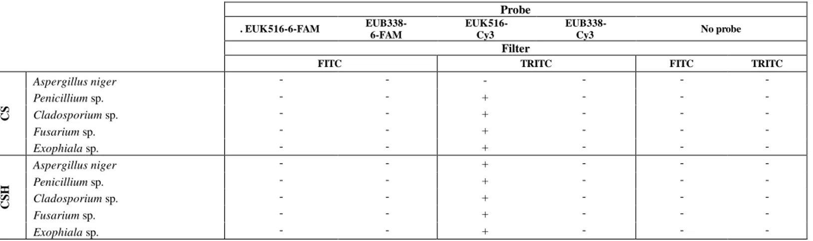
Thus, even if M5 permeabilization method did not allow to detect Aspergillus niger conidia/spores, it was applied, in a second stage of this study, for RNA-FISH detection of conidia, spores and hyphae of the five biodeteriogenic fungal species selected. For considering the aging factor, cells from cultures storaged for 3 months were used. Samples containing conidia and spores only (CS) and also hyphae (CSH) were used in order to evaluate the capacity of the method to detect the various cell types. Five different assays were carried out for each SC and SCH samples: i) two assays with universal probes labelling eukaryotes, which are labelled with different fluorochromes: EUK516-Cy3 and EUK516-6-FAM; ii) two negative controls using the correspondent universal probes for bacteria, EUB338-Cy3 and EUB338-6-FAM and iii) a control for fixative-induced fluorescence, without adding any probe. The results are summarized in Table 3. None of the controls produced fluorescent signals. However, intense fluorescent signals were observed for the hyphae and spores/conidia of all the species tested using EUK516-Cy3 (Figure 2), with the exception of the conidia/spores of Aspergillus niger (as expected). In contrast with the good results found with EUK516-Cy3, no fluorescence was observed with EUK516-6-FAM. This could be associated to the lower quantum yield of 6-FAM in comparison of Cy3.
Table 3. Detected (+) and non-detected (-) fluorescent signals applying the M5 permeabilization method as the first step of RNA-FISH for all the filamentous fungi under study after three months of storage at 4ºC. Results observed for the conventional RNA-FISH assays (with EUK516-Cy3 and -6-FAM), for the negative controls (EUB338-Cy3 and -6-FAM) and for the fixation-induced controls (no probe)
Probe . EUK516-6-FAM EUB338- 6-FAM EUK516- Cy3 EUB338- Cy3 No probe Filter
FITC TRITC FITC TRITC
CS Aspergillus niger - - - - - -Penicillium sp. - - + - - -Cladosporium sp. - - + - - -Fusarium sp. - - + - - -Exophiala sp. - - + - - -C S H Aspergillus niger - - + - - -Penicillium sp. - - + - - -Cladosporium sp. - - + - - -Fusarium sp. - - + - - -Exophiala sp. - - + - -
-9
Thus, the permeabilization method M5 in combination with RNA-FISH staining, using probes labeled with Cy3, allows to detect and identify spores/conidia and hyphae of various fungal taxa with exception of A. niger conidia/spores. This improved the results obtained in previous works using the RNA-FISH technique for analyzing artworks’ biodeteriogenic filamentous fungi. The methodologies already described in the literature in this field only allow to signalized the viable hyphae and some of them involved a time-consuming treatment with cell wall lytic enzymes [11,27]. We have previously investigated the use of 4% w/v solution of fresh paraformaldehyde in PBS as fixative/permeabilizer, detecting only hyphae [12]. However, the effect of using the same concentration of paraformaldehyde in combination with Triton-X-100 in a less concentrated buffer solution notably improved the results obtained by us, allowing to detect also conidia/spores.
Aspergillus niger Penicillium sp. Cladosporium sp. Fusarium sp. Exophiala sp.
CS n.d.
C
S
H
Figure 2. Spores and fluorescent hyphae detected after the application of the best permeabilization method (M5) in combination with RNA-FISH staining using EUK516-Cy3 for the SC and SCH samples; (n.d.: not detected). Scale bar represent 10 µm.
Whereas much work is required to validate the method for its application in real samples from Cultural Heritage it represent an important advance, allowing to broaden the type of cells that can be detected by RNA-FISH.
CONCLUSION
The paraformaldehyde/Triton X-100 permeabilization combined with RNA-FISH allowed the detection of conidia/spores and hyphae of old filamentous fungi, with the exception of A. niger conidia/spores. It represents an important advance for the detection, not only of hyphae but also of spores and conidia of various filamentous fungi taxa by RNA-FISH. This can be an important contribution to Cultural Heritage microbiology considering that conidia and spores can be harmful not only for Cultural Heritage materials but also for the health of visitors, curators and restorers.
ACKNOWLEDGMENTS
This work was co- financed by FCT – Fundação para a Ciência e a Tecnologia through the project "MICROTECH-ART- Microorganisms Thriving on and Endamaging Cultural Heritage -an Analytical Rapid Tool-" (PTDC/BBB-IMG/0046/2014) and by European Union, European Regional Development Fund ALENTEJO 2020 through the project “HIT3CH - HERCULES Interface for Technology Transfer and Teaming in Cultural Heritage” (ALT20-03-0246-FEDER-000004). Marina González-Pérez acknowledges FCT for the economic support through the post-doctoral grant SFRH/BPD/100754/2014.
10 NOMENCLATURE
RNA-FISH: RNA Fluorescence In Situ Hybridization; CS: Conidia/Spores;
CSH: Conidia/Spores/Hyphae.
REFERENCES
1. F. Villa, F. Cappitelli, P. Principi, A. Polo, and C. Sorlini, Lett. Appl. Microbiol. 48, 234 (2009). 2. K. Sterflinger and F. Pinzari, Environ. Microbiol. 14, 559 (2012).
3. K. Sterflinger, Fungal Biol. Rev. 24, 47 (2010).
4. P. Sanmartín, A. DeAraujo, and A. Vasanthakumar, Microb. Ecol. 1 (2016).
5. A. Michaelsen, F. Pinzari, K. Ripka, W. Lubitz, and G. Piñar, Int. Biodeterior. Biodegradation 58, 133 (2006). 6. R. I. Griffiths, A. S. Whiteley, A. G. O’Donnell, and M. J. Bailey, Appl. Environ. Microbiol. 66, 5488 (2000). 7. R. Amann and B. M. Fuchs, Nat. Rev. Microbiol. 6, 339 (2008).
8. A. Moter and U. B. Göbel, J. Microbiol. Methods 41, 85 (2000).
9. Y. Wang, L. Chen, X. Liu, D. Cheng, G. Liu, Y. Liu, S. Dou, D. J. Hnatowich, and M. Rusckowski, Nucl Med Biol 40, 89 (2013).
10. R. M. Da Silva, J. R. Da Silva Neto, C. S. Santos, H. Frickmann, S. Poppert, K. S. Cruz, D. Koshikene, and J. V. B. De Souza, Ann. Clin. Microbiol. Antimicrob. 14, 6 (2015).
11. E. Müller, U. Drewello, R. Drewello, R. Weißmann, and S. Wuertz, J. Cult. Herit. 2, 31 (2001).
12. R. Vieira, P. Nunes, S. Martins, M. González, T. Rosado, A. Pereira, A. Candeias, and A. T. Caldeira, in Sci. Technol. Cult. Herit., edited by A. Rogerio-Candelera (Taylor & Francis Group, London, 2014), pp. 257–262.
13. K. Sterflinger and M. Hain, Stud. Mycol. 1999, 23 (1999).
14. W. R. Teertstra, L. G. Lugones, and H. A. B. Wösten, Fungal Genet. Biol. 41, 1099 (2004).
15. G. Piñar, D. Piombino-mascali, F. Maixner, A. Zink, and K. Sterflinger, FEMS Microbiol. Ecol. 86, 341 (2013). 16. F. Cappitelli, G. Pasquariello, G. Tarsitani, and C. Sorlini, Trends Microbiol. 18, 538 (2010).
17. V. Prigione and V. Filipello Marchisio, J. Microbiol. Methods 59, 371 (2004). 18. C. Urzì and P. Albertano, Methods Enzymol. 336, 340 (2001).
19. K. Sterflinger and G. Piñar, Appl. Microbiol. Biotechnol. 97, 9637 (2013).
20. C. Urzì, V. La Cono, F. De Leo, and P. Donato, Mol. Biol. Cult. Heritage. Lisse, Netherlands, Balkema Publ. 55 (2003).
11
21. H. M. Davey and D. B. Kell, Microbiol. Rev. 60, 641 (1996).
22. N. I. R. Osherov and O. Yarden, Cell. Mol. Biol. Filamentous Fungi 224 (2010). 23. G. Wallner, R. Amann, and W. Beisker, Cytometry 14, 136 (1993).
24. T. Dakal and S. Cameotra, Environ. Sci. Eur. 24, 1 (2012).
25. A. Mihajlovski, D. Seyer, H. Benamara, F. Bousta, and P. Di Martino, Ann. Microbiol. 65, 1243 (2015). 26. K. Sterflinger, W. E. Krumbein, and A. Schwiertz, Int. Microbiol. 1, 217 (1998).