Advance Access publication 31 August 2005
Developmental absence of maxillary lateral incisors in the
Portuguese population
Teresa Pinho*, Purifi cação Tavares**, Patrícia Maciel*** and
Cristina Pollmann**
*Instituto Superior de Ciências da Saúde-Norte (CESPU), Paredes, **Faculty of Dental Medicine, University of
Porto, and***SchoolofHealth Sciences, University of Minho, Braga, Portugal
SUMMARY The aim of this study was to evaluate the prevalence and clinical manifestation of developmental absence of maxillary permanent lateral incisors in the Portuguese population.
The study group comprised 16 771 patients observed between 1993 and 2000 at the Faculty clinic. Two hundred and nineteen patients were found to have missing upper lateral incisors (131 females and 88 males, with ages ranging between 3 and 71 years), a prevalence of 1.3 per cent in this population. Absence of these teeth was bilateral in 44.7 per cent of the patients; of the unilateral cases, 33 per cent occurred on the right side and 21.9 per cent on the left side.
In children under 8 years of age (n = 12), two (0.26 per cent) also had developmental absence of the primary lateral incisors, one unilateral and the other bilateral.
Among the 121 subjects with unilateral developmental absence, the contralateral maxillary lateral incisor was found to be microdont in 57.1 per cent (right lateral 23 per cent, left lateral 34.1 per cent), suggesting the possibility that microdontia represents a variable expression of the same developmental defect that results in absence.
Introduction
Developmental lack of a tooth results from disturbances during the early stages of tooth development. A tooth is defi ned as developmentally missing if it has not erupted in the oral cavity and is not visible radiographically.
Hypodontia is a relatively frequent anomaly of the permanent dentition, with a reported incidence of between 3.5 and 8.8 per cent, when third molars are excluded (Horowitz, 1966; Helm, 1968; Muller et al., 1970; Thilander and Myrberg, 1973; Maklin et al., 1979; Rølling, 1980; Pilo et al., 1987; Aasheim and Øgaard, 1993; Leitão, 1993; Johannsdottir et al., 1997; Baccetti, 1998). It is seen more frequently in females, although gender distribution has shown some local variations when studies are compared (Helm, 1968; Magnusson, 1977; Rølling, 1980; Aasheim and Øgaard, 1993; Leitão, 1993; Johannsdottir et al., 1997; Tavajohi-Kermani et al., 2002).
Although there is some variation in the reported frequency of developmental dental absence (Table 1), the majority of studies indicate that the most frequently absent teeth are: the lower second premolar, followed by the upper second premolar, the upper lateral incisor and the lower central incisor (Horowitz, 1966; Thilander and Myrberg, 1973; Magnusson, 1977; Rølling, 1980; Johannsdottir et al., 1997). In contrast, Muller et al. (1970) suggested that the most commonly absent teeth are: the upper lateral incisor, followed by the lower second premolar, the upper second premolar and the lower central incisor, and Tavajohi-Kermani et al. (2002) stated that the order was: the lower second premolar, the upper lateral incisor, the upper second premolar and the
lower central incisor (Table 1). Developmental absence of the fi rst and second molars, lower canines and upper central incisors are considered relatively rare (Maklin et al., 1979; Dermaut et al., 1986; Schalk-van der Weide et al., 1992; Guckes et al., 1998).
Absence of one or two teeth is the most common fi nding (Muller et al., 1970; Magnusson, 1977; Maklin et al., 1979; Rølling, 1980; Leitão, 1993) and unilateral absence of an upper lateral incisor is often associated with malformation (e.g. microdontia) of the other lateral incisor (Magnusson, 1977; Nieminem et al., 1995) (Figure 1).
Hypodontia in the primary dentition is a rare occurrence (0–0.9 per cent or < 0.9 per cent) and, when present, occurs more frequently in the incisor region, generally including the upper lateral incisor and the lower central or lateral incisor (Ravn, 1971; Bennett and Ronk, 1980; Järvinen and Lehtinen, 1981; Johannsdottir et al., 1997). When hypodontia occurs in the primary dentition, (Figure 2), most authors report absence of the permanent successor in 100 per cent of cases (Ravn, 1971; Bennett and Ronk, 1980; Järvinen and Lehtinen, 1981; Johannsdottir et al., 1997). However, this is not always the case (Nick-Hussein and Majid, 1996; Canut-Brussola, 2000; Daugaard-Jensen et al., 1997).
Estimates of the frequency of absence of upper lateral incisors are not uniform and vary according to the population being studied (Horowitz, 1966; Muller et al., 1970; Thilander and Myrberg, 1973; Magnusson, 1977; Rølling, 1980; Aasheim and Øgaard, 1993; Johannsdottir et al., 1997; Tavajohi-Kermani et al., 2002). Values have
the tooth epithelium, initial error of the mesenchyme, or disturbances in the development of the embryonic fusion of the maxilla and the medial nasal processes (a similar situation occurs in cleft lip and palate) (Woodworth et al., 1985; Svinhufvud et al., 1988). It is known that the structure and development of the human dentition are rigidly controlled by genetics. Several studies in the last decades have aimed to identify those genes whose mutations may be responsible for, or associated with, hypodontia, some of which have generated important new information (Lyngstardaas et al., 1996; Pirinen et al., 1996; Thesleff, 1996; Vastardis et al., 1996; Stimson et al., 1997; Vastardis, 2000; Lidral and Reising, 2002). It is conceivable that the genes may be involved in one or more of the mechanisms proposed above for the absence of lateral incisors.
It has been hypothesized that, if hypodontia is primarily genetic, then bilateral absence would be expected to occur relatively commonly; and when hypodontia occurs unilaterally, it may be associated with structural anomalies of the contralateral tooth (such as microdontia or conical shaped teeth).
Table 1 The prevalence of hypodontia in different samples and populations according to previous publications.
Total prevalence Upper lateral Lower second Upper second Lower fi rst and gender (%) incisor (%) premolar (%) premolar (%) incisor (%)
Aasheim and Øgaard, 1993 6.5 2
Norway, n = 1953 M 5.8, F 7.2 Baccetti, 1998 5.8 Italy, n = 980 Horowitz, 1966 6.5 1.11 2.23 1.41 USA, n = 1000 Helm, 1968 5 Denmark, n = 1240 M 4.8, F 7.3 Johannsdottir et al., 1997 5 0. 8 2.5 Iceland, n = 396 M 4, F 6 Leitão, 1993 6.3 Portugal, n = 666 M < F Magnusson, 1977 7.9 0.95 4.2 1.46 Iceland, n = 1116 M 6.7, F 8.9 Maklin et al., 1979 4.34 USA, n = 847 Muller et al., 1970 3.5 1.65 1.24 0.7 0.23 USA, n = 14 940 Pilo et al., 1987 8 Israel, n = 702 M 9.7, F 6.4 Rølling, 1980 7.8 1.7 4.1 2.15 Denmark, n = 3325 M 7.7, F 7.8 Tavajohi-Kermani et al., 2002 8.8 1.28 1.38 0.7 0.6 USA, n = 1016 M 3, F 6
Thilander and Myberg, 1973 6.1 1.2 2.46 2 0.45
Sweden, n = 5459
Figure 1 Microdontia of the upper right and developmental absence of
the upper left lateral incisor in a 12-year-old patient.
been reported between 0.8 and 2 per cent for the permanent dentition and between 0.1 and 0.7 per cent for the primary dentition.
Various aetiological factors have been suggested for the failure of development of the permanent tooth germ, thus leading to its absence: physical obstruction, rupture of the dental lamina, limitation of space, functional anomalies of
The aims of the present study were to investigate hypodontia involving missing upper lateral incisors in the Portuguese population, and to test the above hypothesis. Material and methods
The research was performed on material from the archives of the Dentistry Clinic of the Instituto Superior de Ciências da Saúde-Norte, Portugal, using 16 771 panoramic radiographs, taken between 1993 and 2000. The patients were between 3 and 71 years of age.
The radiographs were analysed by trained observers who followed a pre-established protocol, similar to that existing in the clinic: general observation and comparison of symmetrical teeth, followed by a systematic analysis of the erupted and unerupted teeth in each quadrant, and fi nally of the bone adjacent to each quadrant. Radiographs showing recent extractions, bone defects, evidence of surgery, trauma and fractures were excluded.
The selection of the study sample was undertaken in two stages: fi rst, radiographs corresponding to individuals with probable developmental absence of the upper lateral permanent incisors (n = 267) were selected; secondly, confi rmation of developmental absence was obtained through a telephone interview or by clinical observation (n = 219). Demographic data including gender and age were also collected.
The study also included investigation of the following parameters: unilateral or bilateral absence, developmental absence of other teeth, supernumerary teeth, and microdontia of the contralateral maxillary lateral incisor in those
subjects presenting with unilateral absence. The terminology suggested by Proffi t (1995), relating to the morphological microdontic classifi cation of the upper lateral incisor was followed, i.e. the upper lateral incisors were considered microdontic when their mesiodistal dimension was less than that of the lower lateral incisors.
The sample was sub-divided into three groups according to age: (1) individuals younger than 8 years (n = 12) in whom absence of the primary upper lateral incisor was studied (data was gathered through clinical confi rmation); (2) individuals who were 8 years of age and older (n = 207) in whom persistence of the primary upper lateral incisor related to absence of the corresponding permanent tooth was noted (data was gathered based only on radiographs, regardless of the level of root resorption of primary teeth); and (3) individuals 15 years and older (n = 182) in whom persistence of the primary upper lateral incisor was found.
The data was analysed using the Chi-square test. A value of P < 0.05 was considered signifi cant.
Results
Of the 16 771 radiographs (8712 females and 8059 males) studied, 267 were identifi ed as showing probable developmental absence of the maxillary upper lateral incisors. Of these, 147 (67.1 per cent) were confi rmed by clinical observation and 72 (32.9 per cent) through telephone interview. Overall 219 cases were confi rmed, corresponding to a prevalence of 1.3 per cent (Figure 3).
Developmental absence of the upper lateral incisor
Of the 219 patients identifi ed as having developmental absence of the upper lateral incisor, 131 (59.8 per cent) were female and 88 (40.2 per cent) male. The prevalence was 1.5 and 1.1 per cent in females and males, respectively. This fi nding was signifi cantly different (P < 0.02). One hundred and twenty one patients (55.2 per cent) were found to have unilateral absence and 98 (44.7 per cent) had bilateral absence (P = 0.2). Of those presenting with unilateral absence, 73 (60.3 per cent) were on the right and 48 (39.7 per cent) on the left side (P = 0.06).
Only two patients (0.26 per cent) presented with developmental absence of the primary upper lateral incisors; one had unilateral absence affecting the right side and the other had bilateral absence.
In those patients aged 8 years or over with developmental absence of the upper lateral incisors (n = 207), the primary tooth was retained in 14 cases (6.7 per cent). This retention was bilateral in seven individuals (3.4 per cent), and unilateral in the other seven, with the upper right being retained in four (1.9 per cent), and the upper left in three (1.4 per cent). However, in the group of patients who were aged 15 years and over (n = 182), only two (1.1 per cent)
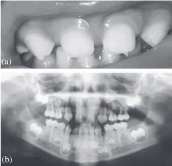
Figure 2 (a) Developmental absence of the primary upper lateral incisors in a 3-year-old child. (b) Panoramic radiograph showing the absence of the upper lateral incisors as well as of one lower incisor.
individuals had bilateral retained primary teeth and three (1.6 per cent) retained upper left lateral primary incisors. Other tooth anomalies associated with developmental absence of the upper lateral incisor
Microdontia of the contralateral incisor. Among the 121 patients presenting with unilateral absence, microdontia of the contralateral upper lateral incisor was found in 73 subjects (59.5 per cent), of which 29 (24 per cent) were on the right and 43 (35.5 per cent) on the left side (P > 0.05). Other developmentally absent or supernumerary teeth Of the 219 subjects identifi ed, in only 164 was it possible to confi rm the existence of other associated developmentally absent teeth. The remaining 55 individuals were excluded from this part of the analysis. Of the 164 subjects for which it was possible to confi rm this information, the majority (n = 143, 87.2 per cent), had no other developmentally absent teeth (Table 2). For the other 21 subjects (12.8 per cent), the dental anomaly most frequently associated with absence of the upper lateral incisor was absence of the upper or lower second premolar (n = 14, 8.5 per cent). No supernumerary teeth were found.
Discussion
In this study, a large sample of the Portuguese population was studied in order to obtain epidemiological and clinical information related to the absence of upper lateral incisors. It is likely that data provided for this study were accurate as patients recall the extraction of teeth in this site due to the aesthetic effects. The use of panoramic radiographs allowed access to a source of reliable, extensive and easily
accessible data. However, a further clinical inquiry and/or examination was needed to confi rm the absence.
Although clinical observation of all individuals would have allowed more accurate data regarding the morphology of the other incisors, as well as any other dental anomalies (Nick-Hussein and Majid, 1996; Zhu et al.,1996b), this was not feasible in the current study. Therefore, as the data was mainly radiographic, the possibility of errors due to image distortion can not be excluded.
The prevalence of developmental absence of the permanent upper lateral incisors varies considerably between studies (Horowitz, 1966; Helm, 1968; Magnusson, 1977; Muller et al., 1970; Rølling, 1980; Moyers, 1988; Leitão, 1993; Johannsdottir et al., 1997; Baccetti, 1998; Canut-Brussola, 2000; Laskaris, 2000). In the present investigation, a prevalence of 1.3 per cent was found. This compares well with other reported values of between 0.8 and 2 per cent. Johannsdottir et al. (1997) quoted a prevalence of 0.8 per cent, in a group of 396 six-year old children from Iceland (average age 6 years 7 months). Horowitz (1966) found a prevalence of 1.11 per cent, in a population of 1000 individuals, with ages ranging between 7 and 16 years. Aasheim and Øgaard (1993), found one of the highest values of around 2 per cent, in a Nordic sample of 1953 nine-year-old children undergoing orthodontic examination. These differences could be related to sample selection, but it is also possible that different populations vary due to genetic variability and/or different exposure to environmental factors.
In one of the very few studies undertaken in Portugal, Leitão (1993) examined a population of 666 twelve-year-old children from the city of Lisboa and estimated a prevalence of 1.4 per cent for upper right lateral incisor absence and 1.7 per cent for upper left. In this age group, it is clearly easier to determine whether the incisor is absent or whether extraction of the incisor has occurred. In the present study, in order to overcome this limitation, individual patients Table 2 Associated developmental absence of other teeth found in the present study.
Other associated congenital absences n Percentage
None 143 87.2 25 1 0.6 31 2 1.2 32 2 1.2 35 1 0.6 41 1 0.6 42 1 0.6 15, 25 3 1.8 31, 41 1 0.6 25, 35 1 0.6 35, 45 3 1.8 15, 25, 35, 45 3 1.8 15, 23, 25, 34, 35, 44 1 0.6 16, 25, 26, 35, 32, 45 1 0.6 Total 164 100.0
Figure 3 Analysis of 16 771 radiographs showed probable congenital
absence of the upper lateral incisor in 267 subjects and of those 219 were confi rmed. Ninety eight had bilateral congenital absence and 121 unilateral; 73 of these were on the right and 48 on the left side. In subjects with unilateral congenital absence, microdontia of the contralateral incisor was found in 29 subjects on the right side and in 43 on the left side.
were contacted by telephone, so that those subjects where the tooth had been extracted could be excluded. The present fi ndings cannot be compared with the results obtained by Leitão (1993), since the prevalence of upper lateral incisor agenesis was determined only for a subset of the population and, in addition, unilateral versus bilateral absence was not investigated, nor the association of upper lateral incisor absence with other dental anomalies. However, for the data that can be compared, the prevalence values found were within the same age range, and the gender distribution also appears similar. The only difference is that the upper left lateral inicisor was noted to be absent more often in Leitão’s study. The sample obtained in Lisboa is likely to be more variable in terms of ethnicity than in the present sample and this could explain some of the differences.
Muller et al. (1970) reported a prevalence of 1.65 per cent for agenesis of the upper lateral incisor, this value being similar to that in the present study, in a sample of 14 940 North Americans (13 459 Caucasians and 1481 Afro-Americans), aged between 6 and 14 years. Those authors also found, interestingly, that when one or two teeth were absent, the upper lateral incisor was the most frequently absent, but when more than two teeth where absent, the second premolar was the most frequently absent.
Developmental absence of the upper lateral incisors has been shown to be more common in females (Horowitz, 1966; Helm, 1968; Magnusson, 1977; Muller et al., 1979; Rølling, 1980; Dermaut et al., 1986; Leitão, 1993; Johannsdottir et al., 1997; Laskaris, 2000), which is in agreement with the results of the present study, where an important difference in gender distribution (P < 0.02) was found. The reasons for this consistent gender difference are not clear, and may be related to gender-determined features, i.e. sexual dimorphism in general skeletal growth and in tooth eruption (Proffi t, 1995).
Of the patients under 8 years of age, only two (0.26 per cent) had developmental absence of the primary lateral incisors, one being bilateral. This confi rms that the occurrence of hypodontia in the primary dentition is rare (Ravn, 1971; Bennett and Ronk, 1980; Järvinen and Lehtinen, 1981; Johannsdottir et al., 1997) and, when verifi ed, the permanent successors were also absent (Ravn, 1971; Järvinen and Lehtinen, 1981). It must be noted, however, that other studies (Nick-Hussein and Majid, 1996; Canut-Brussola, 2000; Daugaard-Jensen et al., 1997) have shown that absence of a primary incisor is not always followed by that of the succedaneous tooth.
In the patients aged 8 years or over, the primary tooth was retained in 6.7 per cent. However, among patients aged 15 years or over, only 2.7 per cent presented with retention of the primary tooth. Therefore, retention of the primary lateral incisors as an aesthetic replacement in the absence of the permanent lateral incisors is not feasible in many cases.
The results of this study are in agreement with those of other authors (Magnusson, 1977; Rølling, 1980; Aasheim and Øgaard, 1993), who also found unilateral absence was more common than bilateral absence, with a trend for the upper right lateral incisor to be absent more often than the left, although the difference was not statistically signifi cant. In subjects with clefts of the lip/palate, an asymmetrical distribution has been described, but with the left side more often affected (Tsai et al., 1998; Shapira et al., 2000); this has weakened the hypothesis that developmental tooth absence and cleft lip/palate could result from similar mechanisms (Ranta et al., 1983; Ranta and Tulensalo, 1988). In addition, studies of the relatives of children with cleft lip/palate did not show an increased frequency of developmental absence (Anderson and Moss, 1996).
In the present study all teeth where the mesiodistal dimensions were reduced were classifi ed as microdont. In agreement with Horowitz (1966), Svinhufvud et al. (1988), Baccetti (1998) and Nieminen et al. (1995), microdontia was the anomaly most frequently associated with hypodontia. Of the 121 patients in this study who had unilateral absence, 72 (59.5 per cent) were found to have a microdont lateral incisor on the other side. Nieminen et al. (1995) proposed that microdontia of one or more upper lateral incisors represented variable expression of the same anomaly. The data in the present study supports their theory, as does the investigation by Garn and Lewis (1970), who stated that microdontia is associated with missing teeth, and is more evident in cases of multiple agenesis and occurs more frequently in females than in males. Schalk-van Der Weide et al. (1994), stated that in patients presenting with developmentally missing teeth, the mesiodistal dimensions and the labiolingual dimensions are diminished for all teeth. In a later study, Schalk-van der Weide and Bosman (1996) also noted a reduction in the size of some teeth in relatives of patients presenting with hypodontia.
The terms ‘hypodontia’ and ‘concomitant hyperdontia’ (Segura and Jimenez-Rubio, 1998) and ‘oligo-pleiodontia’ (Nathanil, 1970) have been used to describe a situation in which developmental absence and supernumerary teeth are present in the same individual. These situations are rare (Ranta and Tulensalo, 1988; Zhu et al., 1996 a,b), and were not found in the present study. This confi rms the results of Baccetti (1998), who stated that supernumerary teeth appear to be of a separate aetiological entity from the absence of upper lateral incisors. As the present sample was larger than those quoted, it may be concluded that this situation occurs even less frequently in the Portuguese population. However, the possibility of these results being distorted by possible extraction of the supernumerary teeth cannot be completely excluded.
The aetiology of hypodontia remains unknown in spite of recent progress. It is interesting that the teeth that erupt in critical terminal areas of the dental lamina (such as the upper lateral incisor, second premolars, third molars) and those
located in the embryonic fusion areas, (the latter growing within the tissue corresponding to each ‘family’ of teeth) are the teeth that are most frequently affected, the so-called ‘end of series’ anomaly (Svinhufvud et al., 1988; Thesleff, 1996). Sejrsen et al. (1995) and Kjær (1997) explained agenesis as a product of the lack of innervation in the fi nal stages of development on the three distinct bilateral neural fi elds, thus altering the development of the teeth furthest from the innervation of the fi eld. In agreement with this, there is a close association between developmental absence of the second premolars and absence of the upper lateral incisors. Molecular factors that control neural development may also have an impact on tooth formation and therefore their malformation may lead to failure of development. The continued search for the genetic basis of hypodontia may help to elucidate these mechanisms.
Conclusion
The fi ndings showed that 1.3 per cent of the individuals studied had developmental absence of the upper lateral incisors, with females being more frequently affected. The most common presentation was that of unilateral absence of the upper right lateral incisor associated with microdontia of the contralateral incisor, suggesting the possibility of the existence of a variant expression of the same characteristic.
Address for correspondence Professor Teresa Pinho Rua Central de Gandra 1317 4585-116 Gandra
Paredes Portugal
E-mail: [email protected] References
Aasheim B, Øgaard B 1993 Hypodontia in 9-year-old Norwegians related to need of orthodontic treatment. Scandinavian Journal of Dental Research 101: 257–260
Anderson P J, Moss A L 1996 Dental fi ndings in parents of children with cleft lip and palate. Cleft Palate-Craniofacial Journal 33: 436–439 Baccetti T 1998 A controlled study of associated dental anomalies. Angle
Orthodontist 68: 267–274
Bennett C G, Ronk S L 1980 Congenitally missing primary teeth: report of case. Dentistry for Children 47: 346–348
Canut-Brussola J A 2000 Ortodoncia Clinica. Salvat Editores, Barcelona Daugaard-Jensen J, Nodal M, Skovgaard L T, Kjær I 1997 Comparison
of the pattern of agenesis in the primary and permanent dentitions in a population characterized by agenesis in the primary dentition. International Journal of Paediatric Dentistry 7: 143–488
Dermaut L R, Goeffers K R, De Smit A A 1986 Prevalence of tooth agenesis correlated with jaw relationship and dental crowding. American Journal of Orthodontics and Dentofacial Orthopedics 90: 204–210
Garn S M, Lewis A B 1970 The gradient and the pattern of crown-size reduction in simple hypodontia. Angle Orthodontist 40: 51–58
Guckes A D, Roberts M W, McCarthy G R 1998 Pattern of permanent teeth present in individuals with ectodermal dysplasia and severe hypodontia suggests treatment with dental implants. Pediatric Dentistry 20: 278–280 Helm S 1968 Malocclusion in Danish children with adolescent dentition: an
epidemiologic study. American Journal of Orthodontics 54: 352–366 Horowitz J M 1966 Aplasia and malocclusion: a survey and appraisal.
American Journal of Orthodontics 52: 440–453
Järvinen S, Lehtinen L 1981 Supernumerary and congenitally missing primary teeth in Finnish children. An epidemiologic study. Acta Odontologica Scandinavica 39: 83–86
Johannsdottir B, Wisth P J, Magnusson T E 1997 Prevalence of malocclusion in 6-year-old Icelandic children. Acta Odontologica Scandinavica 55: 398–402
Kjær I 1997 Can the location of tooth agenesis and the location of initial bone loss seen in juvenile periodontitis be explained by neural developmental fi elds in the jaws? Acta Odontologica Scandinavica 55: 70–72 Laskaris G 2000 Color atlas of oral diseases in children and adolescents.
Thieme, New York
Leitão P 1993 Prevalência da má oclusão em crianças de 12 anos da cidade de Lisboa. Parte I. Revista Portuguesa de Estomatologia e Cirurgia Maxilofacial 33: 193–201
Lidral A C, Reising B C 2002 The role of MSX1 in human tooth agenesis. Journal of Dental Research 81: 274–278
Lyngstardaas S P, Norbo H, Gedde-Dahl Jr T, Thrane P S 1996 On the genetics of hypodontia and microdontia: synergism or allelism of major genes in a family with six affected members. Journal of Medical Genetics 33: 137–142
Magnusson T E 1977 Prevalence of hypodontia and malformations of permanent teeth in Iceland. Community Dentistry and Oral Epidemiology 5: 173–178
Maklin M, Dummett Jr C O, Weinberg R 1979 A study of oligodontia in a sample of New Orleans children. Journal of Dentistry for Children 46: 478–482
Moyers R 1988 Handbook of orthodontics. Year Book Medical Publishers, Chicago
Muller T P, Hill I N, Peterson A C, Blayney J R 1970 A survey of congenitally missing permanent teeth. Journal of the American Dental Association 81: 101–107
Nathanil P 1970 Letter to Editor. British Dental Journal 129: 95
Nick-Hussein N N, Majid Z A 1996 Dental anomalies in the primary dentition: distribution and correlation with the permanent dentition. Journal of Clinical Pediatric Dentistry 21: 15–19
Nieminen P, Arte S, Pirinen S, Peltonen L, Thesleff I 1995 Gene defect in hypodontia; exclusion of Msx1 and Msx2 as candidate genes. Human Genetics 96: 305–308
Pilo R, Kaffe I, Amir E, Sarnat H 1987 Diagnosis of developmental dental anomalies using panoramic radiographs. Journal of Dentistry for Children 54: 267–272
Pirinen S, Arte S, Apajalahti S 1996 Palatal displacement of canine is genetic and related to congenital absence of teeth. Journal of Dental Research 75: 1742–1746
Proffi t W R 1995 Ortodontia contemporânea. Guanabara Koogan, Rio de Janeiro
Ranta R, Stegars T, Rintala A E 1983 Correlations of hypodontia in children with isolated cleft palate. Cleft Palate Journal 20: 163–165
Ranta R, Tulensalo T 1988 Symmetry and combinations of hypodontia in non-cleft and cleft palate children. Scandinavian Journal of Dental Research 96: 1–8
Ravn J J 1971 Aplasia, supernumerary teeth and fused teeth in the primary dentition. An epidemiologic study. Scandinavian Journal of Dental Research 79: 1–6
Rølling S 1980 Hypodontia of permanent teeth in Danish schoolchildren. Scandinavian Journal of Dental Research 88: 365–369
Schalk-van der Weide Y, Bosman F 1996 Tooth size in relatives of individuals with oligodontia. Archives of Oral Biology 41: 469–472
Schalk-van der Weide Y, Steen W H, Beemer F A, Bosman F 1994 Reductions in size and left-right asymmetry of teeth in human oligodontia. Archives of Oral Biology 39: 935–939
Schalk-van der Weide Y, Steen W H, Bosman F 1992 Distribution of missing teeth and tooth morphology in patients with oligodontia. Journal of Dentistry for Children 59: 133–140
Segura J J, Jimenez-Rubio A 1998 Concomitant hypohyperdontia: simultaneous occurrence of a mesiodens and agenesis of a maxillary lateral incisor. Oral Surgery, Oral Medicine, Oral Pathology, Oral Radiology and Endodontics 86: 473–475
Sejrsen B, Kjær I, Jakobsen J 1995 Agenesis of permanent incisors in a mediaeval maxilla and mandible: aetiological aspects. European Journal of Oral Sciences 103: 65–69
Shapira Y, Lubit E, Kuftinec M M 2000 Hypodontia in children with various types of clefts. Angle Orthodontist 70: 16–21
Stimson J M, Sivers J E, Hlava G L 1997 Features of oligodontia in three generations. Journal of Clinical Pediatric Dentistry 21: 269–275 Svinhufvud E, Myllarniemi S, Norio R 1988 Dominant inheritance of
tooth malposition and their association to hipodontia. Clinical Genetics 34: 373–381
Tavajohi-Kermani H, Kapur R, Sciote J J 2002 Tooth agenesis and craniofacial morphology in an orthodontic population. American Journal of Orthodontics and Dentofacial Orthopedics 122: 39–47
Thesleff I 1996 Two genes for missing teeth. Nature Genetics 13: 379–380
Thilander B, Myrberg N 1973 The prevalence of malocclusion in Swedish schoolchildren. Scandinavian Journal of Dental Research 81: 12–21
Tsai T P, Huang C S, Huang C C, See L C 1998 Distribution patterns of primary and permanent dentition in children with unilateral complete cleft lip and palate. Cleft Palate-Craniofacial Journal 35: 154–160 Vastardis H 2000 The genetics of human tooth agenesis: new discoveries
for understanding dental anomalies. American Journal of Orthodontics and Dentofacial Orthopedics 117: 650–656
Vastardis H, Karimbux N, Guthua S W, Seidman J G, Seidman C E 1996 A human MSX1 homeodomain missense mutation causes selective tooth agenesis. Nature Genetics 13: 417–421
Woodworth A, Sinclair P M, Alexander R G 1985 Bilateral congenital absence of maxillary lateral incisors: a craniofacial and dental cast analysis. American Journal of Orthodontics 87: 280–293
Zhu J F, Crevoisier R, Henry R J 1996a Congenitally missing permanent lateral incisors in conjunction with a supernumerary tooth: case report. Pediatric Dentistry 18: 64–66
Zhu J F, Marcushamer M, King D L, Henry R J 1996b Supernumerary and congenitally absent teeth: a literature review. Journal of Clinical Pediatric Dentistry 20: 87–95