Volume 2013, Article ID 630683,7pages http://dx.doi.org/10.1155/2013/630683
Research Article
Bovine Papillomavirus Clastogenic Effect
Analyzed in Comet Assay
R. P. Araldi,
1,2T. C. Melo,
1,2N. Diniz,
1,3J. Mazzuchelli-de-Souza,
1,3R. F. Carvalho,
1W. Beçak,
1,4and R. C. Stocco
1,21Laborat´orio de Gen´etica, Instituto Butantan, Avenida Vital Brasil, 1500, Butant˜a, 05503-900 S˜ao Paulo, SP, Brazil 2Programa de P´os-graduac¸˜ao Interunidades em Biotecnologia, Instituto de Ciˆencias Biom´edicas, Universidade de S˜ao Paulo,
Avenida Prof. Lineu Prestes, 2415 Edif´ıcio ICB-III-Cidade Universit´aria, 05508-900 S˜ao Paulo, SP, Brazil
3Programa de P´os-graduac¸˜ao em Biologia Estrutural e Funcional, Universidade Federal de S˜ao Paulo, Rua Botucatu, 740,
04023-900 S˜ao Paulo, SP, Brazil
4Departamento de Biologia, Universidade Federal da Integrac¸˜ao Latino-Americana (UNILA), Avenida Tancredo Neves, 6731 bloco 4,
85867-970 Foz do Iguac¸´u, PR, Brazil
Correspondence should be addressed to R. C. Stocco; [email protected]
Received 26 March 2013; Accepted 8 May 2013
Academic Editor: Franco Roperto
Copyright © 2013 R. P. Araldi et al. his is an open access article distributed under the Creative Commons Attribution License, which permits unrestricted use, distribution, and reproduction in any medium, provided the original work is properly cited.
Bovine papillomavirus (BPV) is an oncogenic virus related to serious livestock diseases. Oncoproteins encoded by BPV are involved in several steps of cellular transformation and have been reported as presenting clastogenic efects in peripheral lymphocytes and primary culture cells. he aim of this study was to evaluate the clastogenic potential of BPV types 1, 2, and 4 by comet assay. Peripheral blood was collected from 37 bovines, 32 infected with diferent levels of papillomatosis (12 animals have no afection) and ive calves, virus free (negative control). he viral identiication showed presence of more than one virus type in 59.375% of the infected animals. Comet assay was performed according to alkaline technique. he Kruskal-Wallis test showed statistical diference between the negative control group and infected animals (� = 0.0015). he Dunn post hoc test showed diference comparing the infected animals with calves. Mann-WhitneyUtest veriied no diference between animals infected with only one viral type and animals presenting more than one viral type. he comet assay is considered an eicient tool for assessment of damage in the host chromatin due to viral action, speciically highlighting viral activity in blood cells.
1. Introduction
Bovine papillomavirus (BPV) is a widespread oncogenic virus found worldwide belonging to the Papillomaviridae family, which displays tropism for squamous epithelial and mucosal tissues. hese viruses are associated with benign and malignant epithelial lesions. Speciically, BPV presents a double-stranded circular DNA, not coiled, with approxi-mately 8 kb, surrounded by an icosahedral capsid consisting of 360 copies of the L1 protein of 55 kDa, 72 capsomeres arranged in approximately 12 copies of the L2 protein, 39 kDa [1–8]. he papillomavirus genome is divided into three regions: early, late, and noncoding long control region (LCR), separated by two polyadenylation sites [3]. he early control region occupies 50% of the viral genome and encodes E1,
E2, E3, E4, E5, E6, and E7 proteins. he late control region occupies 40% of the genome and contains the genes that codify L1 and L2 capsid proteins and LCR, which comprises 10% of the genome, with 850 bp. However, it also contains the origin of replication and the binding sites of multiple transcription factors [3]. Oncoproteins encoded by BPV are involved in several steps of the cell transformation [1,9].
Currently, there are 13 BPV virus types described in the literature, although this number may be greater than twenty [7,9]. he virus types are divided into three genres:
Delta-papillomavirus (BPV-1, -2, and -13), Epsilonpapillomavirus
(BPV-5 and -8), andXipapillomavirus(BPV-3, -4, -6, -8, -9, -10, and -12), as well as the BPV-7 that remains not ranked in any genre [7]. Beside these, there are 16 new putative BPVs (BAA-1 to -4, BAPV-2 to -5, BAPV-7 to -10, BAPPV11MY and BPV/BR-UEL-2 to -5) [13]. According to Zhu et al. [13], BAA1 was detected in tongue epithelial papilloma, being designated BPV-12, and BPV/BR-UEL-4 described in ear cutaneous lesions was designated BPV-13 [9].
According to Stocco dos Santos et al. [1], papillomavirus can act on host chromatin causing cytogenetic alterations, such as changes in ploidy, chromatin gaps and breaks, dicentric chromosomes and rings. Signiicant increase of chromosomal aberrations was detected in animals infected with BPV, afecting genomic stability [4]. However, to date, there are no studies evaluating the BPV clastogenic potential in peripheral blood cells analyzed by comet assay.
Comet assay or single cell gel electrophoresis was intro-duced by ¨Ostling and Johanson [14] and later modiied by Singh et al. [15]. he comet assay is a simple and versatile technique that requires few eukaryotic cells, as well as having a vast DNA damage spectrum detection capacity [16–19]. In the test, cells are engulfed by agarose gel and spread over the slide, and then subjected to an electric ield that promotes free DNA fragment migration, with the appearance of a comet [15]. he nuclear region causes the head of the comet to fragment, and the length of the tail is directly related to the intensity of the damage.
he objective of this work was to evaluate the clastogenic potential of BPV types 1, 2, and 4 through comet assay in infected animals presenting cutaneous papillomatosis symp-toms (hyperproliferative lesions-papillomas), asymptomatic (without papillomas) and calves, not infected, as negative control. he eicacy of the comet assay in clastogenic eval-uations justiies this study.
2. Material and Methods
2.1. Ethics Statements. he protocols used in this study was
approved by the Ethical Committee in Research of the Uni-versidade Federal de S˜ao Paulo (Protocol number 1829/09) assigned by the President of this committee. All eforts were made to minimize any sufering in the animals.
2.2. BPV Diagnosis
2.2.1. Animal Selection. 37 bovines (Bost aurus) were selected:
32 adults, 12 asymptomatic (without visible cutaneous papil-lomas) and 20 symptomatic (with visible cutaneous papillo-mas) and 5 newborn calves that were separated from their mother ater birth and did not receive breast milk. he presence of papillomas in others organs was not evaluated due the absence of clinical characteristics that suggest bladder and/or esophageal cancer. he farm does not have the pres-ence of bracken fernPteridium aquilinumthat is involved in
oncogenic and mutagenic processes in function of quercetin presence [20]. hese calves were isolated in order to obtain virus negative controls.Blood sample.he peripheral blood samples were collected using the EDTA vacutainer. Blood
DNA extraction. he extraction of DNA from peripheral
blood cells was performed using the GenomicPrep Blood Mini Kit Illustra Spin (GE Healthcare, Buckinghamshire, UK), which uses enzymatic digestion method with protease K, according to the manufacturer. he quality of obtained DNA was veriied using PCR by amplifying a bovine�-globin gene fragment [21].
2.2.2. Viral Identiication. Viral identiication was performed
using speciic primers for BPV-1 (forward: 5� -GGAGCG-CCTGCTAACTATAGGA-3� and reverse: 5� -ATCTGT-TGTTTGGGTGGTGAC-3�), which ampliies the L1 gene, resulting in a 301 bp amplicon, BPV-2 (forward: 5� -GTT-ATACCACCCAAAGAAGACCCT-3�and reverse: 5� -CTG-GTTGCAAC¸ AGCTCTCTTTCTC-3�), which ampliies the L2 gene, resulting in a 164 amplicon, and BPV-4 (forward; 5�-GCTGACCTTCCAGTCTTAAT-3�and reverse; 5� -CAG-TTTCAATCTCCTCTTCA-3�), which ampliies the E7 gene, resulting in a 170 bp amplicon. We choose these primers because the virus types are oten observed in Brazil and in function of the association with esophageal (BPV-4) and bladder cancer (BPV-1 and -2). In detail, the ampliication reactions were performed in a Veriti 96-well thermal cycler (Applied Biosystems, Singapore), with Master Mix (Promega, Madison, USA), under the following conditions: 5 minutes at 95∘C, followed by 35 cycles of 1 minute and 30 seconds at 98∘C, 2 minutes at 52∘C, and 1 minute and 30 seconds at 72∘C and a inal extension step of 5 minutes at 72∘C, for� -globin and speciic primers. he PCR products were analyzed in 2% agarose gel electrophoresis stained with GelRed in TAE bufer, visualized under UV light. he images were captured through the sotware Kodak 1D 3.6.5. Cloned BPV-1, -2 and -4 genomes inEscherichia coliD5H�were used as positive controls. hese clones form part of the biological collection of Genetic Laboratory of Butantan Institute. he fragments were puriied using Illustra GFX PCR DNA and Gel Band Puriication Kit (GE Healthcare, Buckinghamshire, UK). DNA concentration and purity were determined in a spectrophotometer BioPhotometer plus (Eppendorf, Ham-burg, Germany) and submitted to sequencing reactions.
Sequencing. he puriied ampliied products were sequenced
in an ABI377 PRISM Genetic Analyzer. he quality of DNA sequences was checked, the overlapping fragments were assembled using the BioEdit package sotware BioEdit pack-age 7.0.9.0 [22], and the nucleotide sequences were compared through BLAST (http://blast.ncbi.nlm.nih.gov/Blast.cgi).
2.3. Comet Assay. Comet assay was performed according
normal-melting-point agarose (1.5% in PBS) at 60∘C and dried at 22∘C overnight. Ater the agarose had solidiied (4∘C for 10 min), the coverslips were carefully removed, and the slides were immersed in lysis solution (2.5 M NaCl, 100 mM Na2EDTA, 10 mM Tris-HCl; pH 10; 1% Triton X-100, and 10% DMSO) for 1 hour at 4∘C and then placed into a horizontal electrophoresis apparatus illed with bufer (1 mM Na2EDTA, 300 mM NaOH) at 4∘C. he slides were incubated for 40 min in this bufer to unwind the DNA. he electrophoresis was run for 20 min at ixed voltage of 25 V (0.83 V/cm) and 300 mA. At the end of electrophoresis, the slides were washed three times with neutralization bufer (0.4 M Tris-HCl, pH 7.5) and once in 100% ethanol. All steps described above were carried out with the lights out to avoid DNA damage. he slides were stained with 20�L of propidium iodide (4�g/mL) and visualized in Carl Zeiss Axio Scope A1 luorescent microscopy. Nucleoids were classiied from 0 (without lesion) to 2 (major damage), and the number of nucleoids observed per class was multiplied to the class value, resulting in a comet score. Statistical analysis. he data were analyzed using the Kruskal-Wallis test, followed by post hoc Dunn test and Mann-WhitneyUtest, both with signiicance level of 5%, through BioEstat 5.3. sotware.
3. Results
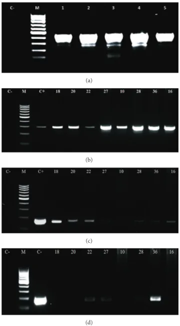
3.1. Viral Genotyping. PCR using speciic primers to�-globin
revealed DNA quality enough for further PCR procedures: all samples resulted in a 450 bp amplicon. We selected the speciic primers for BPV-1, -2, and -4 due to their prevalence in the herd, and we could detect the virus sequences in peripheral blood cells collected from the adult animals, with and without skin papillomas (Figure 1(a)). he resulting bands were puriied and sequenced to conirm the genotyping of the ampliication products. he sequences were aligned through the BioEdit 7.0.9.0, and the nucleotide comparison was done through the BLAST tool, conirming the speciicity of the primers employed. Using these primers, coinfection was reported in 59.375% of the infected bovines—19 animals
(Table 1(a)). he use of these primers did not detect the
presence of BPV in calf peripheral blood samples, with this group being considered a negative control.
3.2. Comet Assay. he samples of peripheral blood cells
collected from infected animals and calves (negative control) were evaluated through comet assay, counting 100 nucleoids per sample that were evaluated and classiied as 0 (without damage), 1 (medium damage), and 2 (maximum damage) according to Figure 2. he nucleoid value per class was multiplied by the respective class value, resulting in the comet score (Table 1(a)).
Based on these data, a Kruskal-Wallis test was used, with 5% signiicance level, through BioEstat 5.3. sotware to compare the diferent groups (infected and not infected group). he test reveled statistical diferences between the groups (� = 12.9714 and �value = 0.0015). he Dunn post hoc test showed diference in score values among calf and infected animals (asymptomatic and symptomatic)
(a)
(b)
(c)
(d)
Figure 1: Electrophoresis’s images of: (a) (�-globin ampliication, resulting in an amplicon of 450 bp), (b) (ampliication of L1 gene, using primer to BPV-1, showing an amplicon of 301 bp), (c) (ampli-ication of L2 gene, using primer to BPV-2, showing an amplicon of 164 bp) and (d) (ampliication of E7 gene, using primer to BPV-4, showing an amplicon of 170 bp), being C−(negative control) and C+ (positive control).
(Table 1(b)). Mann-WhitneyUtest was done using the same
sotware to compare the level of clastogenicity between animals, which showed just one virus type, and coinfected cattle. he results did not reveal statistical diferences between animals infected with only one viral type and animals presenting more than one viral type (Table 1(c)).
4. Discussion
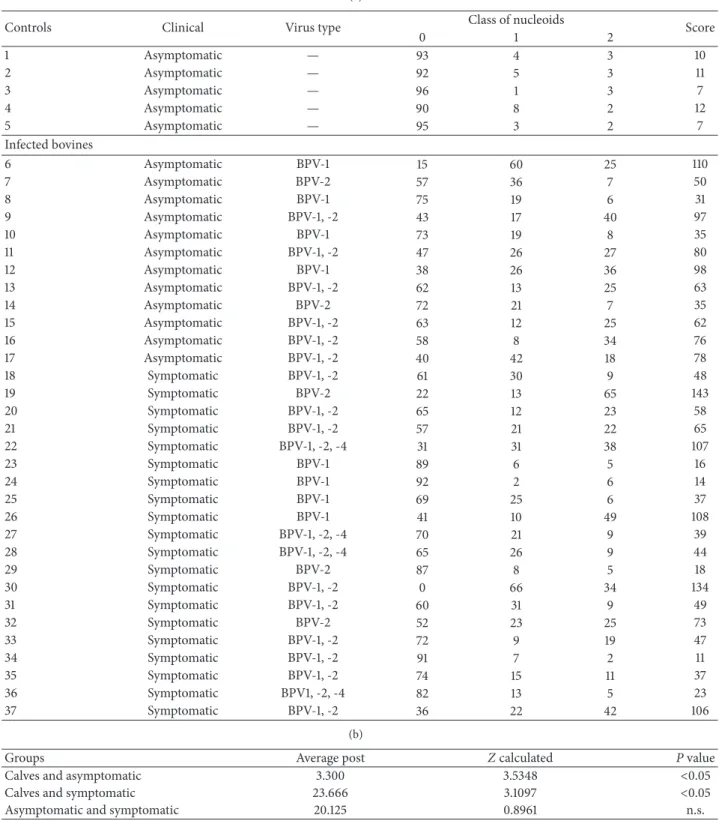
Table 1: (a) Results of molecular diagnosis of calves (negative control) and asymptomatic and symptomatic adult cattle, showing the BPV type, the frequency of nucleoids observed per class, and the comet score. (b) Kruskal-Wallis statistic to compare the clastogenicity among calves, asymptomatic and symptomatic cattle, from the comet score. (c) Comparison between comet score observed in monoinfected and coinfected bovines through Mann-WhitneyUtest.
(a)
Controls Clinical Virus type Class of nucleoids Score
0 1 2
1 Asymptomatic — 93 4 3 10
2 Asymptomatic — 92 5 3 11
3 Asymptomatic — 96 1 3 7
4 Asymptomatic — 90 8 2 12
5 Asymptomatic — 95 3 2 7
Infected bovines
6 Asymptomatic BPV-1 15 60 25 110
7 Asymptomatic BPV-2 57 36 7 50
8 Asymptomatic BPV-1 75 19 6 31
9 Asymptomatic BPV-1, -2 43 17 40 97
10 Asymptomatic BPV-1 73 19 8 35
11 Asymptomatic BPV-1, -2 47 26 27 80
12 Asymptomatic BPV-1 38 26 36 98
13 Asymptomatic BPV-1, -2 62 13 25 63
14 Asymptomatic BPV-2 72 21 7 35
15 Asymptomatic BPV-1, -2 63 12 25 62
16 Asymptomatic BPV-1, -2 58 8 34 76
17 Asymptomatic BPV-1, -2 40 42 18 78
18 Symptomatic BPV-1, -2 61 30 9 48
19 Symptomatic BPV-2 22 13 65 143
20 Symptomatic BPV-1, -2 65 12 23 58
21 Symptomatic BPV-1, -2 57 21 22 65
22 Symptomatic BPV-1, -2, -4 31 31 38 107
23 Symptomatic BPV-1 89 6 5 16
24 Symptomatic BPV-1 92 2 6 14
25 Symptomatic BPV-1 69 25 6 37
26 Symptomatic BPV-1 41 10 49 108
27 Symptomatic BPV-1, -2, -4 70 21 9 39
28 Symptomatic BPV-1, -2, -4 65 26 9 44
29 Symptomatic BPV-2 87 8 5 18
30 Symptomatic BPV-1, -2 0 66 34 134
31 Symptomatic BPV-1, -2 60 31 9 49
32 Symptomatic BPV-2 52 23 25 73
33 Symptomatic BPV-1, -2 72 9 19 47
34 Symptomatic BPV-1, -2 91 7 2 11
35 Symptomatic BPV-1, -2 74 15 11 37
36 Symptomatic BPV1, -2, -4 82 13 5 23
37 Symptomatic BPV-1, -2 36 22 42 106
(b)
Groups Average post Zcalculated Pvalue
Calves and asymptomatic 3.300 3.5348 <0.05
Calves and symptomatic 23.666 3.1097 <0.05
Asymptomatic and symptomatic 20.125 0.8961 n.s.
(c)
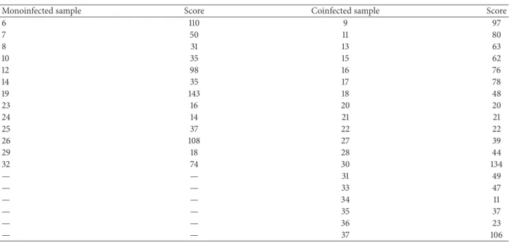
Monoinfected sample Score Coinfected sample Score
6 110 9 97
7 50 11 80
8 31 13 63
10 35 15 62
12 98 16 76
14 35 17 78
19 143 18 48
23 16 20 20
24 14 21 21
25 37 22 22
26 108 27 39
29 18 28 44
32 74 30 134
— — 31 49
— — 33 47
— — 34 11
— — 35 37
— — 36 23
— — 37 106
Mann-WhitneyUtest results:� = 102.5,�(�) = 0.80,Pvalue = 0.21.
(a) (b)
Figure 2: (a) Images of class 0 nucleoids, observed in calves (negative control) and (b) class 2 nucleoids, observed in symptomatic adult bovine, showing DNA fragmentation that is indicative of clastogenicity.
by mutations induced by the virus, associated with the action of viral protein E6, which accelerates the degradation of p53 and E7, which degrades the tumor suppressor protein pRb. hese processes change the transcriptional pathway through degradation of transcription factors, activating telomerase, afecting the DNA repair system, and leading to an increase of damage in host genetic material [6, 23–25]. According to You [6], the protein E7 interacts with microtubules in mitosis, causing defects in the alignment of chromosomes during pre-metaphase, resulting in cytogenetic alteration [1,
26]. Wade et al. [25] discussed that the BPV oncoproteins can act on the signal transduction, allowing the return of interphase spinous epithelial cells to synthesis phase, resulting in mutated cell proliferation. Oncoproteins E6 and E7 induce immortalization of transformed cells [3]. According to Primrose and Twyman [27], these oncoproteins
are required in the process of viral replication and act in the process of oncogenic transformation of the host cell. Furthermore, the accelerated p53 proteasomal degradation has a prominent role in the carcinogenic action of the virus, since the p53 protein function is to check the integrity of the genome, preventing the proliferation of mutated cells. he p53 accumulates in the cell nucleus, keeping the mitotic cycle in early G1 phase by activating p21 gene, whose gene product inhibits the action of cycling-dependent kinases (CDKs) and activates genes related to repair system [28].
but the virus or its DNA sequences, detected in peripheral blood, can represent a potential risk to carcinogenesis.
In this study, we veriied BPV clastogenic action by comet assay indicating chromatin instability. he comet assay results demonstrated that the BPV is able to induce severe DNA damages, which hinder the repair system, this is because the assay allows to evaluate the DNA double-strand breaks (DSBs) and critical lesions that are involved in genomic instability [29–31].
he DSB is associated with the homologous recombina-tion from the formarecombina-tion of DNA simple strand that invades the template strand, originating a Holliday junction, which migrates to the resolution of heteroduplex [29,31]. However, unrepaired DSBs leave to apoptosis or cell-cycle arrest, resulting in carcinogenesis [29]. here are lines of evidence that unrepaired DSBs could leave to telomeric breaks and fusion events, also associated with oncogenic process [29,31]. he viral oncoproteins afect the repair system, allowing an accumulation of stochastic mutations and resulting in increased genomic instability [17]. According to Duensing and M¨unger [23], breaks in DNA afect the cell-cycle check-point that is associated with genomic instability, leading to hyper proliferation, featuring an oncogenic process. Accord-ing to the European Study Group on Health’s Biomarkers, cytogenetic indings, as a high frequency of chromosomal aberrations, including breaks in single or double-stranded, are associated to the carcinogenesis [32]. So, at the same time the presence of BPV is causing DSBs, the virus afects the repair system, favoring oncogenic process associated with unrepaired DSBs.
he virus presence in the blood can suggest one alter-native pathway to infection, in which asymptomatic but infected cattle could turn symptomatic from a tissue injury, considering that a lesion causes an inlammatory process with lymphocyte iniltration [33]. he presence of BPV in leuko-cytes was demonstrated, had been observed the BPV presence in peripheral blood mononuclear cells [33] and the presence of L1 protein in CD4+ and CD8+ leukocytes, representing a potential infection sites to BPV-2 [34]. he possibility of the existence of endogenous pathway of infection has been discussed by Wobeser et al. [33], who suggested that the mononuclear cells act as a source of infection for inlam-mation sites, as inlamed areas become more susceptible to infection by BPV. Furthermore, it is known that lymphocytes express heparan sulfate, being cells susceptible to infection by papillomavirus [33]. Another observation that supports the possibility of infection was reported by Hartl et al. [35], who found that the spontaneous regression of papillomavirus in transient infections in humans and cattle is accompanied by an accumulation of active lymphocytes CD4+ and CD8+. So, in this pathway, the iniltration of BPV infected cell could develop a tumorigenic process from a clonal evolution started in a histologically normal tissue [35,36].
5. Conclusion
his study presents direct evidence of DNA damage related to bovine papillomavirus in blood cells, indicating a viral
activity in peripheral blood. he levels of damage were analyzed in order to verify if the presence of more than one viral type could increase the clastogenic viral action, but no signiicant diferences could be detected. he results showed the same DNA damage both in presence or absence of cutaneous papillomas, indicating that the presence of bovine papillomatosis just represents clinical symptoms due the BPV presence; however, the BPV presence in peripheral blood leaves to double-stranded breaks, which is associated to carcinogenesis, afecting the healthy animal, as previously reported [1,26]. Comet assay can be discussed as an inter-esting technique to evaluate DNA damage which, in this special situation, is related to viral action, demonstrating viral activity in diferent sites as blood cells.
Conflict of Interests
All the authors declare that there is no conlict of interests regarding the research, authorship, and/or publication of this paper. he authors inform that all materials and trademarks mentioned were used just as a part of experimental protocol and there are no inancial gains or favoritism of business.
Acknowledgments
he authors thank the Minist´erio de Ciˆencia, Tecnologia e Inovac¸˜ao/Conselho Nacional de Desenvolvimento Cient´ıico e Tecnol´ogico (CNPq Proc. 402539/2011-7), Coordenac¸˜ao de Aperfeic¸oamento Pessoal de N´ıvel Superior (CAPES), Fundac¸˜ao do Desenvolvimento Administrativo (PAP-FUNDAP) for the inancial support, Carolina da Paz Sabino for the editorial support, and Martin W. Breuer for all the eforts and structural support.
References
[1] R. C. Stocco dos Santos, C. J. Lindsey, O. P. Ferraz et al., “Bovine papillomavirus transmission and chromosomal aberrations: an experimental model,”Journal of General Virology, vol. 79, no. 9, pp. 2127–2135, 1998.
[2] A. C. de Freitas, C. de Carvalho, O. Brunner et al., “Viral DNA sequences in peripheral blood and vertical transmission of the virus: a discussion about BPV-1,”Brazilian Journal of Microbiology, vol. 34, no. 1, pp. 76–78, 2003.
[3] Z.-M. Zheng and C. C. Baker, “Papillomavirus genome struc-ture, expression, and post-transcriptional regulation,”Frontiers in Bioscience, vol. 11, no. 1, pp. 2286–2302, 2006.
[4] A. C. Freitas, M. A. R. Silva, C. C. R. Carvalho et al., “Papillomavirus DNAdetectionin non-epithelial tissue: adis-cussion about bovine papillomavirus,” inCommunicating Cur-rent Research and Educational Topics and Trends in Applied Microbiology, pp. 697–704, 2007.
[5] S. Roperto, R. Brun, F. Paolini et al., “Detection of bovine papillomavirus type 2 in the peripheral blood of cattle with urinary bladder tumours: possible biological role,”Journal of General Virology, vol. 89, no. 12, pp. 3027–3033, 2008.
[6] J. You, “Papillomavirus interaction with cellular chromatin,”
[7] A. C. Freitas, A. R. M. Silva, A. L. S. Jesus et al., “Recent insights into bovine papillomavirus,”African Journal of Microbiology Research, vol. 5, no. 33, pp. 6004–6012, 2011.
[8] C. de Carvalho, A. C. de Freitas, O. Brunner et al., “Bovine papillomavirus type 2 in reproductive tract and gametes of slaughtered bovine females,”Brazilian Journal of Microbiology, vol. 34, no. 1, pp. 82–84, 2003.
[9] M. Lunardi, A. A. Alieri, R. A. A. Otonel et al., “Genetic characterization of a novel bovine papillomavirus member of the Deltapapillomavirus genus,”Veterinary Microbiology, vol. 162, no. 1, pp. 207–213, 2013.
[10] J. DeMasi, M. C. Chao, A. S. Kumar, and P. M. Howley, “Bovine papillomavirus E7 oncoprotein inhibits anoikis,” Journal of Virology, vol. 81, no. 17, pp. 9419–9425, 2007.
[11] C. J. Lindsey, M. E. Almeida, C. F. Vicari et al., “Bovine papillomavirus DNA in milk, blood, urine, semen, and sperma-tozoa of bovine papillomavirus-infected animals,”Genetics and Molecular Research, vol. 8, no. 1, pp. 310–318, 2009.
[12] S. R. C. Campos, C. Trindade, O. P. Ferraz et al., “Can established cultured papilloma cells harbor bovine papillomavirus?” Genet-ics and Molecular Research, vol. 7, no. 4, pp. 1119–1126, 2008. [13] W. Zhu, J. Dong, E. Shimizu et al., “Characterization of novel
bovine papillomavirus type 12 (BPV-12) causing epithelial papilloma,”Archives of Virology, vol. 157, no. 1, pp. 85–91, 2012. [14] O. ¨Ostling and K. Johanson, “Microeletrophoretic study of
radiation-induced DNA damages in individual mammalian cells,”Biochemical and Biophysical Research Communications, vol. 123, pp. 291–298, 1984.
[15] N. P. Singh, M. T. McCoy, R. R. Tice, and E. L. Schneider, “A simple technique for quantitation of low levels of DNA damage in individual cells,”Experimental Cell Research, vol. 175, no. 1, pp. 184–191, 1988.
[16] P. Escobar, “New applications of the Comet Assay in genetic toxicology testing,” in Proceedings of the Genetic Toxicology Association Meeting, September 2008.
[17] S. A. S. Langie, K. M. Cameron, K. J. Waldron, K. P. R. Fletcher, T. von Zglinicki, and J. C. Mathers, “Measuring DNA repair incision activity of mouse tissue extracts towards singlet oxygen-induced DNA damage: a comet-based in vitro repair assay,”Mutagenesis, vol. 26, no. 3, pp. 461–471, 2011.
[18] S. Khoei, S. Delfan, A. Neshasteh-Riz, and S. R. Mahdavi, “Evaluation of the combined efect of 2ME2 and60Co on the inducement of DNA damage by IUdR in a spheroid model of the U87MG glioblastoma cancer cell line using alkaline comet assay,”Cell Journal, vol. 13, no. 2, pp. 83–90, 2011.
[19] R. Fabiani, P. Rosignoli, A. de Bartolomeo, R. Fuccelli, and G. Morozzi, “Genotoxicity of alkene epoxides in human peripheral blood mononuclear cells and HL60 leukaemia cells evaluated with the comet assay,”Mutation Research, vol. 747, pp. 1–6, 2012. [20] J. D. C. Dias, M. D. C. Sgnacchiti, P. G. G. Giuriato, L. C. Nunes, and O. S. Pereira-J´unior, “Detecc¸˜ao do papilomav´ırus bovino tipo 2 em bexigas de bovinos com hemat´uria enzo´otica pela t´ecnica de reac¸˜ao de cadeia de polimerase no Sul do Esp´ırito Santo,”Revista Brasileira de Medicina Veterin´aria, vol. 34, pp. 146–151, 2012.
[21] A. Yaguiu, M. L. Z. Dagli, E. H. Birgel Jr. et al., “Simultaneous presence of bovine papillomavirus and bovine leukemia virus in diferent bovine tissues: in situ hybridization and cytogenetic analysis,”Genetics and Molecular Research, vol. 7, no. 2, pp. 487– 497, 2008.
[22] T. A. Hall, “BioEdit: a user-friendly biological sequence align-ment editor and analysis program for Windows 95/98/NT,”
Nucleic Acids Symposium Series, vol. 41, pp. 95–98, 1999. [23] S. Duensing and K. M¨unger, “he human papillomavirus type
16 E6 and E7 oncoproteins independently induce numerical and structural chromosome instability,”Cancer Research, vol. 62, no. 23, pp. 7075–7082, 2002.
[24] Y. Liu and J. D. Baleja, “Structure and function of the papil-lomavirus E6 protein and its interacting proteins,”Frontiers in Bioscience, vol. 13, no. 1, pp. 121–134, 2008.
[25] R. Wade, N. Brimer, and S. Vande Pol, “Transformation by bovine papillomavirus type 1 E6 requires paxillin,”Journal of Virology, vol. 82, no. 12, pp. 5962–5966, 2008.
[26] T. C. Melo, N. Diniz, S. R. C. Campos et al., “Cytogenetic studies in peripheral blood of bovines alicted by papillomatosis,”
Veterinary and Comparative Oncology, vol. 9, no. 4, pp. 269–274, 2011.
[27] S. B. Primrose and R. M. Twyman,Principles of Gene Manipu-lation and Genomics, Blackwell Scientiic Publications, Oxford, UK, 7th edition, 2006.
[28] G. B. Cavalcanti-J´unior, C. E. Klumb, and R. C. Maia, “p53 e as hemopatias malignas,”Revista Brasileira de Cancerologia, vol. 48, pp. 419–442, 2002.
[29] M. O’Driscoll and P. A. Jeggo, “he role of double-strand break repair—insights from human genetics,”Nature Reviews Genetics, vol. 7, no. 1, pp. 45–54, 2006.
[30] R. Kanaar, J. H. J. Hoeijmakers, and D. C. van Gent, “Molecular mechanisms of DNA double-strand break repair,”Trends in Cell Biology, vol. 8, no. 12, pp. 483–489, 1998.
[31] M. Shrivastav, L. P. de Haro, and J. A. Nickolof, “Regulation of DNA double-strand break repair pathway choice,”Cell Research, vol. 18, no. 1, pp. 134–147, 2008.
[32] A. Collins, D. Anderson, E. Coskun et al., “Launch of the ComNet (Comet-Network) project on the comet assay in human population studies during the International Comet Assay Workshop Meeting in Kusadasi, Turkey (September 13– 16, 2011),”Mutagenesis, vol. 27, pp. 385–386, 2012.
[33] B. K. Wobeser, J. E. Hill, M. L. Jackson et al., “Localization of Bovine papillomavirus in equine sarcoids and inlammatory skin conditions of horses using laser microdissection and two forms of DNA ampliication,”Journal of Veterinary Diagnostic Investigation, vol. 24, no. 1, pp. 32–41, 2012.
[34] S. Roperto, S. Comazzi, E. Ciusani et al., “PBMCS are additional sites of productive infection of bovine papillomavirus type 2,”
Journal of General Virology, vol. 92, no. 8, pp. 1787–1794, 2011. [35] B. Hartl, E. K. Hainisch, S. Shati-Keramat et al., “Inoculation of
young horses with bovine papillomavirus type 1 virions leads to early infection of PBMCS prior to pseudo-sarcoid formation,”
Journal of General Virology, vol. 92, no. 10, pp. 2437–2445, 2011. [36] C. T. Leung and J. S. Brugge, “Outgrowth of single oncogene-expressing cells from suppressive epithelial environments,”
Submit your manuscripts at
http://www.hindawi.com
Stem Cells
International
Hindawi Publishing Corporationhttp://www.hindawi.com Volume 2014
Hindawi Publishing Corporation
http://www.hindawi.com Volume 2014
INFLAMMATION
Hindawi Publishing Corporation
http://www.hindawi.com Volume 2014
Behavioural
Neurology
Endocrinology
International Journal ofHindawi Publishing Corporation
http://www.hindawi.com Volume 2014
Hindawi Publishing Corporation
http://www.hindawi.com Volume 2014
Disease Markers
Hindawi Publishing Corporation
http://www.hindawi.com Volume 2014
BioMed
Research International
Oncology
Journal of Hindawi Publishing Corporationhttp://www.hindawi.com Volume 2014
Hindawi Publishing Corporation
http://www.hindawi.com Volume 2014 Oxidative Medicine and Cellular Longevity Hindawi Publishing Corporation
http://www.hindawi.com Volume 2014
PPAR Research
The Scientiic
World Journal
Hindawi Publishing Corporationhttp://www.hindawi.com Volume 2014
Immunology Research
Hindawi Publishing Corporation
http://www.hindawi.com Volume 2014
Journal of
Obesity
Journal ofHindawi Publishing Corporation
http://www.hindawi.com Volume 2014
Hindawi Publishing Corporation
http://www.hindawi.com Volume 2014
Computational and Mathematical Methods in Medicine
Ophthalmology
Journal ofHindawi Publishing Corporation
http://www.hindawi.com Volume 2014
Diabetes Research
Journal ofHindawi Publishing Corporation
http://www.hindawi.com Volume 2014
Hindawi Publishing Corporation
http://www.hindawi.com Volume 2014
Research and Treatment
AIDS
Hindawi Publishing Corporation
http://www.hindawi.com Volume 2014
Gastroenterology Research and Practice
Hindawi Publishing Corporation
http://www.hindawi.com Volume 2014
Parkinson’s
Disease
Evidence-Based Complementary and Alternative Medicine
Volume 2014