ORIGINAL ARTICLE
METABOLIC PROFILE OF FOOT AND MOUTH DISEASE
STRESSED SHEEP IN SEMI ARID REGION
Anil Gattani
1*, Koshal K Gupta
2, Gurudutt Joshi
1, Sita R Gupta
11
Apollo College of Veterinary Medicine, Agra Road, Jamdoli, Jaipur (Raj.), Postal Code-
302031, INDIA
2
Veterinary Hospital, K. Patan (Raj), INDIA
*
E-mail:[email protected]
Received April 20, 2011
The present study was designed to evaluate serum biochemical parameters in twenty local bred sheep infected with Foot-and-Mouth disease virus (FMDV) serotype O. Ten healthy sheep were used as controls. Peripheral blood was collected from both diseased and control group and serum was separated which was further used to estimate the concentration of glucose, total protein, albumin, urea, calcium, phosphorus, cholesterol and activity of AST, ALT and ALP. It was found that there was a significant increase in glucose, AST and phosphorus in FMD affected sheep (p<0.01). Total protein, albumin, calcium, cholesterol and urea level were significantly lower (p<0.05) in FMD group compared to those in the control group. The biochemical alteration indicates the development of pancreatic dysfunction in Foot and Mouth disease affected sheep with FMDV serotype O.
Key words: Sheep/ Glucose/ AST/ ALT/ FMD
Foot-and-mouth disease (FMD) is an OIE listed
and one of the most feared viral diseases of the
livestock. The disease is highly contagious in
cloven-footed animals, most prevalent in cattle and
buffaloes followed by sheep and goats, whereas pigs
acts as amplifiers. The FMD virus (FMDV) belongs
to genus Aphthovirus of the family Picornaviridae
and is classified into seven antigenically distinct
serotypes i.e. O, A, C, SAT1, SAT2 SAT3, and
Asia1; and innumerable subtypes. In domestic
animals it causes economic losses by decreasing the
production, cost of treatment etc. Lameness is
usually the first indication of FMD in sheep and
goats. An affected animal develops fever, is
reluctant to walk, vesicles develop in the interdigital
cleft, on the heel bulb and on the coronary band.
Vesicles also form in the mouth (on dental pad, hard
(Kitching & Hughes, 2002). Some strains of the
virus cause necrosis of heart muscle and may result
in death before lesions develop in more common
and visible locations such as the mouth or foot
(Rodostitis, 1994).Most of the work being carried
out on FMD is concerned with the cattle. However,
there is lack of published reports related to small
ruminants. The level of certain metabolites in blood
may indicate various types of stress, resulting in an
animal or a herd under a given management system
possibly becoming susceptible to certain diseases
(Payne 1973). The aim of the present study was to
determine the possible alteration in biochemical
parameters in naturally infected sheep with FMD.
MATERIALS AND METHODS
Experimental animals
Two groups of sheep were used, one group
comprises of 20 naturally infected sheep with FMD
virus and other group with 10 healthy sheep was
kept as control. The animals were of local breed and
both groups comprises of both Ram and Ewe. The
animals were kept under free-range system with
optimum maintenance requirement. Their feed
consisted of a concentrate mixture provided for their
optimal maintenance and Jowar (Sorgum) Kadbi or
Green (Lucern or Maize) accordingly to the
availability of fodder.
Collection of blood and separation of serum
Peripheral blood was collected by puncturing the
jugular vein with the least stress to the animal under
aseptic condition directly into sterile tubes. The
serum was separated out on the same day by
centrifuging at 2000 rpm for 15 min. The serum
samples were stored at -20°C till further use.
Virus isolation:
FMD virus was isolated from the tongue
epithelial samples taken in 50% buffered glycerol
and was triturated and centrifuged at centrifuged at
3000 rpm for 15 min. The supernatent was collected
and filtered through 0.22µm membrane filter and
was inoculated in Baby hamster kidney cell line,
BHK-21 CT (Clone Tubingen) grown in MEM
(GIBCO) for the presence of cytopathic effect.
Further, Serotyping was done by sandwitch ELISA
using reference FMDV reference serotypes (vaccine
strains) O, A and Asia1 procured from Central
Laboratory of the Project Directorate on FMD,
IVRI, Mukteswar-Kumaon, Uttrakhand, India.
Biochemical tests:
Blood glucose, total protein, albumin, urea,
calcium, phosphorus, cholesterol levels and activity
of AST, ALT and ALP of both experimental and
controlled group was analyzed on IDEXX VetTest
automated chemistry analyzer.
Statistical Analysis:
Statistical analysis was carried out by using an
unpaired “t” test (Panse & Sukhatme, 1965).
RESULTS
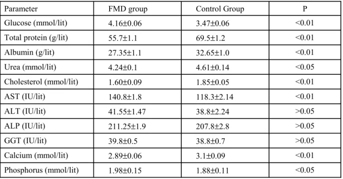
The mean values of the parameters studied are
presented in the table- 1. FMD affected sheep had
significantly increased concentration of glucose and
phosphorus, the activity of AST was significantly
higher. Further the concentration of total protein,
albumin, urea, cholesterol and calcium was
significantly lower. The activity of ALT, ALP and
GGT was non significantly higher in FMD affected
Table 1 Comparison of various Serum biochemical parameters in the FMD affected sheep and Control groups
Parameter FMD group Control Group P
Glucose (mmol/lit) 4.16±0.06 3.47±0.06 <0.01
Total protein (g/lit) 55.7±1.1 69.5±1.2 <0.01
Albumin (g/lit) 27.35±1.1 32.65±1.0 <0.01
Urea (mmol/lit) 4.24±0.1 4.61±0.14 <0.05
Cholesterol (mmol/lit) 1.60±0.09 1.85±0.05 <0.01
AST (IU/lit) 140.8±1.8 118.3±2.14 <0.01
ALT (IU/lit) 41.55±1.47 38.8±2.24 >0.05
ALP (IU/lit) 211.25±1.9 207.8±2.8 >0.05
GGT (IU/lit) 39.8±0.5 38.8±0.7 >0.05
Calcium (mmol/lit) 2.89±0.06 3.1±0.09 <0.01
Phosphorus (mmol/lit) 1.98±0.15 1.88±0.11 <0.05
DISCUSSION
Comparison of the mean values for blood
glucose between the two groups showed a
significant higher (p<0.01) glucose level in FMD
affected sheep. The researches has suggests that interleukin (IL)-1, IL-6 and tumor necrosis factor-α functions as mediators of the host inflammatory
responses in innate immunity. The biological effects
of these proinflammatory cytokines depend on the
quantity of cytokine released. For example, at low
concentration the principal function of IL-1 is as a
mediator of local inflammation. In larger quantities,
IL-1 enters the blood streams and exerts endocrine
effects. This systemic IL-1 has the ability to cause
fever, to induce the synthesis of acute phase protein
by the liver, and to initiate metabolic wasting. There are 2 principal forms of 1, called 1α and IL-1β. Most of the IL-1 activity found in the circulation is IL-1β (Abbas et.al. 1977). Stith & Templer (1994) have stated that a marked decreased in hepatic
glycogen and a concomitant increase in plasma
glucose were observed following central injection of IL-1. Johnson (1997) stated that central IL-1β has been shown to modulate the intermediary
metabolism of carbohydrates in periphery. These
effects could be attributed to the glycolytic action of
glucocorticoids, which are notably elevated by
central IL-1 (Berkenbosch 1987). Kaneko (1997)
also reported that the increase in blood glucose
concentration was in response to hypocalcemia
because of interference with the secretion of insulin
from the pancreas. Elevated blood glucose levels
were reported by Sahal, 1994 in cows recovering
from FMD. The significant decrease in total protein,
albumin and serum urea observed in the study may
be associated with lesions on the oral mucosa and
interdigital regions. It is known that protein
requirement increase in the presence of any lesions
on the body (Kaneko 1997). It is also known that
protein consumption increase in animals with
diabetes mellitus (Feldmann, 1987). Therefore
hyperglycemia is another reason for the decrease in
albumin and protein concentration may also be due to alteration in pancreatic β-cell function that might have developed during the clinical course of FMD
as reported by Barboni, 1980.
The cholesterol level in FMD affected sheep was
lower in present study. This could have been due to
the cytokine induced alteration in energy
metabolism, with lipids being released from adipose
tissue. In this situation, a reduction in cholesterol
rich very low density lipoprotein (VLDL) and low
density lipoprotein (LDL)would be expected, with
an increased ratio of VLDL and LDL to high density
lipoprotein (HDL) (Kaneko 1997).
Thyroid hormone increases both the rate of
cholesterol synthesis and the rate of its catabolism
by the liver. In hypothyroidism condition, lipids and
cholesterol catabolism are decreased to a lesser
degree than the synthesis of the cholesterol. The
effect of these changes, results in an increase in
serum cholesterol (Kaneko 1997). In contrast to this,
however, Lal 1981, found evidence indicative of
hypothyroid activity in FMD affected cows; this
author reported decreased protein bound iodine
(PBI) values in FMD affected Thrparkar cows
compared with the levels in healthy lactating cows.
The low serum calcium level in the present study
may be associated with inappetance and
hypoproteinemia as reported by Kaneko, 1997.
Therefore, in the study there was a significant
decrease in serum protein level and severe anorexia
in FMD sheep, which is the possible explanation for
hypocalcemia observed. The higher phosphorus
level in infected sheep might be due to rapid
respiration, higher pulse rate, tissue oxidation and
acidosis due to lack of excretion. Mullick 1949,
reported high phosphorus and low calcium in FMD
affected cattle than normal. The serum AST values
significantly increase, whereas there was
nonsignificant increase in ALT, ALP, and GGT
activity in FMD affected sheep. The transaminase
activity intimately related with the protein
catabolism and subsequent production of ketoacids.
Georgie 1973 suggested that corticosteroid
accelerates the transaminase reaction thereby
augment the process of gluconeogenesis. Higher
rectal temperature due to fever in infected animal
induces stress condition which might have
accelerated the transaminase activity. Elitok 1999
reported that the blood glucose and AST were
higher in infected cattle than in control.
CONCLUSION
The biochemical alteration indicates the
development of pancreatic dysfunction in Foot and
Mouth disease affected sheep with FMDV serotype
O.
REFERENCES
Abbas, A.K., Leichtman, A.H. & Pober, J.S., 1997.
Cytokines: In Cellular and Molecular
Immunology. 3rd
Edn. Philadelphia, W.B.
Saunders. pp: 250-276
Barboni, E., Mannocchio, I. & Asdrubali, G., 1966.
The development of diabetes mellitus in cattle
experimentally infected with virus of foot and
mouth disease. Vet. Ital. 17, 339.
Berkenbosch, F., Vanoers, J., Derley, A., Tilders, F.
& Besedovsky, H., 1987. Corticotropin
releasing factor producing neuron in the rat
activated by interleukin-1. Science, 238, 524.
Elitok, B., Balikci, E., Kececi, H. & Yilmaz, K.,
1999. Creatinine Phosphokinase (CPK)
Lactate Dehydrogenase (LDH) Aspartate
Aminotransferase (AST) activities, Glucose
level and ECG findings in cattle with foot and
Veterinary Fakultesi Dergisi. 5,161.
Feldman, E.C. & nelson, R.W., 1987. Canine and
feline endocrinology and reproduction. W.B.
Saunders, Philadelphia. Pp: 275.
Georgie, G.C., Chand, D. & Rajdan, M.N., 1973.
Seasonal changes in plasma cholesterol &
serum ALP & Transaminase activities in
crossbred cattle. Ind. J. Experimental Biol. 11,
448.
Johanson, R.W., 1997. Inhibition of growth by
pro-inflammatory cytokines. An integrated view.
Jou. Anim. Sci. 75, 125.
Kitching, R.P. & Hughes, G.J., 2002. Clinical
variation in foot and mouth disease: sheep and
goats. Rev. Sci. tech. Off. Int. Epi. 21(3), 505.
Kaneko, J.J., Harvey, J.W. & Bruss, M.L., 1997.
Clinical biochemistry of domestic animals. 5th
Edn. Academic press, New York. Pp: 661-668
Lal, L.G.S., 1981. Relation between serum protein
bound iodine (PBI) with disease stress (FMD)
in tharparkar cows. Ind. Vet. J. 58, 646.
Mullick, D.N., 1949. Panting in cattle- a sequel to
foot and mouth disease-II. Biochemical
observation. Amer. Jour. Vet. Res. 10, 49.
Panse, V.G. & Sukhatme, P.V., 165. Statistical
methods for agricultural workers. 4th Edn.
New Delhi. Indian Council of Agricultural
Research.
Payne, J.M., Rowlands, G.J., Manstan, M.R., Dew,
S.K. & Bryn, M. P., 1973. The use of
metabolic profiles in dairy herd management
and also an aid in the selection of superior
stock. Britain Cattle Breeder Club Digest, 28,
55.
Rodostits, O.M., 1994. Veterinary Medicine. 8th
Edn. W.B. Saunders, London. pp: 965-974.
Sahal, M.M., Ozlem, M.B., Imren, H.Y. & Tanyel,
B., 1994. Relationship between diabetes
mellitus and foot & mouth disease in dairy
cattle. Veteriner Fakultesi Dergisi Ankara
Universitesi, 41, 169.
Stith, R.D. & Templer, L.A., 1994. Peripheral
endocrine and metabolic responses to
centrally administered interleukin-1.