Research Article
Homozygous Inactivating Mutation in
NANOS3
in Two Sisters
with Primary Ovarian Insufficiency
Mariza G. Santos,
1Aline Z. Machado,
1Conceição N. Martins,
1Sorahia Domenice,
1Elaine M. F. Costa,
1Mirian Y. Nishi,
1Bruno Ferraz-de-Souza,
2Soraia A. C. Jorge,
3Carlos A. Pereira,
3Fernanda C. Soardi,
4Maricilda P. de Mello,
4Andrea T. Maciel-Guerra,
5Gil Guerra-Junior,
6and Berenice B. Mendonca
11Unidade de Endocrinologia do Desenvolvimento, Laborat´orio de Hormˆonios e Gen´etica Molecular/LIM-42,
Hospital das Cl´ınicas da Faculdade de Medicina da Universidade de S˜ao Paulo, Avenida Dr. Eneas de C Aguiar 155, 2 andar Bloco 6, 05403-900 S˜ao Paulo, SP, Brazil
2Laborat´orio de Carboidratos e Radioimunoensaios/LIM-18, Hospital das Cl´ınicas da Faculdade de Medicina da Universidade de
S˜ao Paulo, Avenida Dr. Arnaldo 455, 01246-903 S˜ao Paulo, SP, Brazil
3Laboratorio de Imunologia Viral, Instituto Butantan, Avenida Vital Brasil 1500, 05503-900 S˜ao Paulo, SP, Brazil
4Centro de Biologia Molecular e Engenharia Gen´etica/CBMEG, Faculdade de Ciˆencias M´edicas, Universidade Estadual de Campinas,
13083-970 Campinas, SP, Brazil
5Departamento de Gen´etica M´edica, Faculdade de Ciˆencias M´edicas, Universidade Estadual de Campinas, Rua Tessalia Vieira de
Camargo 126, 13083-970 Campinas, SP, Brazil
6Departamento de Pediatria, Faculdade de Ciˆencias M´edicas, Universidade Estadual de Campinas, Rua Tessalia Vieira de
Camargo 126, 13083-970 Campinas, SP, Brazil
Correspondence should be addressed to Berenice B. Mendonca; beremen@usp.br
Received 10 March 2014; Revised 30 May 2014; Accepted 3 June 2014; Published 26 June 2014 Academic Editor: Svetlana Lajic
Copyright © 2014 Mariza G. Santos et al. his is an open access article distributed under the Creative Commons Attribution License, which permits unrestricted use, distribution, and reproduction in any medium, provided the original work is properly cited.
Despite the increasing understanding of female reproduction, the molecular diagnosis of primary ovarian insuiciency (POI) is seldom obtained. he RNA-binding protein NANOS3 poses as an interesting candidate gene for POI since members of the Nanos family have an evolutionarily conserved function in germ cell development and maintenance by repressing apoptosis. We performed mutational analysis ofNANOS3in a cohort of 85 Brazilian women with familial or isolated POI, presenting with primary or secondary amenorrhea, and in ethnically-matched control women. A homozygous p.Glu120Lys mutation inNANOS3was identiied in two sisters with primary amenorrhea. he substituted amino acid is located within the second C2HC motif in the conserved zinc inger domain of NANOS3 andin silicomolecular modelling suggests destabilization of protein-RNA interaction.In vitroanalyses of apoptosis through low cytometry and confocal microscopy show that NANOS3 capacity to prevent apoptosis was impaired by this mutation. he identiication of an inactivating missense mutation inNANOS3suggests a mechanism for POI involving increased primordial germ cells (PGCs) apoptosis during embryonic cell migration and highlights the importance of NANOS proteins in human ovarian biology.
1. Introduction
Primary ovarian insuiciency (POI) is characterized by ovarian failure in women under the age of 40 years [1, 2]. POI may present as primary amenorrhea (PA) in severe cases
with prepubertal onset or postpubertally as secondary amen-orrhea (SA) associated with infertility, hypoestrogenism, and elevated gonadotropins (FSH> 40U/L). his complex spec-trum of progressive ovarian abnormality is largely related to the size of primordial germ cells (PGCs) pool, where
prepubertal onset might relect a complete lack of germ cells since birth causing a failure in the maintenance of ovarian somatic structure and postpubertal onset would relect a variably insuicient pool of oocytes. his disorder is associated with female infertility and afects 1 to 2% of all women [3–5].
Several genetic mechanisms may lead to POI, including chromosomal abnormalities of the X chromosome or auto-somes and autoimmune disorders [6]. Despite the increas-ing understandincreas-ing of female reproduction, deined causes or mechanisms resulting in primary ovarian insuiciency remain largely unknown [7]. Persani et al. have estimated the prevalence of known genetic alterations in POI patients orig-inally classiied as idiopathic to be around 20 to 25% of cases [8], whereas others have found this prevalence to be lower, around 10%. Rare mutations have been described in genes involved in ovarian development and/or function such as
FSHR(MIM 136435),LHCGR(MIM 152790),BMP15(MIM 300247), POF1B (MIM 300603), NOBOX (MIM 610934),
INHA (MIM 147380), GDF9(MIM 601918), NR5A1 (MIM 184757), and FIGLA (MIM 608697) and in meiotic genes [9–23]. Nevertheless, mutations in these genes account for a minority of cases of ovarian dysfunction, indicating that additional factors remain to be identiied.
Nanoswas irst identiied inDrosophila, where it represses the translation of target mRNAs through binding to their 3� UTR and has a conserved function in germ cell development across species. Members of the evolutionarily conserved
Nanosgene family are preferentially expressed in the ovaries and are known to play an important role in germ cell devel-opment, maintenance, and survival [24–30]. In Drosophila, the singleNanosgene (Nos) is required for development of the abdomen as well as for germ line maintenance [31,32]. hree Nanos homologues exist in mouse, with Nanos2 and Nanos3 functioning primarily in male germ cell development and maintaining PGCs viability, respectively [33,34]. In mice,
Nanos3is expressed in the primordial germ cells (PGCs) from their formation until shortly ater their appearance in the gonads (E13.5 in female and E14.5 in male embryos) [24]. Male and female mice deicient inNanos3are infertile, and female�����3−/− mice have atrophic ovaries in which no germ cells are detectable due to loss of migrating PGCs during embryogenesis [24]. PGCs are lost by apoptosis in the absence ofNanos3, establishing an essential function ofNanos3as a repressor of apoptosis in the germ cell [34].
A similar conserved function for NANOS proteins has been shown in humans. NANOS1 and NANOS2 seem to be mainly involved with male germ cell development and maintenance [35], and indeed NANOS1 mutations have been associated with male infertility manifested as nonobstructive azoospermia or oligozoospermia [36]. NANOS3, on the other hand, is expressed in both male and female fetal and adult gonads, andin vitroevidence suggests a pivotal function in human germ cell development [37].
Despite this compelling evidence for the importance of NANOS3 in germ cell maintenance, initial eforts to identify
NANOS3mutations in women with POI were not successful: in 2007, Qin et al. analyzed 168 infertile Caucasian and Chinese women and failed to identify pathogenic mutations
[38]. More recently, however, Wu et al. performed mutational analysis of the coding regions of NANOS1, NANOS2, and
NANOS3 in 100 Chinese POI patients, identifying a het-erozygous p.Arg153TrpNANOS3mutation in a 23-year-old woman [39]. he mutant NANOS3 protein was shown to have decreased stabilityin vitro, leading the authors to postulate that reduced expression of NANOS3 in the ovaries would result in decreased PGC population and POI [39]. Herein we report a homozygous missense mutation in Nanos3 found in two sisters from our cohort of 85 Brazilian women with POI and present supportingin vitrofunctional data.
2. Materials and Methods
2.1. Subjects. his study was approved by the ethics commit-tee of Hospital das Clinicas, University of Sao Paulo School of Medicine, Brazil (Protocol number 1226/07).
Eighty-ive patients with POI were referred to the Devel-opmental Endocrinology Unit of Hospital das Clinicas, Sao Paulo, or to the Pediatric Department of University of Campinas, in Sao Paulo, Brazil, for evaluation of primary amenorrhea (45 patients, 10 familial cases) or secondary amenorrhea (40 unrelated patients). he age at diagnosis of amenorrhea was29.2 ± 5.9years (18 to 39 yrs). Parental consanguinity was present in 80% of familial cases, and no other relevant familial history was reported. Each family had 2 afected individuals, and age at diagnosis of familial cases was19.4 ± 6.2years (14 to 36 yrs). FSH levels were elevated in all patients, ranging from 27 to 150 U/L in patients with primary amenorrhea and from 32 to 158 U/L in patients with secondary amenorrhea. Mutations inFSHR,NR5A1,BMP15,
and GDF9, premutations in FMR1, and thyroid, adrenal, or ovarian autoimmune disorders had been excluded in all patients. Ethnically and age-matched (23 to 39 years old) women with normal menarche and menstrual cycles were invited to participate as controls. Institutional review board approval and written informed consent were obtained from all subjects before sample collection for DNA analysis.
2.2. Mutational Analysis of NANOS3. Two protein-coding transcripts of Nanos3 are described in the Ensembl database: a 581 bp transcript (ENST00000339133) resulting in a 192-residue protein and a 776 bp transcript (ENST00000397555) resulting in a 173-residue protein; only transcript ENST00000339133 is part of the human consensus coding sequence set (CCDS).
Following genomic DNA extraction from peripheral blood leukocytes, the entire exonic region and at least 15 bp of exon/intron boundaries of transcript ENST00000339133 (RefSeq NM 001098622.2) were PCR-ampliied and submit-ted to direct automasubmit-ted sequencing in an ABI PRISM 310 (Applied Biosystems, Foster City, CA).
Wild type
c.358G>A
(a)
1 192
2nd C2HC motif
112 128
Zinc finger p.Glu120Lys (p.E120K)
D. melanogaster Macaca mulatta Homo sapiens Pan troglodytes Bos taurus Rattus norvegicus Mus musculus
(b)
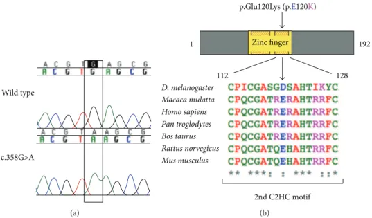
Figure 1: A Mutational analysis ofNANOS3revealed a homozygous c.358G>A substitution in exon 1 in two sisters with primary amenorrhea. he GAG to AAG change in codon 120 results in a substitution from glutamic acid to lysine in the codiied protein (p.Glu120Lys). B, Cartoon representation of the NANOS3 protein (192 amino acids), with the zinc inger domain (amino acid 76 to 130) depicted in yellow, showing the location of the p.Glu120Lys mutation. Glutamic acid 120 (shown as E120) is localized within one of two highly conserved Cys-Cys-His-Cys (C2HC) motifs (shaded red and yellow boxes). Conservation among species of the second C2HC motif sequence (human residues 112 to 128) is shown in detail; glutamic acid 120 (E) is highly conserved. Alignment was performed in Clustal Omega using protein sequences obtained from UniProt.
(F: 5� GGC GCC ACA CGT AAG CGC GCC CAC AC
3�; R: 5� GTG TGG GCG CGC TTA CGT GTG GCG
CC 3�). African green monkey kidney COS-1 cells were transiently transfected using Lipofectamine 2000 (Invitrogen, Carlsbad, CA) and analyzed 48 hours later with or without treatment with 5 mM sodium butyrate (Calbiochem, San Diego, CA). Cells were stained with Annexin-V conjugates (Molecular Probes Invitrogen, Carlsbad, CA) according to manufacturer’s protocol. GFP and Annexin-V luorescence were excited at 488 nm and 650 nm and emission measured using 530/30 nm and 660/30 band pass ilters, respectively.
2.4. Confocal Microscopy. Confocal microscopy micrographs were obtained using a 488 nm laser line and light emitted between 500 and 600 nm for GFP detection and a 650 nm laser line and light emitted between 660 and 700 nm for red Annexin-V detection. Images were collected on a META LSM 510 laser scanning confocal microscope equipped with a 63.0 × 1.2W objective (Carl Zeiss, Jena, Germany).he same settings for image acquisition and processing were applied for all samples to allow comparison of the luorescence intensities among diferent samples.
2.5. Molecular Modeling of NANOS3. A model for mutated human NANOS3 was built using the resolved structure of L3MBTL1 (PDB-ID 2RI7) as template since NANOS3 structure has not yet been resolved. In order to ind the best model for NANOS3, a Blast alignment with proteins available in PDB was performed. Highest homology was found with the crystal structure of Nanos from zebraish (PDB-ID 3ALR) with 58% identity. However, this structure
did not include the region where the p.Glu120Lys mutation is located. herefore, we looked for another protein bearing similarities with NANOS3. Such protein was L3MBTL1. Its X-ray resolved structure presented the second highest homology with NANOS3 (33% of identity) and included the p.Glu120Lys mutant region. Molecular modeling was performed with the SwissModel web-served program and images examined and edited using the web-based BlueStar STING (CNPTIA-Embrapa, Brazil) and PyMOL sotwares (freely available athttp://www.pymol.org).
3. Results
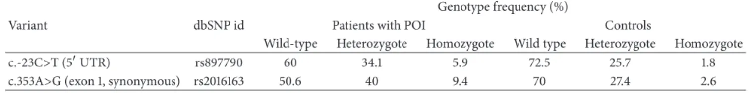
3.1. Identiication of NANOS3 Mutation. Mutational analysis ofNANOS3revealed that two sisters carried a homozygous c.358G>A, p.Glu120Lys mutation (Figure1), which was not detected in 113 ethnically matched control women and has not been reported in the 1000 Genome Project or dbSNP databases. heir mother was heterozygous and the father’s DNA was not available for study. In the remaining 83 women composing our POI cohort, no new variants were identiied inNANOS3. Two previously described SNPs were identiied in our cohort, rs897790 and rs2016163 (Table1).
Table 1: Polymorphic variants inNANOS3identiied in POI patients and controls. Genotype frequency (%)
Variant dbSNP id Patients with POI Controls
Wild-type Heterozygote Homozygote Wild type Heterozygote Homozygote
c.-23C>T (5�UTR) rs897790 60 34.1 5.9 72.5 25.7 1.8
c.353A>G (exon 1, synonymous) rs2016163 50.6 40 9.4 70 27.4 2.6
FSH = 150and 61 U/L, resp.). Estradiol levels were within the prepubertal range in both girls. Measured heights were 145 and 156 cm, and weights were 38 and 46 Kg, respectively. Pelvic ultrasonography showed no masses, and ovaries were not visualized. Treatment with conjugated estrogens followed by progesterone replacement resulted in complete breast development and normal cycling.
heir heterozygous mother has a history of menarche at 16 years old and diiculties to conceive. Her irst pregnancy was at 27 years old ater 4 years of attempts to conceive and her second and last gestation occurred at 30. Menopause occurred at 51 years of age.
3.2. Increased Apoptosis In Vitro with p.Glu120Lys Mutant NANOS3. Considering that Nanos3 has been shown to maintain germ cell development in model organisms through repression of apoptosis [34],in vitrostudies were performed to assess the efects of the p.Glu120Lys substitution on the apoptotic rate of cultured cells. COS-1 cells were transfected with GFP-tagged wild-type or mutant NANOS3 and viability assessed by Annexin-V staining. Flow cytometry analysis showed that the percentage of Annexin-V stained GFP-positive cells was signiicantly higher in cells transfected with mutant NANOS3 in comparison to wild type, in untreated or sodium butyrate-treated cells (� < 0.05) (Figure2), indicat-ing that the p.Glu120Lys substitution profoundly impairs cell viability.
Since NANOS3 has been shown to act in the cell nucleus [37], studies were performed to investigate whether the p.Glu120Lys mutation would afect intracellular localization. Transfected COS-1 cells were analyzed by confocal laser scan-ning microscopy for cellular localization of GFP-tagged wild type or mutant NANOS3 and Annexin-V staining (Figure3). Nuclear localization was observed in cells transfected with both wild type and mutant cDNA. However, Annexin-V staining in the plasma membrane, indicating apoptosis, was only detected in cells expressing the p.Glu120Lys mutant NANOS3.
3.3. In Silico Mutant NANOS3 Modelling. Glutamic acid120 is a negatively charged amino acid located within the zinc inger domain. his residue is found at the surface of NANOS3 within the second Cys-Cys-His-Cys (C2HC) motif, which is important for its ability to bind RNA [40]. In general, substitution of amino acids that share similar hydrogen binding capabilities and tridimensional requirements such as glutamic acid and lysine has limited structural consequences. However, lysine has a positive charge on the aliphatic side chain that can afect DNA binding activity when inserted
0 1 2 3 4 5
Fl
uo
re
sc
en
t cells (Annexin-V) (%)
NT WT MUT
Apoptosis detection
NaBu− NaBu+
Figure 2: Detection of apoptosis using low cytometry and Annexin-V staining in COS-1 cells transfected with wild type (WT) or p.Glu120Lys mutant (MUT) NANOS3-GFP, before and ater induction of apoptosis with sodium butyrate (NaBu). Expression of mutant NANOS3 results in signiicantly higher apoptosis in comparison to wild type (� < 0.05). Error bars represent SEM for nine replicates; NT: nontransfected control.
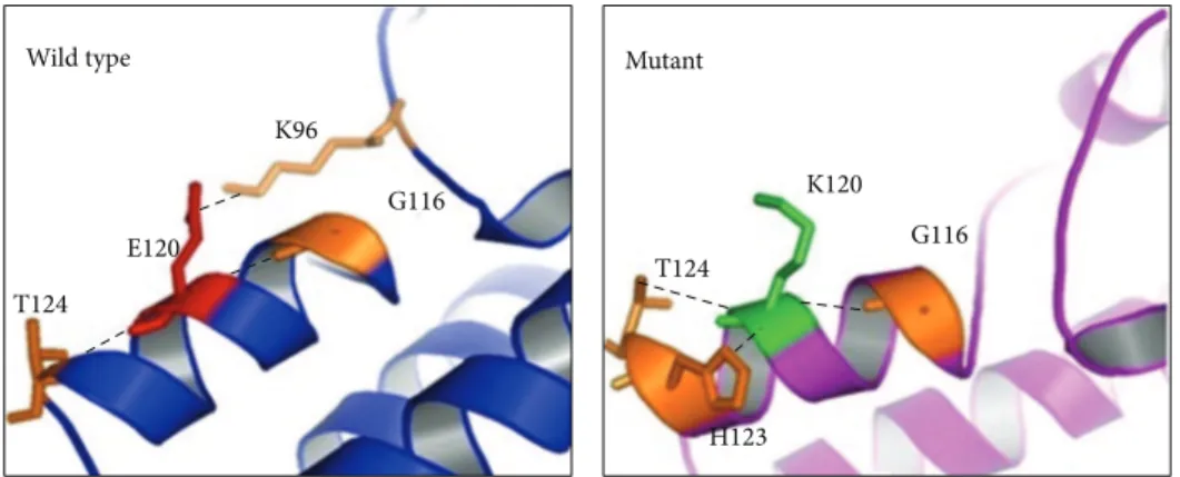
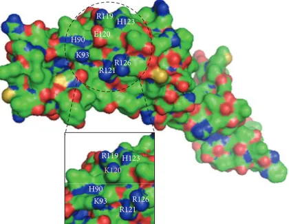
into the zinc inger domain. In silico analysis shows that lysine 120 establishes a new contact with Lys96 and abolishes the original contact with His123 (Figure 4). Consequently, these changes could lead to internal contact conformational modiications resulting in impaired protein function. In addition, 3D structure indicates that the negatively charged glutamic acid 120 residue is surrounded by negatively charged residues (Figure5). herefore, the presence of the positively charged lysine 120 residue in the protein surface might cause strong electrostatic repulsion among the side chains and lead to destabilization upon protein-RNA interaction.
4. Discussion
he present study aimed to investigate the contribution of variants in NANOS3 to human POI. We have identiied a nonsynonymous homozygous NANOS3mutation in two sisters with primary amenorrhea.
Nanos3-GFP Annexin-V Merge
WT
5 𝜇m 5 𝜇m 5 𝜇m
(a)
Mut
5 𝜇m 5 𝜇m 5 𝜇m
(b)
Figure 3: Confocal microscopy analysis of apoptosis in COS-1 cells following 48 h induction of apoptosis with sodium butyrate. In cells transfected with wild type (WT, upper row) or p.Glu120Lys mutant (Mut, lower row) GFP-tagged NANOS3, expression of green NANOS3-GFP is seen predominantly in the nucleus. Red Annexin-V staining in the cytoplasm and plasma membrane, indicating apoptosis, is seen in cells expressing p.Glu120Lys mutant NANOS3 but not in those expressing WT NANOS3.
Wild type Mutant
T124 E120
K96
G116
T124
H123 K120
G116
H123
H90 R119
K93
R121R126
E120
H123
H90 R119
K93 R121
R126
K120
Figure 5: Bulk representation of wild-type NANOS3, with p.Glu120Lys mutant shown in details box. he dashed circle highlights a protein surface region rich in basic residues, shown in blue. At the center of this region, lies the acidic residue glutamic acid 120 (E120), shown in red. In the details, substitution by lysine 120 (K120) disturbs the electrostatic interactions among adjacent residues.
in infertility due to apoptosis of migrating PGCs during fetal life, resulting in atrophic gonads where germ cells are absent [24, 34]. Furthermore, the contribution ofNanos3 to PGC maintenance and survival seems to be dosage-dependent in mice, as a model with attenuated Nanos3 transcript levels displayed markedly reduced numbers of PGCs [39].
In humans, NANOS3 is expressed in fetal and adult ovaries during multiple stages of oogenesis andin vitro stud-ies of human embryonic stem cell-derived germ cells have shown coexpression of NANOS3 in nuclei with important germ cell factors such as BLIMP1, VASA, and STELLA in the cytoplasm [37]. In these cells, reduction of NANOS3 expression resulted in altered gene expression patterns and reduced number of cells in active division, suggesting that in humans too NANOS3 has an important function in germ cell maintenance and survival. Recently, a heterozygous p.Arg153TrpNANOS3mutation was reported in a 23-year-old Chinese woman out of a cohort of 100 women with POI [39]. Familial segregation was not deined, but the mutation was not found in 200 ethnically-matched controls.
In vitrostudies suggested decreased protein stability due to the p.Arg153>Trp mutation, leading the authors to postulate a mechanism of reduced dosage of NANOS3 expression in the ovaries leading to decreased PGC population and resulting in POI [39].
he p.Glu120Lys mutation identiied in our patients lies within the NANOS3 zinc inger domain, which is composed of two C2HC motifs located between amino acids 77 and 131. he diferences in chemical properties between amino acids might lead to impaired binding of NANOS3 to its mRNA targets, as suggested by protein modeling. In vitro experi-ments show that the mutant NANOS3 identiied in the two sisters with POI was associated with increased apoptosis in
transfected cells, suggesting that loss of NANOS3-mediated protective efect against apoptosis in primordial germ cells may be the mechanism underlying POI in these patients.
Primordial germ cell pool size in the developing ovary and maintenance of these cells throughout life are at the cen-ter of the pathogenesis of POI. It is remarkable that our two patients with homozygousNANOS3mutation have presented with primary amenorrhea, perhaps indicating more severely PGC-depleted ovaries, while their heterozygous mother had diiculty to conceive, which could represent a manifestation of insuicient PGC pool size. Indeed, the patient described by Wu et al. carried a heterozygous missense change and presented with secondary, rather than primary, amenorrhea [39], supporting the concept of a dosage efect for NANOS3-related POI.
In conclusion, we report a rare homozygous missense mutation inNANOS3in two Brazilian sisters with primary amenorrhea from a cohort of 85 Brazilian women with POI. In vitro and in silico functional studies support that this inactivating mutation abolishes NANOS3 ability to pre-vent apoptosis, suggesting a mechanism for POI involving increased PGC apoptosis during embryonic cell migration.
Conflict of Interests
he authors declare that there is no conlict of interests regarding the publication of this paper.
Acknowledgments
Junior (Flow Cytometry Laboratory, Instituto Butantan) and Toshie Kawano and Alexsander Seixas de Souza (Parasitology Laboratory, Instituto Butantan) for technical assistance. his work was supported by Conselho Nacional de Desenvolvi-mento Cient´ıico e Tecnol´ogico (CNPq, Grant 301339/2008-9) and the S˜ao Paulo Research Foundation (FAPESP, Grants 05/04726-0, 07/512156, and 10/51102-0).
References
[1] C. K. Welt, “Primary ovarian insuiciency: a more accurate term for premature ovarian failure,”Clinical Endocrinology, vol. 68, no. 4, pp. 499–509, 2008.
[2] L. M. Nelson, “Clinical practice. Primary ovarian insuiciency,”
he New England Journal of Medicine, vol. 360, no. 6, pp. 606–
614, 2009.
[3] D. Goswami and G. S. Conway, “Premature ovarian failure,”
Human Reproduction Update, vol. 11, no. 4, pp. 391–410, 2005.
[4] L. M. Nelson, S. N. Covington, and R. W. Rebar, “An update: spontaneous premature ovarian failure is not an early menopause,”Fertility and Sterility, vol. 83, no. 5, pp. 1327–1332, 2005.
[5] P. Beck-Peccoz and L. Persani, “Premature ovarian failure,”
Orphanet Journal of Rare Diseases, vol. 1, no. 1, article 9, 2006.
[6] D. Toniolo, “X-linked premature ovarian failure: a complex disease,”Current Opinion in Genetics and Development, vol. 16, no. 3, pp. 293–300, 2006.
[7] M. M. Matzuk and D. J. Lamb, “he biology of infertility: research advances and clinical challenges,”Nature Medicine, vol. 14, no. 11, pp. 1197–1213, 2008.
[8] L. Persani, R. Rossetti, and C. Cacciatore, “Genes involved in human premature ovarian failure,” Journal of Molecular
Endocrinology, vol. 45, no. 5, pp. 257–279, 2010.
[9] P. Laissue, G. Vinci, R. A. Veitia, and M. Fellous, “Recent advances in the study of genes involved in non-syndromic pre-mature ovarian failure,”Molecular and Cellular Endocrinology, vol. 282, no. 1-2, pp. 101–111, 2008.
[10] R. Rossetti, E. D. Pasquale, A. Marozzi et al., “BMP15 mutations associated with primary ovarian insuiciency cause a defective production of bioactive protein,”Human Mutation, vol. 30, no. 5, pp. 804–810, 2009.
[11] Y. Qin, Y. Choi, H. Zhao, J. L. Simpson, Z. Chen, and A. Rajkovic, “NOBOX homeobox mutation causes premature ovarian failure,”he American Journal of Human Genetics, vol. 81, no. 3, pp. 576–581, 2007.
[12] D. Toniolo and F. Rizzolio, “X chromosome and ovarian failure,”
Seminars in Reproductive Medicine, vol. 25, no. 4, pp. 264–271,
2007.
[13] A. Lacombe, H. Lee, L. Zahed et al., “Disruption of POF1B bind-ing to nonmuscle actin ilaments is associated with premature ovarian failure,”American Journal of Human Genetics, vol. 79, no. 1, pp. 113–119, 2006.
[14] A. N. Shelling, K. A. Burton, A. L. Chand et al., “Inhibin: a candidate gene for premature ovarian failure,”Human
Repro-duction, vol. 15, no. 12, pp. 2644–2649, 2000.
[15] A. Marozzi, C. Porta, W. Vegetti et al., “Mutation analysis of the inhibin alpha gene in a cohort of Italian women afected by ovarian failure,”Human Reproduction, vol. 17, no. 7, pp. 1741– 1745, 2002.
[16] H. Dixit, L. K. Rao, V. V. Padmalatha et al., “Missense mutations in the BMP15 gene are associated with ovarian failure,”Human
Genetics, vol. 119, no. 4, pp. 408–415, 2006.
[17] P. Laissue, S. Christin-Maitre, P. Touraine et al., “Mutations and sequence variants in GDF9 and BMP15 in patients with premature ovarian failure,”European Journal of Endocrinology, vol. 154, no. 5, pp. 739–744, 2006.
[18] E. Kovanci, J. L. Simpson, P. Amato et al., “Oocyte-speciic G-protein-coupled receptor 3 (GPR3): no perturbations found in 82 women with premature ovarian failure (irst report),”Fertility
and Sterility, vol. 90, no. 4, pp. 1269–1271, 2008.
[19] X. X. Zhao, N. Suzumori, M. Yamaguchi, and K. Suzumori, “Mutational analysis of the homeobox region of the human NOBOX gene in Japanese women who exhibit premature ovarian failure,”Fertility and Sterility, vol. 83, no. 6, pp. 1843– 1844, 2005.
[20] D. Louren¸o, R. Brauner, L. Lin et al., “Mutations in NR5A1 associated with ovarian insuiciency,”he New England Journal
of Medicine, vol. 360, no. 12, pp. 1200–1210, 2009.
[21] B. Mandon-P´epin, P. Touraine, F. Kuttenn et al., “Genetic investigation of four meiotic genes in women with premature ovarian failure,”European Journal of Endocrinology, vol. 158, no. 1, pp. 107–115, 2008.
[22] H. Zhao, Z. Chen, Y. Qin et al., “Transcription factorFIGLAis mutated in patients with premature ovarian failure,”American
Journal of Human Genetics, vol. 82, no. 6, pp. 1342–1348, 2008.
[23] Z. Z. Zhao, J. N. Painter, J. S. Palmer et al., “Variation in bone morphogenetic protein 15 is not associated with spontaneous human dizygotic twinning,”Human Reproduction, vol. 23, no. 10, pp. 2372–2379, 2008.
[24] M. Tsuda, Y. Sasaoka, M. Kiso et al., “Conserved role of nanos proteins in germ cell development,”Science, vol. 301, no. 5637, pp. 1239–1241, 2003.
[25] L. Mosquera, C. Forristall, Y. Zhou, and M. L. King, “A mRNA localized to the vegetal cortex of Xenopus oocytes encodes a protein with a nanos-like zinc inger domain,”Development, vol. 117, no. 1, pp. 377–386, 1993.
[26] M. Pilon and D. A. Weisblat, “A nanos homolog in leech,”
Development, vol. 124, no. 9, pp. 1771–1780, 1997.
[27] K. Subramaniam and G. Seydoux, “nos-1 and nos-2, two genes related toDrosophila nanos, regulate primordial germ cell devel-opment and survival inCaenorhabditis elegans,”Development, vol. 126, no. 21, pp. 4861–4871, 1999.
[28] A. Forbes and R. Lehmann, “Nanos and Pumilio have critical roles in the development and function of Drosophila germline stem cells,”Development, vol. 125, no. 4, pp. 679–690, 1998. [29] M. K¨oprunner, C. hisse, B. hisse, and E. Raz, “A zebraish
nanos-related gene is essential for the development of primor-dial germ cells,”Genes & Development, vol. 15, no. 21, pp. 2877– 2885, 2001.
[30] H. Kurokawa, Y. Aoki, S. Nakamura, Y. Ebe, D. Kobayashi, and M. Tanaka, “Time-lapse analysis reveals diferent modes of primordial germ cell migration in the medaka Oryzias latipes,”
Development Growth and Diferentiation, vol. 48, no. 3, pp. 209–
221, 2006.
[31] S. Kobayashi, M. Yamada, M. Asaoka, and T. Kitamura, “Essential role of the posterior morphogen nanos for germline development in Drosophila,”Nature, vol. 380, no. 6576, pp. 708– 711, 1996.
germ-line progenitors of Drosophila melanogaster embryos,”
Development Growth & Diferentiation, vol. 43, no. 5, pp. 545–
552, 2001.
[33] A. Suzuki and Y. Saga, “Nanos2 suppresses meiosis and pro-motes male germ cell diferentiation,”Genes & Development, vol. 22, no. 4, pp. 430–435, 2008.
[34] H. Suzuki, M. Tsuda, M. Kiso, and Y. Saga, “Nanos3 maintains the germ cell lineage in the mouse by suppressing both Bax-dependent and -inBax-dependent apoptotic pathways,”
Develop-mental Biology, vol. 318, no. 1, pp. 133–142, 2008.
[35] K. M. Kusz, L. Tomczyk, M. Sajek et al., “he highly conserved NANOS2 protein: Testis-speciic expression and signiicance for the human male reproduction,”Molecular Human Reproduc-tion, vol. 15, no. 3, pp. 165–171, 2009.
[36] K. Kusz-Zamelczyk, M. Sajek, A. Spik et al., “Mutations of NANOS1, a human homologue of the Drosophila morphogen, are associated with a lack of germ cells in testes or severe oligo-astheno-teratozoospermia,”Journal of Medical Genetics, vol. 50, no. 3, pp. 187–193, 2013.
[37] V. T. Julaton and R. A. Reijo Pera, “NANOS3function in human germ cell development,”Human Molecular Genetics, vol. 20, no. 11, pp. 2238–2250, 2011.
[38] Y. Qin, H. Zhao, E. Kovanci, J. L. Simpson, Z. Chen, and A. Rajkovic, “Mutation analysis of NANOS3 in 80 Chinese and 88 Caucasian women with premature ovarian failure,”Fertility &
Sterility, vol. 88, no. 5, pp. 1465–1467, 2007.
[39] X. Wu, B. Wang, Z. Dong et al., “A NANOS3 mutation linked to protein degradation causes premature ovarian insuiciency,”
Cell Death and Disease, vol. 4, article e825, 2013.
[40] H. Hashimoto, K. Hara, A. Hishiki et al., “Crystal structure of zinc-inger domain of Nanos and its functional implications,”
Submit your manuscripts at
http://www.hindawi.com
Stem Cells
International
Hindawi Publishing Corporation
http://www.hindawi.com Volume 2014
Hindawi Publishing Corporation
http://www.hindawi.com Volume 2014 INFLAMMATION
Hindawi Publishing Corporation
http://www.hindawi.com Volume 2014
Behavioural
Neurology
Endocrinology
International Journal ofHindawi Publishing Corporation
http://www.hindawi.com Volume 2014
Hindawi Publishing Corporation
http://www.hindawi.com Volume 2014
Disease Markers
Hindawi Publishing Corporation
http://www.hindawi.com Volume 2014
BioMed
Research International
Oncology
Journal ofHindawi Publishing Corporation
http://www.hindawi.com Volume 2014
Hindawi Publishing Corporation
http://www.hindawi.com Volume 2014
Oxidative Medicine and Cellular Longevity
Hindawi Publishing Corporation
http://www.hindawi.com Volume 2014
PPAR Research
The Scientiic
World Journal
Hindawi Publishing Corporation
http://www.hindawi.com Volume 2014
Immunology Research
Hindawi Publishing Corporation
http://www.hindawi.com Volume 2014
Journal of
Obesity
Journal ofHindawi Publishing Corporation
http://www.hindawi.com Volume 2014
Hindawi Publishing Corporation
http://www.hindawi.com Volume 2014 Computational and Mathematical Methods in Medicine
Ophthalmology
Journal ofHindawi Publishing Corporation
http://www.hindawi.com Volume 2014
Diabetes Research
Journal ofHindawi Publishing Corporation
http://www.hindawi.com Volume 2014
Hindawi Publishing Corporation
http://www.hindawi.com Volume 2014
Research and Treatment
AIDS
Hindawi Publishing Corporationhttp://www.hindawi.com Volume 2014
Gastroenterology Research and Practice
Hindawi Publishing Corporation
http://www.hindawi.com Volume 2014
Parkinson’s
Disease
Evidence-Based Complementary and Alternative Medicine
Volume 2014