Although the biological properties of mesenchymal stem cells (MSC) are well-characterized
in vitro
, MSC clinical application is still far away to be achieved, mainly due to the need of
xenogeneic substances for cell expansion, such as fetal bovine serum (FBS). FBS presents
risks regarding pathogens transmissions and internalization of animal’s proteins, which can
unleash antigenic responses in patients after MSC implantation. A wide range of venous blood
derivatives (VBD) has been reported as FBS substitutes showing promising results. Thus, the
aim of this study was to conduct a systematic scoping review to analyze whether VBD are
effective FBS substitutes for MSC
ex vivo
expansion. The search was performed in SciVerse
Scopus
TM, PubMed, Web of Science
TM, BIREME, Cochrane library up to January 2016. The
keywords were selected using MeSH and entry terms. Two independent reviewers scrutinized
the records obtained considering specific inclusion criteria. The included studies were evaluated
in accordance with a modified Arksey and O’ Malley’s framework. From 184 found studies, 90
were included. Bone marrow mesenchymal stem cells (BMMSC) were presented in most of these
studies. Overall, VBD allowed for either, maintenance of MCS’s fibroblast-like morphology,
high proliferation, high colony-formation ability and maintenance of multipotency. Besides.
MSC expanded in VBD supplements presented higher mitogen activity than FBS. VBD seems to
be excellent xeno-free serum for
ex vivo
expansion of mesenchymal stem cells. However, an
accentuated heterogeneity was observed between the carried out protocols for VBD isolation
did not allowing for direct comparisons between the included studies.
Venous Blood Derivatives as
FBS-Substitutes for Mesenchymal Stem
Cells: A Systematic Scoping Review
Luiz A. Chisini
1, Marcus C.M. Conde
2, Guillermo Grazioli
3, Alissa S. San
Martin
1, Rodrigo Varella de Carvalho
4, Jacques E. Nör
5, Flávio F. Demarco
11Graduate Program in Dentistry,
Dental School, UFPel - Universidade Federal de Pelotas, Pelotas, RS, Brazil
2Graduate Program in Dentistry,
School of Dentistry, UNIVATES - Universidade do Vale do Taquari, Lajeado, Brazil
3School of Dentistry, Universidad de
la República, Montevideo, Uruguay
4School of Dentistry, IMED
- Faculdade Meridional, Passo Fundo, RS, Brazil
5Department of Cariology, Restorative
Sciences and Endodontics, School of Dentistry, University of Michigan, Ann Arbor, MI, USA
Correspondence: Flávio Fernando Demarco, Rua Gonçalves Chaves 457, 5° and., 96015-560 Pelotas, RS, Brasil. Tel: +55-53-98112-1141. e-mail: ffdemarco@gmail.com
Key Words: human serum,
mesenchymal stem cells,
platelet, venous blood
derivatives, xeno-free.
Review Article
Introduction
Mesenchymal stromal/stem cells (MSC) have been
exhaustively investigated
in vitro
and due to high
proliferative, self-renewal, immunomodulatory properties
and multipotency, MSC present a high therapeutic potential
to be applied in Stem Cell-Based Therapies (SC-BT) (1).
Several strategies and approaches to use regenerative
therapies in dentistry have been investigated (1-3), since
that materials employed by the clinicians are, basically,
synthetic (4-6) and can present limited ability to induce
regeneration (1,3,7). Thus, the use of MSC could improve the
regenerative potential of bone, periodontal and dental pulp
regenerative approaches. However, a recent scoping review
evaluating the capacity of dental pulp tissue regeneration
by strategies to revascularization of root canal has shown
limited ability to promote regeneration (3) and this could
be improved with the application of MSC. Although MSC’s
biological properties have been well-characterized
in vitro
,
MSC clinical application is still far away to be achieved (8).
To be clinically applied, MSC must be previously isolated
and expanded
ex vivo
in order to obtain a needed amount
of cells, which will be replanted in patients (8,9).
Ex vivo
MSC expansion relies on solutions composed by a basal
medium, basically amino acids, vitamins and inorganic
salts, which must be supplemented by Fetal Bovine Serum
(FBS) (10,11). FBS is the most applied supplement for
MSC culture comprising a complex mixture of growth
factors (GF), proteins, carbohydrates and cytokines
indispensable for cell development and survival
in vitro
(12-14). However, FBS presents risks regarding pathogens
transmissions and internalization of animal’s proteins,
which can unleash antigenic responses in the patient after
MSC implantation (9,15). Animal-derived (or xenogeneic)
proteins can be detected in human MSC expanded in FBS,
even after consecutive cell washings (9). Additionally, FBS
induces changes in MSC surface markers and due to such
characteristics. FBS must not be applied in humans (16).
L.A. Chisini et al.
Material and Methods
This study was designed following the modified
five-stage framework proposed by Arksey and O’Malley (26)
denominated scoping review. Recently, several studies
applied scoping review to state the current knowledge
in a particular area when a systematic review cannot be
conducted (3,27). The scoping review design is indicated
when the methodologies of studies present a considerable
heterogeneity performing a qualitative analysis while a
systematic review provides a quantitative analysis. A scoping
review presents an exploratory research to respond a broader
question through a systematized research, aiming to define
concepts and mapping the methodologies used to define
gaps in the literature to indicate the need for new studies.
A scoping review was carried out to perform a knowledge
synthesis regarding VBD as FBS substitutes for
ex vivo
MSC
expansion. A complementary search was performed to
identify the studies using MSC in humans expanded in VBD.
Conceptual Definition
According to MeSH database (http://www.ncbi.nlm.nih.
gov/mesh), serum is defined as “The clear portion of blood
that is left after blood coagulation to remove blood cells
and clotting proteins by centrifugation”. In the meantime,
plasma is defined as “the residual portion of blood that is
left after removal of blood cells by centrifugation without
prior blood coagulation”. Unlike plasma, serum naturally
contains platelet-derived molecules, such as
α
-granules-derived growth factors, which become available exclusively
after platelet-activation. For Human Serum (HS) the whole
blood is collected in an anticoagulant-free plastic bag
and stored overnight (4 °C), or at the end of shelf-life,
to allow for blood coagulation. Right after, the formed
clot must be centrifuged (3000 rpm for 5 min) in order to
obtain a supernatant, corresponding to HS (21).
Platelet-Rich Plasma (PRP) is stated as “A preparation consisting
of platelets concentrated in a limited volume of plasma”.
While the natural-formed blood clot contains 95% of red
blood cells, 5% platelets, less than 1% white blood cells and
fibrin strands, PRP holds 4% red blood cells, 95% platelets
and 1% white blood cells (28). PRP should be chemically
activated by the addition of human/bovine thrombin or
Calcium Chloride - CaCl
2, affording activated PRP -aPRP
(24,25,29). To obtain PRP, whole anticoagulated blood,
must be submitted to a double-centrifugation; the first
one (soft spin) results in a three-layer suspension where
the red blood cells are found at the bottommost layer.
Both, topmost, named platelet-poor plasma (PPP), and
intermediate (PRP) layers should be transferred to another
tube without anticoagulant. Thus, the second spinning
(hard spin) is performed, to allows platelets settle the
bottom of tube. Then, superficial layer is discarded and the
remaining material (PRP) is shaken. To release its platelet
content, PRP must be activated (aPRP) by the addition
of human/bovine thrombin or Calcium Chloride (CaCl
2)
(24,25). Human Platelet Lysate (HPL) results from a lysis of
high platelet concentrate (traditionally the PRP), being the
platelet mechanical lysis induced by susceptive freeze-thaw
cycles at -80 or -20 °C (typically 2 or 3 cycles). Thus, the
platelet debris must be separated from the clear portion
containing platelet released by centrifugation.
Information Sources, Literature Search and Inclusion
Criteria
A structured search was performed in SciVerse Scopus
TM,
PubMed/Medline, ISI Web of Science
TM, and BIREME up to
January 2016. The relevant MeSH terms and entry terms
(Table 1) were selected based on the PICO-structured
Table 1. Structured search strategy carried out in MEDLINE/PubMed database. The search followed structural of each database
Search Syntaxes
#1
“mesenchymal stromal cells”[MeSH Terms] OR (“mesenchymal”[All Fields] AND “stromal”[All Fields] AND “cells”[All Fields]) OR “mesenchymal stromal cells”[All Fields] OR (“mesenchymal”[All Fields]
AND “stem”[All Fields] AND “cells”[All Fields]) OR “mesenchymal stem cells”[All Fields]
#2
“Culture Media, Serum-Free”[All Fields] OR “Culture Media, Serum Free”[All Fields] OR (“culture media, serum-free”[MeSH Terms] OR (“culture”[All Fields] AND “media”[All Fields] AND “serum-free”[All Fields]) OR “serum-free culture media”[All Fields]
OR (“media”[All Fields] AND “serum”[All Fields] AND “free”[All Fields] AND “culture”[All Fields])) OR “Serum-Free Culture Media”[All Fields] OR “Serum-Free Media”[All Fields] OR “Media, Serum-Free”[All Fields] OR “Serum Free Media”[All Fields] OR “Protein-Free Media”[All Fields] OR (“culture media, serum-free”[MeSH Terms] OR (“culture”[All Fields] AND “media”[All Fields] AND “serum-free”[All Fields]) OR “serum-free culture media”[All Fields] OR (“media”[All Fields] AND “protein”[All Fields]
AND “free”[All Fields])) OR “Protein Free Media”[All Fields] OR “Low-Serum Media”[All Fields] OR “Low Serum Media”[All Fields] OR (“culture media, serum-free”[MeSH Terms] OR (“culture”[All Fields] AND “media”[All Fields] AND “serum-free”[All
Fields]) OR “serum-free culture media”[All Fields] OR (“media”[All Fields] AND “low”[All Fields] AND “serum”[All Fields]))
#3
“Platelet lysate”[All Fields] OR “thrombin activated platelet”[All Fields] OR (allogenic[All Fields] AND pooled[All Fields] AND (“humans”[MeSH Terms] OR “humans”[All Fields] OR “human”[All Fields]) AND (“serum”[MeSH Terms] OR “serum”[All Fields])) OR “pooled human serum”[All Fields] OR “autologous serum”[All Fields] OR “human serum”[All Fields] OR “thrombin
V
enous blood derivatives
question “Could human venous blood derivatives be applied
as FBS substitutive for MSC
ex vivo
expansion?”, where:
•
P: Human MSC
•
I: human serum; Platelet Rich Plasm; Activated
Platelet-Rich Plasm ; Human Platelet lysate; Plasma; Platelet
Poor Plasma; Human Plasm
•
C: Fetal Bovine Serum
•
O: MSC biological properties: Cell viability, cell
proliferation, multipotency, population doubling time,
senescence, telomere shortening
The retrieved records were uploaded into the EndNote
TMsoftware, aiming to delete duplicates and to build up a
virtual library (VL). Two independent reviewers (LAC and
MCMC) read the titles and abstracts of all reports, under
predefined inclusion criteria (Table 2). To confirm if the
selected studies met the inclusion criteria, the same
reviewers independently judged each full text. If any
disagreement was found, the reviewers attempted to reach
a consensus through discussions. Persistent disagreements
have been decided by an intervention from the third
reviewer (FFD). Thus, manual evaluation of references from
each evaluated study was performed. Twenty percent of
the studies were randomly raffled, and the data have been
again checked.
Search to identify clinical application of cell therapy
using blood derivate serums: The literature was investigated
using the keywords: “clinical study”, “mesenchymal stem
cell”, “serum-free medium”, “Platelet lysate”, “human
serum” and “autologous serum” for identify studies
employing cell therapy in humans with
ex vivo
expansion
in medium supplemented with VBD, to provide an overview
of clinical application.
Results
The initial search yielded 272 records corresponding to
184 studies (Table 3). After preliminary titles and abstracts
evaluation, 102 studies were select for full-text assessment
(Fig. 1). Ninety papers were designated for data extraction.
Excluded studies (10,30-40) and reasons are described in
Table 4. Most of the included studies described protocols
relying on the platelets lyse after freeze-thaw cycles
to obtain VBDs. Bone marrow mesenchymal stem cells
(BMSC) were presented in most of this studies (Table 5);
adipose stem cells - ASC (24,41-46), dental pulp stem
cells - DPSC (47,48), umbilical cord stem cells - UCST
(22,25,49-52) and orbital fat-derived stem cells (53)
were also tested. VBD concentrations added to culture
medium ranged from 0.5 to 30% (54-56). Overall, VBD
allowed for either, maintenance of MCS’s fibroblast-like
morphology (17,43,57), high proliferation (58-60), high
colony-formation ability (52,61,62) and maintenance of
multipotency (63-67). A linear dose-dependent response
regarding medium concentration was not observed for
evaluated VBD (54,56,68).
Human platelet lysate (HPL): HPL showed a higher
osteogenic potential than MSC in FBS (55,69,70). Besides,
MSC expanded in HPL seems presented immunomodulatory
ability (52,55,71,72) despite a decrease of ability of
inhibition NK and T-cells has also been observed (73).
Platelets lyse was triggered by either, freeze-thaw cycles
(1 to 5), ultrasound (63) or chemical treatment (43). HPL
presented a high growth factors and cytokines content
(74-77). HPL seemed to be better than FBS for
ex vivo
MSC
expansion (52,72,78,79) since HPL-expanded MSC did not
present telomerase shortening (68).
Human Serum (HS): Overall, HS allowed for the
isolation and expansion of BMSC, ASC, DPSC maintaining
proper cell biological properties both
in vitro
and
in vivo
(48,80-85). Both, 10% HS in DMEM/F-12 or 5%-10% HS
in
α
MEM provided, to ASC and BMMSC, proliferation rates
and multipotency as higher as 10% FBS (24,48,51,86-88).
Platelet-rich plasma: Platelets form PRP has been
activate with trombin or calcium chloride aiming the
increase of bioactive molecules release (24,29,89), however
similar properties were observed in PRP (10% freeze thawed
human PRP (49,91) / with platelet concentration (92,93)/
platelet and leukocytes concentration (90)). PRP (5% or
10%)-supplemented media heat-inactivated (platelet
concentration 79.6 x 10
4/µL) provided similar results for
adipogenic and osteogenic differentiation when compared
to 10% FBS (92). Besides, PRP supplemented
α
MEM also
provides osteogenenic, chondrogenic and adipogenic
differentiation promoting an increase of cell culture
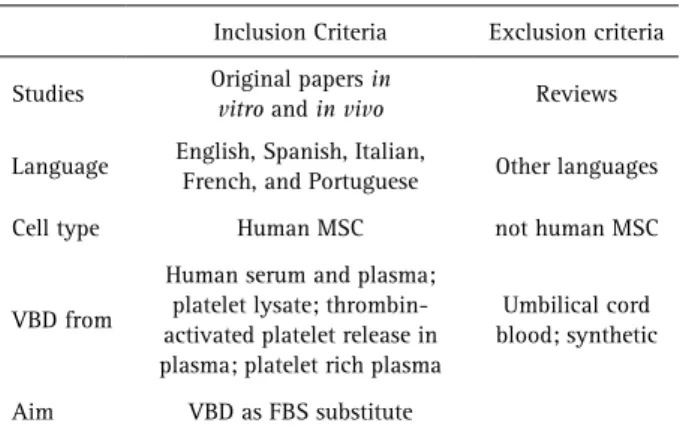
Table 2. Criteria for selection of the studiesInclusion Criteria Exclusion criteria
Studies Original papers in
vitro and in vivo Reviews
Language English, Spanish, Italian,
French, and Portuguese Other languages
Cell type Human MSC not human MSC
VBD from
Human serum and plasma; platelet lysate; thrombin-activated platelet release in plasma; platelet rich plasma
Umbilical cord blood; synthetic
Aim VBD as FBS substitute
Table 3. Records recovered in each database
PubMed® Scopus® ISI web of science®
Cochrane library®
BVS Bireme®
L.A. Chisini et al.
proliferation (22,49). In this way, 10% aPRP (platelets
concentered) has been reported as providing similar (25),
or higher proliferation rates than FBS (89). In addition, only
in one study reported the presence of leukocyte into PRP
(0.3 x 10
4mL) (90). Strategies to decreased leukocyte in the
PRP even as assessment of activation by flow cytometry
did not were reported on included studies. Other VBD: MSC
were also expanded by applying platelet-poor plasma (PPP)
(94), fresh frozen platelet plasma (FFPP) (56,95) and HS
obtained from PPP (42). Either MSC expanded in PPP, FFPP
and in FBS had the same phenotype for antigens CD73,
CD90, CD105, CD14, CD19, CD34, CD45, HLA-DR. Such
MSC presented osteogenic, adipogenic and chondrogenic
differentiation and were able to survive in a fibrin clot.
BMSC expanded in culture medium containing high (20
and 30%) and low (1%) FFPP concentrations, produced
insufficient calcified matrix (56).
Human studies: Seven studies employed MSC expanded
in VBD for tissue regeneration (96-102). HPL and HS were
used for MSC expansion for clinical application. These studies
did not observe neither signal of malign transformation nor
some complication associated with HPL or HS.
Figure 1. Flow Diagram
Table 4. Excluded studies and reasons for exclusion
Studies Reason
Cheng et al. (30); Jung et al. (10); Dolley-Sonneville et al. (32); Mark et al. (33); Reza et al. (34); Sato et al. (35); Tan et al. (36); Trubiani et al. (37)
Not defined venous blood derivative serum
V
enous blood derivatives
Table 5. Included studies and respective VBD supplements and concentrations, concentration of FBS and stem cells utilized
Author Xeno-free evaluated and concentration Contol and
concentration Stem Cells
Stute et al. (21) 1%, 3%, and 10% HS 10% FBS BMSC
Doucet et al. (62) 5% HPL 10% FBS BMSC
Shahdadfar et al. (80) 20% HS FBS 20% BMSC
Gregory et al. (81) 20% HS and 10% HS + grow factors FBS 20% BMSC
Muller et al. (95) 2.5% and 5% HPL 10% FBS BMSC
Vogel et al. (111) 3% PRP FBS 2% BMSC
Le Blanc et al. (82) 10% HS 10% FBS BMSC
Capelli et al. (60) 5% HPL FxBS 10% BMSC
Kocaoemer et al. (89) 10% HS and 10% aPRP 10% FBS ASC
Lange et al. (78) 5% HPL 10% FBS BMSC
Lataillade et al. (102) * 8% HPL - BMSC
Reinisch et al. (72) 10% HPL 10% FBS UCMSC and BMSC
Schallmoser et al. (75) 10% HPL 10% FBS BMSC
Schallmoser et al. (114) 5% and 10% HPL 10% FBS BMSC
Shayesteh et al. (97) * 20% HS - BMSC
Zaky et al. (70) 5% HPL and 5% HPL + 10% FBS 10% FBS and FGF2 BMSC
Behnia et al. (99) 20% HS - BMSC
Bieback et al. (29) 10% aPRP and 10% HPL and 10% HS 10% FBS BMSC
Blande et al. (64) 2.5% or 10% HPL 10% FBS ASC
Lindroos et al. (86) 10% HS 10% FBS or
StemPro® ASC
Kishimoto et al. (84) 8% 4% 2%, 1%, 0.5% HS with FGF2
5 ng/mL (F/P MP-Coated plates) not coated HS BMSC
Pytlik et al. (88) 10% HS 10% FBS BMSC
Schallmoser et al. (20) 10% HPL - MSC
Bieback et al. (24) 10% HS and 10% aPRP 10% FBS ASC
Centeno et al. (101) * 10-20% HPL - BMSC
Chevallier et al. (59) 5% HPL 10% FBS BMSC
Felka et al. (56) FFP with platelet concentrate 0.5, 1%, 5%, 10%, 20% and 30%
and human plasma-enriched with platelets media 5% and 10% 10% FBS BMSC
Hartmann et al. (51) 2% HS FBS UCMSC
Horn et al. (69) 10% HPL 10% FBS MSC
Ichiyanagi et al. (94) Platelet poor plasma 5%, 10% and 20% 10% FBS BMSC
Lindroos et al. (87) 10%, 15% and 20% HS 10% FBS ASC
Lucchini et al. (100) * 5% HPL - MSC
Salvade et al. (77) 5% HPL 10% FBS BMSC
Abdelrazik et al. (73) 10% HPL 10% FBS MSC
Aldahmash et al. (85) 2.5%, 5.0% and 10.0% HS 10% FBS BMSC
L.A. Chisini et al.
Cholewa et al. (41) 1%, 2.5%, 5%, 10%, and 20% HPL FBS 1%, 2.5%, 5%,
10%, and 20% ASC
Crespo-Diaz et al. (109) 5% HPL 10% FBS BMSC and ASC
Flemming et al. (71) 10% HPL 10% FBS BMSC
Goedecke et al. (92) 5% and 10% PRP 10% FBS BMSC
Govindasamy et al. (47) 10% HPL 10% FBS DPSC
Hatlapatka et al. (50) HS 2%, 5%, 10% and HS 2% with 0.5 ng/mL FGF FBS UCMSC
Jenhani et al. (65) 5%, 10% HPL or 5% HPL and 10% FBS or 10%HPL and 10%FBS 10% FBS BMSC and UCMSC
Shih et al. (43) 10% HPL 10% FBS ASC
Venugopal et al. (106) 5% HS and 2 ng/mL bFGF 10% FBS WJSC
Xia et al. (76) 7.5% HPL 10% FBS BMSC
Jenhani et al. (65)
5%,10% HPL 10% FBS
5% HPL
10% FBS 1 ng/mL fibroblast growth factor 2 (FGF2)
BMSC
Behnia et al. (96) * 20% HS - BMSC
Fekete et al. (12) 2.5%, 5%, 10%, 15% and 20% HPL FBS 20% BMSC
Fekete et al. (13) 10% HPL - BMSC
Gottipamula et al. (54) 10% HPL (5 to 20% preliminary screening) 10% FBS BMSC
Kishimoto et al. (93) 4%, 2%, 1% and 0.5% PRP
or 4%, 2%, 1% and 0.5% HS - BMSC and ASC
Kruger et al. (90) 10% HS and 5% PRP - CSSC
Lohmann et al. (61) 10% HPL 10% FBS BMSC
Murphy et al. (22) 1% or 10% PRP 10% FBS UCMSC
Pisciotta et al. (48) 10% HS 10% FBS DPSC
Poloni et al. (113) 5% HPL and HS 10% FBS 20% CVMS and BMSC
Stehlik et al. (11) 10% HS - BMSC
Bernardi et al. (63) 10% HPL 10% FBS BMSC
Griffiths et al. (68) 2%, 5% and 10% HPL FBS 16% BMSC
Hemeda et al. (117) 10 % HPL - BMSC and ASC
Trojahn Kolle et al. (107) 10% HPL 10% FBS ASC
Kyllonen et al. (45) 10% HS 10% FBS ASC
Mojica-Henshaw et al. (118) 10% HPL 10% FBS MSC
Pawitan et al. (83) 5% and 10% PRP
5% and 10% HS - ASC
Rojewski et al. (120) 10% HPL 10% FBS BMSC
Sandor et al. (98) * 15% HS - ASC
Schallmoser et al. (58) 10% HPL 10% FBS MSC
Shanskii et al. (74) 10% HPL
FBS/HPL ratios: 10/0, 7.5/2.5, 5.0/5.0, 2.5/7.5, and 0/10) FBS MSC
Castiglia et al. (23) 10% HPL 10% FBS BMSC
Fekete et al. (14) 5%, 10% and 20% HPL
5%, 10% and 20% HS
FBS 5%, 10%
and 20% BMSC
Gottipamula et al. (104) 2.5 % HS 10% FBS BMSC
V
enous blood derivatives
Koellensperger et al. (42) HS 2%, 10%; PPP 2%, 10%; PL 2%, 10% 10% FBS ASC
Martins et al. (53) 10% HS 10% FBS OFSC
Muraglia et al. (19) 5% HPL 10% FBS BMSC
Pham et al. (25) 2%, 5%, 7% and 10% aPRP 10% FBS UCMSC
Yamauchi et al. (79) 5% HPL 10% FBS BMSC
Antoninus et al. (17) HPL 0.031 mg/mL, 0.063 mg/mL, 0.125
mg/mL, and 0.250 mg/mL 20% FBS WJSC
Budiyanti et al. (49) PRP 10% MesenCult® UCSC
Castren et al. (18) AB-plasma 2.5% + HPL 0.5% 10% FBS BMSC
Jonsdottir-Buch et al. (15) 10% HPL - BMSC
Laitinen et al. (55) 5% and 10% HPL 10% FBS BMSC
Laner-Plamberger et al. (67) 10% HPL 10% HPL BMSC and UCMSC
Li et al. (57) 5% HPL - BMSC and ASC
Luzzani et al. (52) 10% HPL 10% FBS UCMSC and iPS
Oikonomopoulos et al. (46) 10% HPL 10% FBS BMSC and ASC
Paula et al. (44) HS 10% 10% FBS ASC
Pawitan et al. (91) PRP 10% 10% FBS BMSC
Riordan et al. (105) 5%, 7.5% and 10% HPL 10% FBS WJSC
* MSC expanded in VBD for tissue regeneration in humans. MSC: mesenchymal stem cells; UCSC: umbilical cord derived mesenchymal stem cells; ASC: adipocyte stem cells; BMSC: bone marrow stem cells; WJSC: Wharton’s Jelly-derived stem cells; OFSC: orbital fat-derived stem cells; CVMS: chorionic villi-derived stem cells; DPSC: dental pulp stem cells; CSSC: human cortico-spongious stem cells; FFP: fresh frozen plasma.
Discussion
The recent literature has been investigated FBS
substitutes for MSC
ex vivo
expansion aiming to eliminated
the risks inherent to xenogeneic agents for clinical
translation of regenerative therapies (103,104). In this
systematic scoping review, the studies evaluating VBD
as FBS substitutes were summarized. Overall, different
protocols were developed and tested to obtain a FBS
substitute able to maintain MSC
in vitro
, preserving
stemness and multipotency (103,105). VBD were promissors
as FBS substitutes, since senescence (68,106) or Karyotype/
chromosomal alteration were not detected in MSC
expanded in VBD (13,23,107-109). In addition, clinical
studies strengthened such evidence by reporting none
malign transformation
in vivo
(96,99,101,102).
Despite blood of umbilical cord possess a higher amount
of growth factors (platelet-derived growth factors -PDGF,
fibroblast growth factor 2 -FGF-2 and vascular endothelial
growth factor - VEGF) when compared to venous blood, the
application of blood from umbilical cord is restricted due
the difficult to obtain large volumes (12,21-23). Besides,
the growth factors concentration found in different VBD
(12,22,110) was reported as higher than in FBS (22,41,43,111).
Autologous and homologous blood have been presented
a high amount of platelets, which contains the growth
factors responsible VBD therapeutic potential (112). During
platelet activation biomolecules such as insulin-like growth
factor (IGF), PDGF, transforming grow factor
β
(TGF-
β
), and
other molecules such as thrombospondin and fibronectin
are released (112). HPL presented high concentrations of
endothelial growth factor (EGF), PDGF, TGF-
β
, fibroblast
growth factors
β
(FGF-
β
) and VEGF when compared to HS
(110). Furthermore, the low proteic content observed in HPL
may be beneficial by decreasing the risk of immunological
reactions for allogenic blood-derived (110). The platelets
lyse seems to release all platelet-derived growth factors
available, which could not happen during blood coagulation
(103). Nonetheless, Poloni (113) showed HS inducing higher
cell proliferation than HPL, contrary to the expected. Even
so, both were better than FBS.
L.A. Chisini et al.
(48). The studies reported 5% (68) and 10% (12) as being
the optimal HPL concentration range for MSC expansion
(12,54,64,114). Moreover, different VBD concentration
(0.5-30%) have been tested presenting different results,
which have not been necessarily dose-dependent (54-56).
Overall, the selected studies indicated VBD concentrations
ranging from 5% to 10% by presenting good results to be
applied as FBS substitutive (8,21,25,56,94).
DMEM is currently applied for MSC isolation and
expansion (1,5,115,116); DMEM calcium content can
stimulate a polymerization of fibrin present in HPL, thereby
forming a gel into the solution (8). To avoid this, fibrinogen
or heparin has been applied (69). Despite almost studies
use 1 or 2 IU/mL heparin, Hemeda, Giebel (8) demonstrated
that 0.61 IU/mL or 0.024 mg/mL for low-molecular-weight
heparin was sufficient to avoid gel formation. However,
high heparin concentrations seem to reduce adipogenic
and osteogenic differentiation, as well as cell proliferation
(117). In the protocol to obtain the HPL, the Induction of
fibrin clotting formation with calcium chloride (instead of
thrombin) and posterior centrifugation provides a serum
with the same profile of growing factor and cytokines
than conventional HPL activated with thrombin, resulting
in a serum without free of xenogeneic substances such
as porcine thrombin (118). HPL supplemented without
anticoagulants tend to form a translucent and viscous gel
in 1 h providing a natural scaffold for MSC culture and
expansion (61). This matrix, composed by a fibrin network,
was biocompatible and biodegradable. Additionally, MSC
cultured in this fibrin matrix presented higher proliferation
since growth is available in three-dimensional environment,
increasing the culture area (8,61).
Interesting findings have been observed regarding
to trypsin kinetics of MSC supplemented with VBD. MSC
expanded in HS or HPL have been trypsinized with 0.05%
Trypsin/EDTA, instead the conventional 0.25% applied
for MSC culture in FBS. VBD supplementation provides a
decrease in production of adhesion proteins (54,94). An
ASC genome gene expression analysis depicted 102 genes
were commonly expressed in differentiated ASC being 90
genes, including those responsible for MSC adhesion, high
expressed in 10% FBS-supplemented ASC (24), which may
be connected to high sensitivity presented to trypsin by MSC
expanded in VBD. Changes in surface markers expression
has been contradictorily reported in VBD-expanded MSC
(25,49,50,92). HS and aPRP provided maintenance of MSC
immunophenotype (25).
Currently it is clear that the number of passages
reduces MSC differentiation potential and the capacity
of proliferation conducting to function alteration (119).
MSC senescence has been the focus of some selected
studies (41,44,68,106).
β
-galactosidase, a biomarker for cell
senescence, was expressed strongly in FBS cultures when
compared with HPL up to 16 passages (68). On the other
hand, Venugopal, Balasubramanian (106) showed similar
senescence rates for MSC expanded in HS or FBS. Besides,
the age of VBD-donor could influence MSC senescence
however, controversial results have been described (61,110).
MSC expanded in HS from older donors (>45 years old)
presented higher
β
-galactosidase expression than MSC from
younger donors (<35 years old) (54). However, donors’ age
did not influence the GF concentration, hormones content
of MSC expanded in HPL (61). Individual variations are
expected in VBD, principally in plasm components. Blood
contains several components as lipids (HDL, cholesterol),
proteins, metabolites of uric acid, creatinine, and albumin;
even as several ions: calcium, potassium, resulting from
individual diet. Thus, plasm portion may have changes
in biochemical composition (74). Growth factor’s release
profile of HPL and HS were contrasting. HPL seems to
contain higher amounts of PDGF, VEGF, EGF, FGF-
β
, TGF-
β
and less insulin like growth factor 1 (IGF-1) than HS (110).
A high variation between carried out protocols as well as
in the methodologies was detected in the selected studies.
Thus, evaluation of quality of the methodologies thought
available tools is not possible. However, some points can be
highlight about the quality of methodologies. The majority
of studies present a high control of surface markers before
and after the use of VBD showing the differentiation
in almost three cell lineages. Besides, selected studies
present control groups (positive and negative) to compare
statistically the results. However, variables such as, steps
applied to obtain VBD, centrifugation times and culture
medium applied were superficially described in the selected
studies, which did not allow for direct comparisons between
the included studies. In addition, few studies performed
a direct comparison between different VBD in the same
study. In such a context, it is strongly recommended
to perform well-designed randomized controlled trials
comparing different VBD obtaining protocols. Besides,
protocols standardization should be considered to perform
such comparisons.
V
enous blood derivatives
orthopedic conditions. Neoplasic transformations were
not reported after 3 years of follow-up. Besides, Lucchini,
Introna (100) realized an administration of intravenous MSC
(expanded in 5% HPL) in eleven patients. However, such
clinical results corroborate with
in vitro
and
in vivo
data,
showing safe application of expanded MSC in humans. It is
important to highlight that no malignant transformation
was reported in such studies.
Venous blood derivatives seem to be excellent xeno-free
serum for
ex vivo
expansion of mesenchymal stem cells.
The replacement of Fetal Bovine Serum by venous blood
derivatives can be an important step towards the translation
of stem cell-based therapies to the clinic.
Resumo
Embora as propriedades biológicas das células-tronco mesenquimais (MSC) sejam bem caracterizadas in vitro, a aplicação clínica das MSC ainda está longe de ser alcançada, principalmente devido à necessidade de substâncias xenogênicas para expansão celular, como o soro fetal bovino (FBS). O FBS apresenta riscos quanto às transmissões de patógenos e à internalização de proteínas animais, o que pode desencadear respostas antigênicas em pacientes após a implantação das MSC. Uma vasta gama de derivados do sangue venoso (VBD) têm sido relatada como substitutos do FBS mostrando resultados promissores. Assim, o objetivo deste estudo foi conduzir uma revisão de escopo sistemática para analisar se VBD poderiam ser substitutos do FBS eficazes para expansão das MSC em condições ex vivo. A pesquisa foi realizada no SciVerse Scopus, PubMed, Web of Science, BIREME e biblioteca Cochrane até janeiro de 2016. As palavras-chave foram selecionadas usando MeSH e entre termos. Dois revisores independentes examinaram os registros obtidos considerando critérios de inclusão específicos. Os estudos incluídos foram avaliados de acordo com uma estrutura modificada de Arksey e O ‘Malley. Dos 184 estudos encontrados, 90 foram incluídos. As células-tronco da medula óssea (BMMSC) foram utilizadas na maior parte destes estudos. Em geral, o VBD permitiu tanto a manutenção da morfologia semelhante a fibroblastos das MCS, alta proliferação, alta capacidade de formação de colônias e manutenção de multipotêncialidade. Além disso, as MSC expandidas em suplementos derivados do sangue venoso apresentaram uma maior atividade mitogênica do que as expandidas em FBS. Os VBD parecem ser excelentes soro livres de agentes xenogênicos para expansão
ex vivo de MSC. Entretanto, observou-se uma heterogeneidade acentuada entre os protocolos realizados para o isolamento VBD, não permitindo assim comparações diretas entre os estudos incluídos.
References
1. Conde MC, Chisini LA, Demarco FF, Nor JE, Casagrande L, Tarquinio SB. Stem cell-based pulp tissue engineering: variables enrolled in translation from the bench to the bedside, a systematic review of literature. Int Endod J 2016;49:543-550.
2. Chisini L, Karam S, Noronha T, Sartori L, San Martin A, Demarco F, et al.. Platelet-poor plasma as a supplement for fibroblasts cultured in platelet-rich fibrin. Acta stomatol Croat 2017;51:133-140.
3. Conde MC, Chisini LA, Sarkis-Onofre R, Schuch HS, Nor JE, Demarco FF. A scoping review of root canal revascularization: relevant aspects for clinical success and tissue formation. Int Endod J 2016;50:860-874. 4. Chisini LA, Conde MC, Correa MB, Dantas RV, Silva AF, Pappen FG, et
al.. Vital pulp therapies in clinical practice: findings from a survey with dentist in Southern Brazil. Braz Dent J 2015;26:566-571.
5. Demarco FF, Conde MC, Cavalcanti BN, Casagrande L, Sakai VT, Nor JE. Dental pulp tissue engineering. Braz Dent J 2011;22:3-13.
6. De Carvalho RV, Chisini LA, Ferrua CP, Guiraldo RD, Gonini-Junior A, Moura SK, et al.. The influence of concentration of HEMA on degree
of conversion and cytotoxicity of a dental bonding resin. Minerva Stomatol 2016;65:65-71.
7. Alcazar JC, Salas MM, Conde MC, Chisini LA, Demarco FF, Tarquinio SB, et al.. Electrochemical cathodic polarization, a simplified method that can modified and increase the biological activity of titanium surfaces: a systematic review. PLoS One 2016;11:e0155231.
8. Hemeda H, Giebel B, Wagner W. Evaluation of human platelet lysate versus fetal bovine serum for culture of mesenchymal stromal cells. Cytotherapy 2014;16:170-180.
9. Haque N, Kasim NH, Rahman MT. Optimization of pre-transplantation conditions to enhance the efficacy of mesenchymal stem cells. Int J Biol Sci 2015;11:324-334.
10. Jung S, Sen A, Rosenberg L, Behie LA. Human mesenchymal stem cell culture: rapid and efficient isolation and expansion in a defined serum-free medium. J Tissue Eng Regen Med 2012;6:391-403.
11. Stehlik D, Pytlik R, Rychtrmocova H, Kideryova L, Vesela R, Kopecny Z, et al.. Xenogeneic protein-free cultivation of mesenchymal stromal cells - towards clinical applications. Folia Biol (Praha) 2012;58:106-114. 12. Fekete N, Gadelorge M, Furst D, Maurer C, Dausend J, Fleury-Cappellesso S, et al.. Platelet lysate from whole blood-derived pooled platelet concentrates and apheresis-derived platelet concentrates for the isolation and expansion of human bone marrow mesenchymal stromal cells: production process, content and identification of active components. Cytotherapy 2012;14:540-554.
13. Fekete N, Rojewski MT, Furst D, Kreja L, Ignatius A, Dausend J, et al.. GMP-compliant isolation and large-scale expansion of bone marrow-derived MSC. PLoS One 2012;7:e43255.
14. Fekete N, Rojewski MT, Lotfi R, Schrezenmeier H. Essential components for ex vivo proliferation of mesenchymal stromal cells. Tissue Eng Part C Methods 2014;20:129-139.
15. Jonsdottir-Buch SM, Sigurgrimsdottir H, Lieder R, Sigurjonsson OE. Expired and pathogen-inactivated platelet concentrates support differentiation and immunomodulation of mesenchymal stromal cells in culture. Cell Transplant 2015;24:1545-1554.
16. Mannello F, Tonti GA. Concise review: no breakthroughs for human mesenchymal and embryonic stem cell culture: conditioned medium, feeder layer, or feeder-free; medium with fetal calf serum, human serum, or enriched plasma; serum-free, serum replacement nonconditioned medium, or ad hoc formula? All that glitters is not gold! Stem Cells 2007;25:1603-1699.
17. Antoninus AA, Widowati W, Wijaya L, Agustina D, Puradisastra S, Sumitro SB, et al.. Human platelet lysate enhances the proliferation of Wharton’s jelly-derived mesenchymal stem cells. Biomarkers and Genomic Med 2015;7:87-97.
18. Castren E, Sillat T, Oja S, Noro A, Laitinen A, Konttinen YT, et al.. Osteogenic differentiation of mesenchymal stromal cells in two-dimensional and three-two-dimensional cultures without animal serum. Stem Cell Res Ther 2015;6:167.
19. Muraglia A, Ottonello C, Spano R, Dozin B, Strada P, Grandizio M, et al.. Biological activity of a standardized freeze-dried platelet derivative to be used as cell culture medium supplement. Platelets 2014;25:211-220. 20. Schallmoser K, Strunk D. Preparation of pooled human platelet lysate
(pHPL) as an efficient supplement for animal serum-free human stem cell cultures. J Vis Exp 2009;30:1-4.
21. Stute N, Holtz K, Bubenheim M, Lange C, Blake F, Zander AR. Autologous serum for isolation and expansion of human mesenchymal stem cells for clinical use. Exp Hematol 2004;32:1212-1225. 22. Murphy MB, Blashki D, Buchanan RM, Yazdi IK, Ferrari M, Simmons
PJ, et al.. Adult and umbilical cord blood-derived platelet-rich plasma for mesenchymal stem cell proliferation, chemotaxis, and cryo-preservation. Biomaterials 2012;33:5308-5316.
23. Castiglia S, Mareschi K, Labanca L, Lucania G, Leone M, Sanavio F, et al.. Inactivated human platelet lysate with psoralen: a new perspective for mesenchymal stromal cell production in Good Manufacturing Practice conditions. Cytotherapy 2014;16:750-763.
L.A. Chisini et al.
25. Pham PV, Vu NB, Pham VM, Truong NH, Pham TL, Dang LT, et al.. Good manufacturing practice-compliant isolation and culture of human umbilical cord blood-derived mesenchymal stem cells. J Transl Med 2014;12:56.
26. Arksey H, O’Malley L. Scoping studies: towards a methodological framework. Int J Soc Res Methodol 2005;8:19-32.
27. Rizk M, Monaghan M, Shorr R, Kekre N, Bredeson CN, Allan DS. Heterogeneity in studies of mesenchymal stromal cells to treat or prevent graft-versus-host disease: a scoping review of the evidence. Biol Blood Marrow Transplant 2016;22:1416-1423.
28. Marx RE. Platelet-rich plasma (PRP): what is PRP and what is not PRP? Implant Dent 2001;10:225-228.
29. Bieback K, Hecker A, Kocaomer A, Lannert H, Schallmoser K, Strunk D, et al.. Human alternatives to fetal bovine serum for the expansion of mesenchymal stromal cells from bone marrow. Stem Cells 2009;27:2331-2341.
30. Cheng MT, Liu CL, Chen TH, Lee OK. Optimization of culture conditions for stem cells derived from human anterior cruciate ligament and bone marrow. Cell Transplant 2014;23:791-803.
31. Jung S, Sen A, Rosenberg L, Behie LA. Identification of growth and attachment factors for the serum-free isolation and expansion of human mesenchymal stromal cells. Cytotherapy 2010;12:637-657. 32. Dolley-Sonneville PJ, Romeo LE, Melkoumian ZK. Synthetic surface for
expansion of human mesenchymal stem cells in xeno-free, chemically defined culture conditions. PLoS One 2013;8:e70263.
33. Mark P, Kleinsorge M, Gaebel R, Lux CA, Toelk A, Pittermann E, et al.. Human mesenchymal stem cells display reduced expression of cd105 after culture in serum-free medium. Stem Cells Int 2013;2013:698076. 34. Reza AT, Nicoll SB. Serum-free, chemically defined medium with
TGF-beta(3) enhances functional properties of nucleus pulposus cell-laden carboxymethylcellulose hydrogel constructs. Biotechnol Bioeng 2010;105:384-395.
35. Sato Y, Wakitani S, Takagi M. Xeno-free and shrinkage-free preparation of scaffold-free cartilage-like disc-shaped cell sheet using human bone marrow mesenchymal stem cells. J Biosci Bioeng 2013;116:734-739. 36. Tan KY, Teo KL, Lim JF, Chen AK, Choolani M, Reuveny S, et al..
Serum-free media formulations are cell line-specific and require optimization for microcarrier culture. Cytotherapy 2015;17:1152-1165.
37. Trubiani O, Piattelli A, Gatta V, Marchisio M, Diomede F, D’Aurora M, et al.. Assessment of an efficient xeno-free culture system of human periodontal ligament stem cells. Tissue Eng Part C Methods 2015;21:52-64.
38. Schallmoser K, Rohde E, Bartmann C, Obenauf AC, Reinisch A, Strunk D. Platelet-derived growth factors for GMP-compliant propagation of mesenchymal stromal cells. Biomed Mater Eng 2009;19:271-276. 39. Jung S, Panchalingam KM, Rosenberg L, Behie LA. Ex vivo expansion
of human mesenchymal stem cells in defined serum-free media. Stem Cells Int 2012;2012:123030.
40. Jung S, Panchalingam KM, Wuerth RD, Rosenberg L, Behie LA. Large-scale production of human mesenchymal stem cells for clinical applications. Biotechnol Appl Biochem 2012;59:106-120.
41. Cholewa D, Stiehl T, Schellenberg A, Bokermann G, Joussen S, Koch C, et al.. Expansion of adipose mesenchymal stromal cells is affected by human platelet lysate and plating density. Cell Transplant 2011;20:1409-1422.
42. Koellensperger E, Bollinger N, Dexheimer V, Gramley F, Germann G, Leimer U. Choosing the right type of serum for different applications of human adipose tissue-derived stem cells: influence on proliferation and differentiation abilities. Cytotherapy 2014;16:789-799. 43. Shih DT, Chen JC, Chen WY, Kuo YP, Su CY, Burnouf T. Expansion of
adipose tissue mesenchymal stromal progenitors in serum-free medium supplemented with virally inactivated allogeneic human platelet lysate. Transfusion 2011;51:770-778.
44. Paula AC, Martins TM, Zonari A, Frade SP, Angelo PC, Gomes DA, et al.. Human adipose tissue-derived stem cells cultured in xeno-free culture condition enhance c-MYC expression increasing proliferation but bypassing spontaneous cell transformation. Stem Cell Res Ther 2015;6:76.
45. Kyllonen L, Haimi S, Mannerstrom B, Huhtala H, Rajala KM, Skottman
H, et al.. Effects of different serum conditions on osteogenic differentiation of human adipose stem cells in vitro. Stem Cell Res Ther 2013;4:17.
46. Oikonomopoulos A, van Deen WK, Manansala AR, Lacey PN, Tomakili TA, Ziman A, et al.. Optimization of human mesenchymal stem cell manufacturing: the effects of animal/xeno-free media. Sci Rep 2015;5:16570.
47. Govindasamy V, Ronald VS, Abdullah AN, Ganesan Nathan KR, Aziz ZA, Abdullah M, et al.. Human platelet lysate permits scale-up of dental pulp stromal cells for clinical applications. Cytotherapy 2011;13:1221-1233.
48. Pisciotta A, Riccio M, Carnevale G, Beretti F, Gibellini L, Maraldi T, et al.. Human serum promotes osteogenic differentiation of human dental pulp stem cells in vitro and in vivo. PLoS One 2012;7:e50542. 49. Budiyanti E, Liem I, Pawitan J, Wulandari D, Jamaan T, Sumapradja K.
Umbilical cord derived mesenchymal stem cell proliferation in various platelet rich plasma and xeno-material containing medium. Int J Res Pharm Sci 2015;6:7-13.
50. Hatlapatka T, Moretti P, Lavrentieva A, Hass R, Marquardt N, Jacobs R, et al.. Optimization of culture conditions for the expansion of umbilical cord-derived mesenchymal stem or stromal cell-like cells using xeno-free culture conditions. Tissue Eng Part C Methods 2011;17:485-493. 51. Hartmann I, Hollweck T, Haffner S, Krebs M, Meiser B, Reichart B, et al..
Umbilical cord tissue-derived mesenchymal stem cells grow best under GMP-compliant culture conditions and maintain their phenotypic and functional properties. J Immunol Methods 2010;363:80-89. 52. Luzzani C, Neiman G, Garate X, Questa M, Solari C, Fernandez Espinosa
D, et al.. A therapy-grade protocol for differentiation of pluripotent stem cells into mesenchymal stem cells using platelet lysate as supplement. Stem Cell Res Ther 2015;6:6.
53. Martins TM, de Paula AC, Gomes DA, Goes AM. Alkaline phosphatase expression/activity and multilineage differentiation potential are the differences between fibroblasts and orbital fat-derived stem cells--a study in animal serum-free culture conditions. Stem Cell Rev 2014;10:697-711.
54. Gottipamula S, Sharma A, Krishnamurthy S, Majumdar AS, Seetharam RN. Human platelet lysate is an alternative to fetal bovine serum for large-scale expansion of bone marrow-derived mesenchymal stromal cells. Biotechnol Lett 2012;34:1367-1374.
55. Laitinen A, Oja S, Kilpinen L, Kaartinen T, Moller J, Laitinen S, et al.. A robust and reproducible animal serum-free culture method for clinical-grade bone marrow-derived mesenchymal stromal cells. Cytotechnology 2015;68:891-906.
56. Felka T, Schafer R, De Zwart P, Aicher WK. Animal serum-free expansion and differentiation of human mesenchymal stromal cells. Cytotherapy 2010;12:143-153.
57. Li CY, Wu XY, Tong JB, Yang XX, Zhao JL, Zheng QF, et al.. Comparative analysis of human mesenchymal stem cells from bone marrow and adipose tissue under xeno-free conditions for cell therapy. Stem Cell Res Ther 2015;6:55.
58. Schallmoser K, Strunk D. Generation of a pool of human platelet lysate and efficient use in cell culture. Methods Mol Biol 2013;946:349-362. 59. Chevallier N, Anagnostou F, Zilber S, Bodivit G, Maurin S, Barrault A,
et al.. Osteoblastic differentiation of human mesenchymal stem cells with platelet lysate. Biomaterials 2010;31:270-278.
60. Capelli C, Domenghini M, Borleri G, Bellavita P, Poma R, Carobbio A, et al.. Human platelet lysate allows expansion and clinical grade production of mesenchymal stromal cells from small samples of bone marrow aspirates or marrow filter washouts. Bone Marrow Transplant 2007;40:785-791.
61. Lohmann M, Walenda G, Hemeda H, Joussen S, Drescher W, Jockenhoevel S, et al.. Donor age of human platelet lysate affects proliferation and differentiation of mesenchymal stem cells. PLoS One 2012;7:e37839.
62. Doucet C, Ernou I, Zhang Y, Llense JR, Begot L, Holy X, et al.. Platelet lysates promote mesenchymal stem cell expansion: a safety substitute for animal serum in cell-based therapy applications. J Cell Physiol 2005;205:228-236.
V
enous blood derivatives
al.. Production of human platelet lysate by use of ultrasound for ex vivo expansion of human bone marrow-derived mesenchymal stromal cells. Cytotherapy 2013;15:920-929.
64. Blande IS, Bassaneze V, Lavini-Ramos C, Fae KC, Kalil J, Miyakawa AA, et al.. Adipose tissue mesenchymal stem cell expansion in animal serum-free medium supplemented with autologous human platelet lysate. Transfusion 2009;49:2680-2685.
65. Jenhani F, Durand V, Ben Azouna N, Thallet S, Ben Othmen T, Bejaoui M, et al.. Human cytokine expression profile in various conditioned media for in vitro expansion bone marrow and umbilical cord blood immunophenotyped mesenchymal stem cells. Transplant Proc 2011;43:639-643.
66. Iudicone P, Fioravanti D, Bonanno G, Miceli M, Lavorino C, Totta P, et al.. Pathogen-free, plasma-poor platelet lysate and expansion of human mesenchymal stem cells. J Transl Med 2014;12:28.
67. Laner-Plamberger S, Lener T, Schmid D, Streif DA, Salzer T, Oller M, et al.. Mechanical fibrinogen-depletion supports heparin-free mesenchymal stem cell propagation in human platelet lysate. J Transl Med 2015;13:354.
68. Griffiths S, Baraniak PR, Copland IB, Nerem RM, McDevitt TC. Human platelet lysate stimulates high-passage and senescent human multipotent mesenchymal stromal cell growth and rejuvenation in vitro. Cytotherapy 2013;15:1469-1483.
69. Horn P, Bokermann G, Cholewa D, Bork S, Walenda T, Koch C, et al.. Impact of individual platelet lysates on isolation and growth of human mesenchymal stromal cells. Cytotherapy 2010;12:888-898.
70. Zaky SH, Ottonello A, Strada P, Cancedda R, Mastrogiacomo M. Platelet lysate favours in vitro expansion of human bone marrow stromal cells for bone and cartilage engineering. J Tissue Eng Regen Med 2008;2:472-481.
71. Flemming A, Schallmoser K, Strunk D, Stolk M, Volk HD, Seifert M. Immunomodulative efficacy of bone marrow-derived mesenchymal stem cells cultured in human platelet lysate. J Clin Immunol 2011;31:1143-1156.
72. Reinisch A, Bartmann C, Rohde E, Schallmoser K, Bjelic-Radisic V, Lanzer G, et al.. Humanized system to propagate cord blood-derived multipotent mesenchymal stromal cells for clinical application. Regen Med 2007;2:371-382.
73. Abdelrazik H, Spaggiari GM, Chiossone L, Moretta L. Mesenchymal stem cells expanded in human platelet lysate display a decreased inhibitory capacity on T- and NK-cell proliferation and function. Eur J Immunol 2011;41:3281-3290.
74. Shanskii YD, Sergeeva NS, Sviridova IK, Kirakozov MS, Kirsanova VA, Akhmedova SA, et al.. Human platelet lysate as a promising growth-stimulating additive for culturing of stem cells and other cell types. Bull Exp Biol Med 2013;156:146-151.
75. Schallmoser K, Bartmann C, Rohde E, Reinisch A, Kashofer K, Stadelmeyer E, et al.. Human platelet lysate can replace fetal bovine serum for clinical-scale expansion of functional mesenchymal stromal cells. Transfusion 2007;47:1436-1446.
76. Xia W, Li H, Wang Z, Xu R, Fu Y, Zhang X, et al.. Human platelet lysate supports ex vivo expansion and enhances osteogenic differentiation of human bone marrow-derived mesenchymal stem cells. Cell Biol Int 2011;35:639-643.
77. Salvade A, Della Mina P, Gaddi D, Gatto F, Villa A, Bigoni M, et al.. Characterization of platelet lysate cultured mesenchymal stromal cells and their potential use in tissue-engineered osteogenic devices for the treatment of bone defects. Tissue Eng Part C Methods 2010;16:201-214.
78. Lange C, Cakiroglu F, Spiess AN, Cappallo-Obermann H, Dierlamm J, Zander AR. Accelerated and safe expansion of human mesenchymal stromal cells in animal serum-free medium for transplantation and regenerative medicine. J Cell Physiol 2007;213:18-26.
79. Yamauchi T, Saito H, Ito M, Shichinohe H, Houkin K, Kuroda S. Platelet lysate and granulocyte-colony stimulating factor serve safe and accelerated expansion of human bone marrow stromal cells for stroke therapy. Transl Stroke Res 2014;5:701-710.
80. Shahdadfar A, Fronsdal K, Haug T, Reinholt FP, Brinchmann JE. In vitro expansion of human mesenchymal stem cells: choice of serum is a
determinant of cell proliferation, differentiation, gene expression, and transcriptome stability. Stem Cells 2005;23:1357-1366.
81. Gregory CA, Reyes E, Whitney MJ, Spees JL. Enhanced engraftment of mesenchymal stem cells in a cutaneous wound model by culture in allogenic species-specific serum and administration in fibrin constructs. Stem Cells 2006;24:2232-2243.
82. Le Blanc K, Samuelsson H, Lonnies L, Sundin M, Ringden O. Generation of immunosuppressive mesenchymal stem cells in allogeneic human serum. Transplantation 2007;84:1055-1059.
83. Pawitan J, Wulandari D, Suryani D, Damayanti L, Purwoko R, Liem I. Flow cytometry analysis of adipose tissue-derived stem cells that were cultured in various media. Int J Pharm Tech Res 2013;5:1301-1306. 84. Kishimoto S, Hattori H, Nakamura S, Amano Y, Kanatani Y, Tanaka Y,
et al.. Expansion and characterization of human bone marrow-derived mesenchymal stem cells cultured on fragmin/protamine microparticle-coated matrix with fibroblast growth factor-2 in low serum medium. Tissue Eng Part C Methods 2009;15:523-527.
85. Aldahmash A, Haack-Sorensen M, Al-Nbaheen M, Harkness L, Abdallah BM, Kassem M. Human serum is as efficient as fetal bovine serum in supporting proliferation and differentiation of human multipotent stromal (mesenchymal) stem cells in vitro and in vivo. Stem Cell Rev 2011;7:860-868.
86. Lindroos B, Boucher S, Chase L, Kuokkanen H, Huhtala H, Haataja R, et al.. Serum-free, xeno-free culture media maintain the proliferation rate and multipotentiality of adipose stem cells in vitro. Cytotherapy 2009;11:958-972.
87. Lindroos B, Aho KL, Kuokkanen H, Raty S, Huhtala H, Lemponen R, et al.. Differential gene expression in adipose stem cells cultured in allogeneic human serum versus fetal bovine serum. Tissue Eng Part A 2010;16:2281-2294.
88. Pytlik R, Stehlik D, Soukup T, Kalbacova M, Rypacek F, Trc T, et al.. The cultivation of human multipotent mesenchymal stromal cells in clinical grade medium for bone tissue engineering. Biomaterials 2009;30:3415-3427.
89. Kocaoemer A, Kern S, Kluter H, Bieback K. Human AB serum and thrombin-activated platelet-rich plasma are suitable alternatives to fetal calf serum for the expansion of mesenchymal stem cells from adipose tissue. Stem Cells 2007;25:1270-1278.
90. Kruger JP, Hondke S, Endres M, Pruss A, Siclari A, Kaps C. Human platelet-rich plasma stimulates migration and chondrogenic differentiation of human subchondral progenitor cells. J Orthop Res 2012;30:845-852.
91. Pawitan J, Feroniasanti L, Kispa T, Dilogo I, Fasha I, Kurniawati T, et al.. Simple method to isolate mesenchymal stem cells from bone marrow using xeno-free material: A preliminary study. Int J Pharm Tech Re 2015;7:354-359.
92. Goedecke A, Wobus M, Krech M, Munch N, Richter K, Holig K, et al.. Differential effect of platelet-rich plasma and fetal calf serum on bone marrow-derived human mesenchymal stromal cells expanded in vitro. J Tissue Eng Regen Med 2011;5:648-654.
93. Kishimoto S, Ishihara M, Mori Y, Takikawa M, Hattori H, Nakamura S, et al.. Effective expansion of human adipose-derived stromal cells and bone marrow-derived mesenchymal stem cells cultured on a fragmin/ protamine nanoparticles-coated substratum with human platelet-rich plasma. J Tissue Eng Regen Med 2013;7:955-964.
94. Ichiyanagi T, Anabuki K, Nishijima Y, Ono H. Isolation of mesenchymal stem cells from bone marrow wastes of spinal fusion procedure (TLIF) for low back pain patients and preparation of bone dusts for transplantable autologous bone graft with a serum glue. Biosci Trends 2010;4:110-118.
95. Muller I, Kordowich S, Holzwarth C, Spano C, Isensee G, Staiber A, et al.. Animal serum-free culture conditions for isolation and expansion of multipotent mesenchymal stromal cells from human BM. Cytotherapy 2006;8:437-444.
96. Behnia H, Khojasteh A, Soleimani M, Tehranchi A, Atashi A. Repair of alveolar cleft defect with mesenchymal stem cells and platelet derived growth factors: a preliminary report. J Craniomaxillofac Surg 2012;40:2-7.
L.A. Chisini et al.
Ahmadbeigi N. Sinus augmentation using human mesenchymal stem cells loaded into a beta-tricalcium phosphate/hydroxyapatite scaffold. Oral Surg Oral Med Oral Pathol Oral Radiol Endod 2008;106:203-209. 98. Sandor GK, Tuovinen VJ, Wolff J, Patrikoski M, Jokinen J, Nieminen E,
et al.. Adipose stem cell tissue-engineered construct used to treat large anterior mandibular defect: a case report and review of the clinical application of good manufacturing practice-level adipose stem cells for bone regeneration. J Oral Maxillofac Surg 2013;71:938-950. 99. Behnia H, Khojasteh A, Soleimani M, Tehranchi A, Khoshzaban A,
Keshel SH, et al.. Secondary repair of alveolar clefts using human mesenchymal stem cells. Oral Surg Oral Med Oral Pathol Oral Radiol Endod 2009;108:e1-6.
100. Lucchini G, Introna M, Dander E, Rovelli A, Balduzzi A, Bonanomi S, et al.. Platelet-lysate-expanded mesenchymal stromal cells as a salvage therapy for severe resistant graft-versus-host disease in a pediatric population. Biol Blood Marrow Transplant 2010;16:1293-1301. 101. Centeno CJ, Schultz JR, Cheever M, Freeman M, Faulkner S,
Robinson B, et al.. Safety and complications reporting update on the re-implantation of culture-expanded mesenchymal stem cells using autologous platelet lysate technique. Curr Stem Cell Res Ther 2011;6:368-378.
102. Lataillade JJ, Doucet C, Bey E, Carsin H, Huet C, Clairand I, et al.. New approach to radiation burn treatment by dosimetry-guided surgery combined with autologous mesenchymal stem cell therapy. Regen Med 2007;2:785-794.
103. Bieback K. Platelet lysate as replacement for fetal bovine serum in mesenchymal stromal cell cultures. Transfus Med Hemother 2013;40:326-335.
104. Gottipamula S, Ashwin KM, Muttigi MS, Kannan S, Kolkundkar U, Seetharam RN. Isolation, expansion and characterization of bone marrow-derived mesenchymal stromal cells in serum-free conditions. Cell Tissue Res 2014;356:123-135.
105. Riordan NH, Madrigal M, Reneau J, de Cupeiro K, Jimenez N, Ruiz S, et al.. Scalable efficient expansion of mesenchymal stem cells in xeno free media using commercially available reagents. J Transl Med 2015;13:232.
106. Venugopal P, Balasubramanian S, Majumdar AS, Ta M. Isolation, characterization, and gene expression analysis of Wharton’s jelly-derived mesenchymal stem cells under xeno-free culture conditions. Stem Cells Cloning 2011;4:39-50.
107. Trojahn Kolle SF, Oliveri RS, Glovinski PV, Kirchhoff M, Mathiasen AB, Elberg JJ, et al.. Pooled human platelet lysate versus fetal bovine serum-investigating the proliferation rate, chromosome stability and angiogenic potential of human adipose tissue-derived stem cells intended for clinical use. Cytotherapy 2013;15:1086-1097.
108. Capelli C, Gotti E, Morigi M, Rota C, Weng L, Dazzi F, et al.. Minimally manipulated whole human umbilical cord is a rich source of
clinical-grade human mesenchymal stromal cells expanded in human platelet lysate. Cytotherapy 2011;13:786-801.
109. Crespo-Diaz R, Behfar A, Butler GW, Padley DJ, Sarr MG, Bartunek J, et al.. Platelet lysate consisting of a natural repair proteome supports human mesenchymal stem cell proliferation and chromosomal stability. Cell Transplant 2011;20:797-811.
110. Rauch C, Feifel E, Amann EM, Spotl HP, Schennach H, Pfaller W, et al.. Alternatives to the use of fetal bovine serum: human platelet lysates as a serum substitute in cell culture media. ALTEX 2011;28:305-316. 111. Vogel JP, Szalay K, Geiger F, Kramer M, Richter W, Kasten P.
Platelet-rich plasma improves expansion of human mesenchymal stem cells and retains differentiation capacity and in vivo bone formation in calcium phosphate ceramics. Platelets 2006;17:462-469.
112. Harrison P, Cramer EM. Platelet alpha-granules. Blood Rev 1993;7:52-62.
113. Poloni A, Maurizi G, Serrani F, Mancini S, Discepoli G, Tranquilli AL, et al.. Human AB serum for generation of mesenchymal stem cells from human chorionic villi: comparison with other source and other media including platelet lysate. Cell Prolif 2012;45:66-75.
114. Schallmoser K, Rohde E, Reinisch A, Bartmann C, Thaler D, Drexler C, et al.. Rapid large-scale expansion of functional mesenchymal stem cells from unmanipulated bone marrow without animal serum. Tissue Eng Part C Methods 2008;14:185-196.
115. Chisini LA, Conde MC, Alcazar JC, Silva AF, Nor JE, Tarquinio SB, et al.. Immunohistochemical Expression of TGF-beta1 and Osteonectin in engineered and Ca(OH)2-repaired human pulp tissues. Braz Oral Res 2016;30:e93.
116. Conde MC, Chisini LA, Grazioli G, Francia A, Carvalho RV, Alcazar JC, et al.. Does cryopreservation affect the biological properties of stem cells from dental tissues? a systematic review. Braz Dent J 2016;27:633-640. 117. Hemeda H, Kalz J, Walenda G, Lohmann M, Wagner W. Heparin
concentration is critical for cell culture with human platelet lysate. Cytotherapy 2013;15:1174-1181.
118. Mojica-Henshaw MP, Jacobson P, Morris J, Kelley L, Pierce J, Boyer M, et al.. Serum-converted platelet lysate can substitute for fetal bovine serum in human mesenchymal stromal cell cultures. Cytotherapy 2013;15:1458-1468.
119. Dimri GP, Lee X, Basile G, Acosta M, Scott G, Roskelley C, et al.. A biomarker that identifies senescent human cells in culture and in aging skin in vivo. Proc Natl Acad Sci U S A 1995;92:9363-9367.
120. Rojewski MT, Fekete N, Baila S, Nguyen K, Furst D, Antwiler D, et al.. GMP-compliant isolation and expansion of bone marrow-derived MSCs in the closed, automated device quantum cell expansion system. Cell Transplant 2013;22:1981-2000.