Polysaccharide, and Biofilm Formation in
Pseudomonas
aeruginosa
through Tyrosine Phosphatase TpbA
(PA3885)
Akihiro Ueda1, Thomas K. Wood1,2,3*
1Artie McFerrin Department of Chemical Engineering, Texas A & M University, College Station, Texas, United States of America,2Department of Biology, Texas A & M University, College Station, Texas, United States of America,3Zachry Department of Civil Engineering, Texas A & M University, College Station, Texas, United States of America
Abstract
With the opportunistic pathogenPseudomonas aeruginosa, quorum sensing based on homoserine lactones was found to
influence biofilm formation. Here we discern a mechanism by which quorum sensing controls biofilm formation by screening 5850 transposon mutants ofP. aeruginosaPA14 for altered biofilm formation. This screen identified the PA3885 mutant, which had 147-fold more biofilm than the wild-type strain. Loss of PA3885 decreased swimming, abolished swarming, and increased attachment, although this did not affect production of rhamnolipids. The PA3885 mutant also had a wrinkly colony phenotype, formed pronounced pellicles, had substantially more aggregation, and had 28-fold more exopolysaccharide production. Expression of PA3885 intransreduced biofilm formation and abolished aggregation. Whole transcriptome analysis showed that loss of PA3885 activated expression of thepellocus, an operon that encodes for the synthesis of extracellular matrix polysaccharide. Genetic screening identified that loss of PelABDEG and the PA1120 protein (which contains a GGDEF-motif) suppressed the phenotypes of the PA3885 mutant, suggesting that the function of the PA3885 protein is to regulate 3,5-cyclic diguanylic acid (c-di-GMP) concentrations as a phosphatase since c-di-GMP enhances biofilm formation by activating PelD, and di-GMP inhibits swarming. Loss of PA3885 protein increased cellular c-di-GMP concentrations; hence, PA3885 protein is a negative regulator of c-c-di-GMP production. Purified PA3885 protein has phosphatase activity against phosphotyrosine peptides and is translocated to the periplasm. Las-mediated quorum sensing positively regulates expression of the PA3885 gene. These results show that the PA3885 protein responds to AHL signals and likely dephosphorylates PA1120, which leads to reduced c-di-GMP production. This inhibits matrix exopolysaccharide formation, which leads to reduced biofilm formation; hence, we provide a mechanism for quorum sensing control of biofilm
formation through thepellocus and suggest PA3885 should be named TpbA for tyrosine phosphatase related to biofilm
formation and PA1120 should be TpbB.
Citation:Ueda A, Wood TK (2009) Connecting Quorum Sensing, c-di-GMP, Pel Polysaccharide, and Biofilm Formation inPseudomonas aeruginosathrough Tyrosine Phosphatase TpbA (PA3885). PLoS Pathog 5(6): e1000483. doi:10.1371/journal.ppat.1000483
Editor:Frederick M. Ausubel, Massachusetts General Hospital, United States of America
ReceivedDecember 12, 2008;AcceptedMay 22, 2009;PublishedJune 19, 2009
Copyright:ß2009 Ueda, Wood. This is an open-access article distributed under the terms of the Creative Commons Attribution License, which permits unrestricted use, distribution, and reproduction in any medium, provided the original author and source are credited.
Funding:This work was supported by grants from the National Institutes of Health (R01 EB003872) and the Army Research Office (W911NF-06-1-0408) to TKW, and by the Japan Society for the Promotion of Science as a postdoctoral fellowship to AU. The funders had no role in study design, data collection and analysis, decision to publish, or preparation of the manuscript.
Competing Interests:The authors have declared that no competing interests exist. * E-mail: Thomas.Wood@chemail.tamu.edu
Introduction
Pseudomonas aeruginosa, an opportunistic pathogen, is often used to elucidate how biofilms form because persistence of this bacterium is linked to its ability to form biofilms [1]. Biofilms are formed by the attachment of bacteria to submerged surfaces in aquatic environments through their production of microbial products including polysaccharides, proteins, and nucleic acids [1]. InP. aeruginosaPA14, the glucose-rich extracellular polysac-charide (EPS) of the biofilm matrix is formed by proteins encoded by thepeloperon; note the related strainP. aeruginosaPAO1 has two EPS production loci, peland psl [2,3]. Mutations in the pel
locus ofP. aeruginosaPA14 dramatically decrease biofilm formation as well as pellicle formation; pellicles are formed at the interface between the air and liquid medium [3].
Regulation of Pel polysaccharide involves 3,5-cyclic diguanylic acid (c-di-GMP) which is formed by diguanylate cyclases with GGDEF motifs that synthesize this second messenger; phospho-diesterases with EAL motifs catabolize c-di-GMP. Many proteins with GGDEF motifs enhance biofilm formation [4]; for example, c-di-GMP increases cellulose biosynthesis inAcetobacter xylinus[5], and c-di-GMP enhances EPS production by binding the PelD protein that is a c-di-GMP receptor inP. aeruginosaPA14 [6]. Thus, biofilm formation is controlled by a signal cascade mediated by a complex of c-di-GMP and PelD inP. aeruginosaPA14; however, the upstream portions of this cascade have not been elucidated [7].
inP. aeruginosa. Las-based QS is regulated byN -(3-oxododecanoyl)-L-homoserine lactone, produced by LasRI [8], and Rhl-based QS is regulated byN-butyryl homoserine lactone, produced by RhlRI [9]. The third QS molecule, 2-heptyl-3-hydroxy-4-quinolone (PQS), was identified as a regulator for both Las- and Rhl-QS [10]. These cell communication signals regulate several pheno-types including virulence and antibiotic resistance [11]. Although the relationship between QS and biofilm formation has not been fully elucidated, some lines of evidence show the importance of QS for biofilm formation. Cells lacking Las-QS inP. aeruginosaform flat biofilms, and this structural abnormality makes bacteria in biofilms more sensitive to antibiotic treatment [12]. Biofilm architecture is regulated by rhamnolipids whose synthesis is controlled by Rhl-QS [13]. Thus, P. aeruginosa QS seems to participate in the development of biofilm architecture rather than initiation of biofilm formation. In addition, LasR- and RhlR-QS have been shown to influence the pel operons indirectly and another transcriptional regulator that controls pel has been predicted [7].
Protein phosphorylation and dephosphorylation are well-conserved posttranslational modifications in both prokaryotes and eukaryotes [14]. Protein kinases and phosphatases modulate cellular activity by adding and removing phosphate groups at Ser, Thr, or Tyr residues. Phosphorylation also occurs at His and Asp residues by histidine kinases and response regulators in two-component regulatory systems. Although the discovery of protein phosphorylation was delayed in prokaryotes compared to eukaryotes [14], many genome sequences predict the existence of phosphorylation/dephosphorylation systems in prokaryotes. TheP. aeruginosagenome encodes an extraordinary number of the genes for two-component regulatory systems [15], and diverse cellular functions are regulated by His-Asp phosphorylation including chemotaxis, iron acquisition, alginate production, and virulence factors [16]. In contrast, phosphorylation at Ser, Thr, and Tyr residues has not been studied well in P. aeruginosa; although, Fha1 of the Type VI secretion system is posttransla-tionally regulated through Thr phosphorylation by the protein
kinase PpkA and dephosphorylated by the phosphatase PppA [17]. InBacillus subtilis, mutations inprkC, a Ser/Thr kinase, andprpC, a phosphatase, decrease sporulation and biofilm formation [18]. Mutations instk1, a Ser/Thr kinase, andstp1, a phosphatase of Stk1, decrease virulence in Streptococcus agalactiae [19]. These findings show that posttranslational modification via protein phosphorylation at Ser, Thr, and Tyr residues regulates various cellular functions.
In this study, our goal was to explore the complex regulatory cascade that includes detection of QS signals, Pel polysaccharide production, and biofilm formation. By screening 5850 transposon mutants for altered biofilm formation, we identified and characterized a novel protein tyrosine phosphatase, TpbA (tyrosine phosphatase related to biofilm formation), that represses biofilm formation through thepellocus. ThetpbAmutant displays pleiotropic phenotypes such as hyperbiofilm formation, enhanced EPS production, altered colony morphology, increased aggrega-tion, elevated c-di-GMP, and abolished swarming. Loss of an uncharacterized GGDEF protein, PA1120 (TpbB), suppressed these phenotypes, indicating that TpbA controls c-di-GMP production through TpbB. Therefore, the mechanism for QS-control of biofilm formation has been extended to include a novel phosphatase (TpbA), a diguanylate cyclase (TpbB), and c-di-GMP; hence, the predicted additional level of control of the pel
polysaccharide locus has been identified and involves c-di-GMP as controlled by a tyrosine phosphatase.
Results
TpbA negatively regulates biofilm formation and positively regulates swimming and swarming
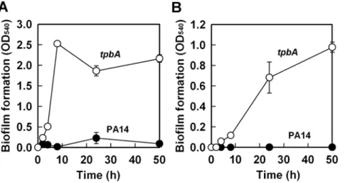
Previously, by screening 5850 transposon mutants for altered biofilm formation, we identified 137 transposon mutants of P. aeruginosaPA14 with over 3-fold enhanced biofilm formation [20]. Among these mutants, thetpbA(PA3885) mutant increased biofilm formation by 147-fold after 8 h in LB medium at 37uC (Fig. 1A). This significant increase in biofilm formation upon inactivatingtpbA
is partially due to enhanced attachment to the polystyrene surface because biofilm formation at the bottom of the plates (solid/liquid interface) increased gradually with thetpbAmutant while PA14 did not form biofilm on the bottom of the plate (Fig. 1B).
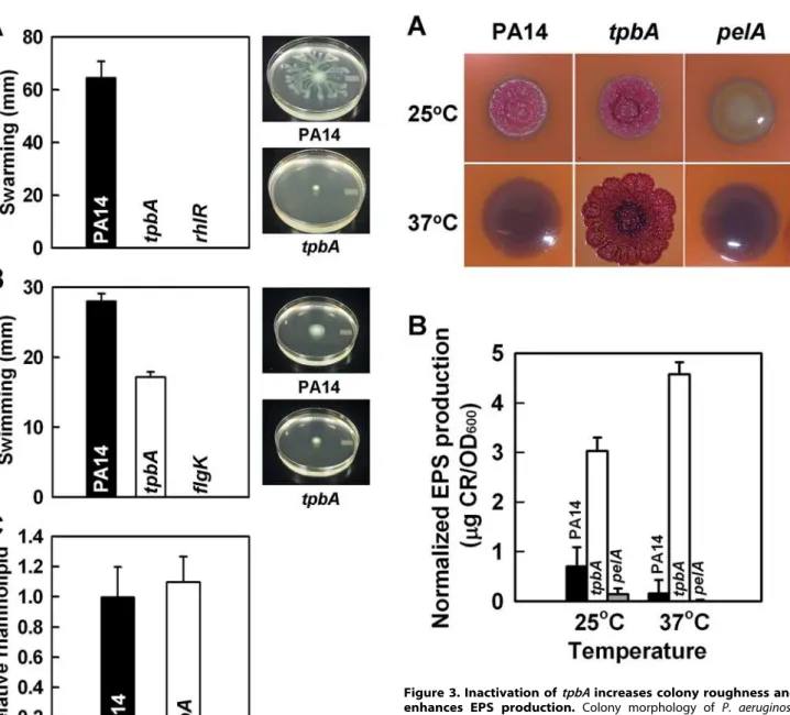
Motility often influences biofilm formation inP. aeruginosa; biofilm formation is inversely influenced by swarming motility [21], and swimming motility increases initial attachment to surfaces during biofilm development [22]. To examine the relationship between enhanced biofilm formation and motility in thetpbAmutant, we examined swimming and swarming motility for this mutant;rhlR
[23] and flgK [22] mutants were used as negative controls for swarming and swimming motility, respectively. Although PA14 swarmed on the surface of plates at 24 h, the tpbA mutations abolished swarming like the rhlR mutation (Fig. 2A). The tpbA
mutation also decreased swimming motility by 40% (Fig. 2B). Swarming is positively influenced by production of the biosurfactant putisolvins inP. putida[24] and rhamnolipids inP. aeruginosa[23]. However, no significant difference was found in the production of rhamnolipids between PA14 and thetpbAmutant (Fig. 2C). This shows thetpbAmutation abolishes swarming in a manner distinct from the production of rhamnolipids.
TpbA affects colony morphology, decreases EPS, and decreases pellicle production
Congo-red is often used to observe colony morphology because it detects EPS production and this impacts biofilm formation; for example, thewspFmutant shows wrinkly colony morphology on
Author Summary
Most bacteria live in biofilms, which are complex communities of microorganisms attached to a surface via polysaccharides; these biofilms are responsible for most
human bacterial diseases. The pathogen Pseudomonas
aeruginosais best-studied for biofilm formation. Currently, it is recognized that cell communication or quorum sensing is important for biofilm formation, but how these external signals are converted into internal signals to regulate the networks of genes that result in biofilm formation is not well understood. Here, by studying 5850 bacterial strains, each of which lacks a single protein, we
identify a new enzyme of P. aeruginosa, a tyrosine
Congo-red plates and has increased biofilm formation [25], while smooth colonies like the pelAmutant [3] form less biofilm. We found that thetpbAmutant formed a red, wrinkly colony when it was grown on Congo-red plates at 37uC, although PA14 and the
pelAmutant formed white smooth colonies (Fig. 3A). When the bacteria were grown at 25uC, both PA14 and the tpbA mutant formed red wrinkly colonies, but the pelAmutant still formed a white smooth colony (Fig. 3A). These observations with thepelA
mutant and wild-type PA14 are identical to the previous report that expression of the pelgenes is induced at room temperature and repressed at 37uC [7]. Therefore, the red wrinkly colony formed by thetpbAmutant at 37uC implies increased production of EPS via Pel.
We also quantified the amount of EPS bound to cells of PA14 and thetpbAmutant at both 37uC and 25uC. As shown in Fig. 3B, thetpbAmutant produced 28-fold more EPS than PA14 at 37uC. ThetpbAmutant also produced 4.3-fold more EPS than PA14 at room temperature. The pelA mutant (negative control) did not form EPS at both temperatures tested. We also found that thetpbA
mutant formed a pronounced pellicle at 37uC after 1 day, but PA14 and thepelAmutant did not form a pellicle (data not shown). At 25uC, both thetpbAmutant and PA14 formed pellicles after 5 days. Taken together with the EPS production data, TpbA reduces pellicle formation by decreasing Pel activity.
Differentially regulated genes in biofilm cells of thetpbA
mutant
To confirm the impact of thetpbAmutation onpelexpression and to investigate its impact on the whole genome, a whole-transcriptome analysis was performed with biofilm cells of thetpbA
mutant at 37uC at 7 h; planktonic cells were not assayed since we were primarily interested in how TpbA controls biofilm formation. Inactivation oftpbAaltered diverse loci including genes related to EPS production (pelACDFinduced approximately 4-fold), transport (PA2204 repressed approximately 5-fold, PA4142–PA4143 in-duced approximately 3-fold), type IV pili (PA4302 to PA4306 repressed approximately 4-fold), and a putative adhesin and its regulator (PA4624–PA4625 induced approximately 4-fold) (Tables S1 and S2). Expression oftpbAwas induced as much as 120-fold in the tpbA mutant, suggesting that TpbA negatively regulates its
transcription. The whole-transcriptome experiments were per-formed twice using independent cultures of PA14 and thetpbA
mutant at 7 h, and most of the differentially regulated genes were consistently altered exceptpelgenes which were induced the most in the samples containing an RNase inhibitor. A whole-transcriptome analysis was also conducted using biofilm cells at 4 h since the mode of growth switched from planktonic to biofilm for thetpbAmutant at this time (Fig. 1A). Similar to the 7 h results, several loci were induced includingpelAEF(1.5- to 1.7-fold),tpbA
(42-fold), PA1168–PA1169 (1.4- to 2.1-fold), PA3886 (3.5-fold), and PA4624–PA4625 (2- to 3.7-fold) (Table S1).
To verify induction of the pel locus, expression of pelA was determined by quantitative real time-PCR (qRT-PCR). Using two independent RNA samples extracted from biofilm cells at 7 h,pelA
was induced 1126100-fold in thetpbA mutant vs. PA14. These results showed EPS production is induced significantly in thetpbA
mutant due to overexpression of pel genes. qRT-PCR also confirmed induction of PA4625 (767-fold) as well as PA4139 (38630-fold) that encodes a hypothetical protein.
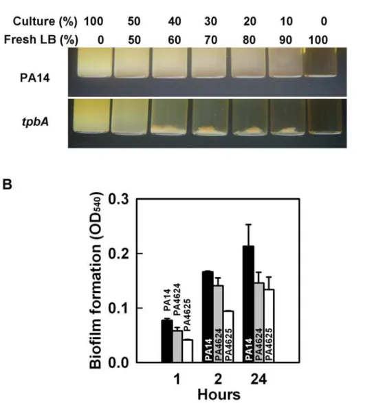
TpbA represses adhesin expression and reduces aggregation
Cell aggregative behavior is also related to biofilm formation so we investigated the role of TpbA on cell aggregation and found the
tpbAmutant causes cell aggregation (Fig. 4A). Autoaggregation of thetpbA mutant was also observed in 96-well polystyrene plates during biofilm formation (data not shown). Our whole-transcrip-tome analysis showed that inactivatingtpbAinduced both PA4624 (encodes for a putative hemolysin activator) and PA4625 (encodes for an adhesin/hemagglutinin) by 2.1- to 4.9-fold. In E. coli, adhesin regulates cell aggregation as well as attachment [26]. To examine whether PA4624–PA4625 control adhesive activity inP. aeruginosa, we investigated biofilm formation with these mutants. Both mutants showed decreased initial biofilm formation; i.e., initial attachment, to polystyrene plates at 1 h and 2 h (Fig. 4B), and final biofilm formation at 24 h was also decreased for both the PA4624 and PA4625 mutants, which suggests that both gene products control attachment to the surface. Therefore, TpbA decreases cell aggregation probably by repressing the PA4624 and PA4625 genes.
Figure 1. Inactivation oftpbAincreases biofilm formation.Total biofilm formation (at the liquid/solid and air/liquid interfaces) (A), and biofilm formation on the bottom of polystyrene plates (B) byP. aeruginosaPA14 and thetpbAmutant at 37uC in LB after 50 h. Six to ten wells were used for each culture. Data show the average of the two independent experiments6s.d.
Complementation of thetpbAmutation
To verify whether the phenotypes observed in thetpbAmutant were caused by loss of function of TpbA, we confirmed transposon insertion in tpbA by PCR at residue 25. Furthermore, biofilm formation for both PA14 and thetpbAmutant were examined with
tpbAexpressed intransunder the control of an arabinose-inducible promoter. tpbAexpression reduced biofilm formation of thetpbA
mutant by 33% (Fig. S1A) and abolished biofilm formation on the bottom of the plates (Fig. S1B). Similar results were found upon expressingtpbAin wild-type PA14 (OD540value was 0.2260.02 for PA14/pMQ70 and 0.0260.01 for PA14/pMQ70-tpbA, Fig. S1A). In addition, the aggregative phenotype of thetpbAmutant was also
complemented by expression of tpbA in trans(Fig. S1C). Taken together, TpbA functions as a negative regulator of biofilm formation and aggregation in PA14.
Genetic screening identified Pel and GGDEF-proteins downstream of TpbA
To investigate how TpbA regulates biofilm formation, EPS production, wrinkly colony morphology, and cell aggregation, genetic screening was conducted using Tn5-luxABtransposon mutagenesis to find suppressive loci for the phenotypes of thetpbA mutation. The double mutant library (tpbA plus random gene inactivations) was screened first for a reduction in aggregation; this step eliminated most cells with unaltered phenotypes by allowing them to aggregate and precipitate at the bottom of the tube. The cells remaining in the supernatant that failed to aggregate like thetpbAmutant were grown on Congo-red plates, incubated at 37uC for 3–4 days, and colonies displaying a white and smooth shape like the wild-type strain were chosen. After that, a third screen was performed by assaying biofilm Figure 2. TpbA regulates swarming, swimming motility, and
production of rhamnolipids. Swarming motility (A), swimming motility (B), and production of rhamnolipids (C) ofP. aeruginosaPA14 and thetpbAmutant at 37uC after 24 h. Five plates were used for each swarming and swimming culture, and data show the average of two independent experiments. For the production of rhamnolipids, data show the average of the two independent experiments6s.d. doi:10.1371/journal.ppat.1000483.g002
Figure 3. Inactivation oftpbAincreases colony roughness and enhances EPS production. Colony morphology of P. aeruginosa
PA14, thetpbAmutant, and thepelAmutant on Congo-red plates after 6 days at 25uC or 37uC (A). EPS production of each strain after 24 h at 37uC or after 48 h at 25uC (B). Data show the average of the two independent experiments6s.d.
formation using 96-well polystyrene plates to identify double mutants that had biofilm formation like the wild-type strain. Twenty-six mutants were identified that showed reduced aggregation, a white smooth colony, and reduced biofilm formation like the wild-type strain, and 19 of these mutations were in the pel locus (Fig. 5A, Table 1). Four of the other mutants have the Tn5-luxABinsertion in the TpbB gene (encodes a GGDEF-motif protein) and in the PA1121 gene (encodes a hypothetical protein). In addition, insertions were found in the PA1678 gene (encodes a putative DNA methylase) and in the promoter of the PA5132 gene (encodes a putative protease) (Table 1). Like the double mutants, all of the single mutants lacking the gene identified by genetic screening were tested for biofilm formation, and all of these mutants formed less biofilm as reported previously (Fig. 5) [3,4].
TpbA negatively controls cellular c-di-GMP concentrations
Results of genetic screening and the whole-transcriptome analysis implied TpbA regulates c-di-GMP concentrations since loss of one of
the GGDEF proteins (TpbB) masked the phenotypes of the tpbA
mutant.tpbBencodes a functional GGDEF protein whose activity was confirmed by overexpressing this gene inP. aeruginosa[4]. We also confirmed that expression of tpbB increases cell aggregation and attachment to tubes so thetpbBmutation may be complemented (Fig. S2). In addition, we measured the cellular c-di-GMP concentrations of PA14 and the tpbA mutant using high performance liquid chromatography (HPLC) as reported previously [4]. The peaks corresponding to c-di-GMP were observed with the extracts of the
tpbAmutant, but not with those of PA14, and the peak was confirmed by comparing the spectrum to purified c-di-GMP as well as by spiking the samples with purified c-di-GMP (Fig. S3). We estimated the cellular c-di-GMP concentration was 1062 pmol/mg cells in the
tpbAmutant. This is comparable to the c-di-GMP concentration found for a small colony variant that showed aggregation (around 2.0 pmol/mg cells) and a mutant with wrinkly colony morphology [27]. Overexpression oftpbBresults in c-di-GMP concentrations of 134 pmol/mg cells in PA14 [4]. Therefore, TpbA reduces c-di-GMP concentrations in the cell and probably does so via TpbB.
TpbA is a tyrosine phosphatase
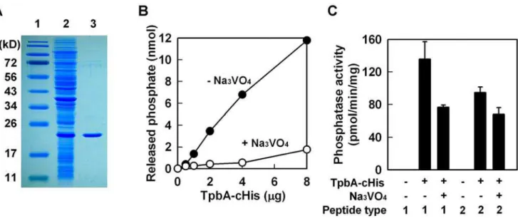
tpbAencodes a 218 aa protein that has the conserved domain for a protein tyrosine phosphatase [28,29] since it has the C(X)5R(S/ T) motif beginning at aa 132 (CKHGNNRT). To confirm it is a tyrosine phosphatase, we purified TpbA by adding a polyhistidine tag at either the N-terminus (TpbA-nHis) or the C-terminus (TpbA-cHis) (note only the C-terminus fusion protein was active). Expression of recombinant TpbA was confirmed inE. coliby clear expression of a band at 24 kD (Fig. 6A). The purified TpbA protein had phosphatase activity with p-nitrophenyl phosphate (pNPP) that is often used as a general phosphatase substrate [29] (Fig. 6B). Further proof that TpbA is a tyrosine phosphatase was found using a tyrosine phosphatase specific inhibitor, trisodium orthovanadate [30], that completely inhibited the phosphatase
activity of TpbA-cHis (Fig. 6B). The third and fourth lines of evidence that TpbA is a tyrosine phosphatase were found using tyrosine specific substrates; TpbA-cHis dephosphorylated both phosphotyrosine peptides, END(pY)INASL (peptide type I) and DADE(pY)LIPQQG (peptide type II) (Fig. 6C), and this activity was inhibited by trisodium orthovanadate. These results show conclusively that TpbA encodes a tyrosine phosphatase.
Tyrosine phosphorylation enhances biofilm formation in PA14
To see the effect of tyrosine phosphorylation on biofilm formation, biofilm formation was examined in PA14 with trisodium orthovanadate at 37uC for 4 h which should reduce dephosphorylation by TpbA. Trisodium orthovanadate increased PA14 biofilm formation 3.6-fold (Fig. S4), showing that cellular tyrosine phosphorylation increases biofilm formation.
TpbA is found in the periplasm
The N-terminal region of TpbA protein has a putative signal peptide, predicted by pSORT [31], that appears necessary for secretion of this protein (28 aa, MHRSPLAWLRLLLAAVL-GAFLLGGPLHA). This implied that processing of N-terminal region of TpbA protein may be essential for full phosphatase activity. To prove that TpbA has an active signal sequence, we expressed TpbA inE. coliand collected the proteins from cytosolic, periplasmic, and membrane fractions. All fractioned proteins were analyzed by SDS-PAGE, and we found that TpbA exclusively localized in the periplasm (data not shown). Hence, TpbA probably dephosphorylates its substrate in the periplasm which explains why phosphatase activity was seen only with the fusion protein with the His tag at the C-terminus.
Y48 and Y62 are responsible for TpbB activity
Since TpbA is a tyrosine phosphatase that is found in the periplasm and since TpbB has three likely periplasmic tyrosines (Y48, Y62, and Y95) [32], we mutated the periplasmic tyrosine residues by converting them to phenylalanine and checked for TpbB activity in thetpbBmutant. Active TpbB, from overexpres-sion oftpbBusing thetpbBmutant and pMQ70-tpbB, always leads to aggregation whereas the empty plasmid does not cause aggregation (Fig. S5); hence, if a necessary tyrosine is mutated, there should be a reduction in aggregation. Aggregation was always observed with Y95F in nine cultures; hence TpbB-Y95F remains active even though it lacks tyrosine 95 so this tyrosine is not phosphorylated/dephosphorylated. In contrast, the Y48F mutation of TpbB decreased aggregation for 43% of the cultures (20 of 46 cultures did not aggregate), and the Y62F mutation decreased aggregation for 24% of the cultures (9 of 37 cultures did not aggregation). Hence, both Y48 and Y62 are likely targets for tyrosine phosphorylation/dephosphorylation of TpbB with Y48 preferred. We confirmed that these mutations did not affect expression level of TpbB protein (data not shown).
TpbA tyrosine phosphatase is unique among bacteria and eukaryotes
Tyrosine phosphorylation and dephosphorylation have crucial roles in cellular signaling and are well-conserved among many organisms [33]. Some bacterial tyrosine phosphorylations have been identified and these regulatory systems control divergent cellular functions [34]. In order to predict whether TpbA function is conserved among other species, we conducted a BLASTP search and found the TpbA protein is highly conserved among P. aeruginosa(PAO1, PA14, C3719, and PA7 with an E value less than Figure 5. Reduction in biofilm formation bytpbAphenotype
reversal mutations. Biofilm formation of double mutants (A) and single mutants (B) identified by genetic screening for thetpbAmutation at 37uC in LB after 24 h. Six to ten wells were used for each culture. Representative data are shown in (A). Biofilm formation of each mutant was calculated relative to that of PA14 (OD540mutant/OD540wild-type).
3e-98) and is well-conserved amongP. fluorescensPf-5, P. fluorescens
Pf0-1, P. mendocina, Burkholderia cepacia, Pelobacter carbinolicus,
Desulfatibacillum alkenivorans, Bacteroides thetaiotaomicron, B. ovatus, B. caccae, Acinetobacter baumannii, Desulfococcus oleovorans, and Geobacter
metallireducens (E values less than 3e-11). All of these conserved tyrosine phosphatases have the C(X)5R(S/T) signature and most are uncharacterized. Even though protein similarity is not very high, some eukaryotes, such asHomo sapiensandArabidopsis thaliana,
Table 1.Phenotype reversal loci for thetpbAmutation.
Strain PAO1 ID PA14 ID
Gene
Name Gene function
Relative biofilm formation
Colony
morphology Aggregation
PA14 - - - - 0.14 white smooth 2
tpbA PA3885 PA14_13660 tpbA Tyrosine phosphatase (this study) 1.00 red wrinkly +
Mutant 1 PA1120 PA14_49890 tpbB c-di-GMP cyclase, GGDEF motif 0.13 white smooth 2
Mutant 19 PA1120 PA14_49890 tpbB c-di-GMP cyclase, GGDEF motif 0.10 white smooth 2
Mutant 4 PA1121 PA14_49880 Hypothetical protein 0.22 white smooth 2
Mutant 11 PA1121 PA14_49880 Hypothetical protein 0.07 white smooth 2
Mutant 2 PA1678 PA14_42790 Putative DNA methylase 0.30 white smooth 2
Mutant 6 PA1678 PA14_42790 Putative DNA methylase 0.28 white smooth 2
Mutant 5 PA3058 PA14_24560 pelG Predicted membrane protein related to EPS production, PelG
0.24 white smooth 2
Mutant 7 PA3058 PA14_24560 pelG Predicted membrane protein related to EPS production, PelG
0.19 white smooth 2
Mutant 9 PA3058 PA14_24560 pelG Predicted membrane protein related to EPS production, PelG
0.27 white smooth 2
Mutant 12 PA3058 PA14_24560 pelG Predicted membrane protein related to EPS production, PelG
0.25 white smooth 2
Mutant 18 PA3058 PA14_24560 pelG Predicted membrane protein related to EPS production, PelG
0.42 white smooth 2
Mutant 10 PA3060 PA14_24530 pelE Sucrose synthase related to EPS production, PelE
0.41 white smooth 2
Mutant 15 PA3060 PA14_24530 pelE Sucrose synthase related to EPS production, PelE
0.45 white smooth 2
Mutant 3 PA3061 PA14_24510 pelD L-lactate permease related to EPS production, PelD
0.20 white smooth 2
Mutant 16 PA3063 PA14_24490 pelB Conserved hypothetical protein related to EPS production, PelB
0.31 white smooth 2
Mutant 21 PA3063 PA14_24490 pelB Conserved hypothetical protein related to EPS production, PelB
0.21 white smooth 2
Mutant 22 PA3063 PA14_24490 pelB Conserved hypothetical protein related to EPS production, PelB
0.17 white smooth 2
Mutant 23 PA3063 PA14_24490 pelB Conserved hypothetical protein related to EPS production, PelB
0.38 white smooth 2
Mutant 25 PA3063 PA14_24490 pelB Conserved hypothetical protein related to EPS production, PelB
0.28 white smooth 2
Mutant 27 PA3063 PA14_24490 pelB Conserved hypothetical protein related to EPS production, PelB
0.23 white smooth 2
Mutant 8 PA3064 PA14_24480 pelA Oligogalacturonide lyase related to EPS production, PelA
0.18 white smooth 2
Mutant 14 PA3064 PA14_24480 pelA Oligogalacturonide lyase related to EPS production, PelA
0.45 white smooth 2
Mutant 17 PA3064 PA14_24480 pelA Oligogalacturonide lyase related to EPS production, PelA
0.29 white smooth 2
Mutant 20 PA3064 Pro-PA14_24480 pelA Oligogalacturonide lyase related to EPS production, PelA
0.25 white smooth 2
Mutant 24 PA3064 PA14_24480 pelA Oligogalacturonide lyase related to EPS production, PelA
0.24 white smooth 2
Mutant 13 PA5132 Pro-PA14_67780 Putative protease 0.15 white smooth 2
Genetic screening identified additional mutations that mask the phenotypes of thetpbAmutant (enhanced biofilm formation, EPS production, wrinkly colony morphology, and aggregation). Relative biofilm formation is shown as a ratio of that of thetpbAmutant.
have TpbA homologs with a C(X)5R(S/T) signature. Therefore, TpbA and TpbA homologs may share important functions in procaryotes and eucaryotes.
Las-QS regulates expression oftpbA
QS regulates many genes inP. aeruginosa via a conserved cis -element in the promoter of each gene. N -(3-oxododecanoyl)-L-homoserine lactone binds to the LasR transcriptional regulator [35], and this complex interacts with thelas-box, defined as CT-(N)12-AG sequence [36]. The Las-box is conserved among the promoters of the Las-QS regulated genes including lasB, rhlAB, and rhlI [36]. Another class of transcriptional regulation is governed by thelys-box, that is defined as a palindromic sequence, T-(N)11-A [37], and MvfR is a LysR-type transcription factor that binds to the lys-box [38]. We found that the promoter of tpbA
(ptpbA) has a putativelas-box 220 bp upstream of the start codon (CTCGCCTCGCTGAAAG) and a putative lys-box 90 bp upstream of the start codon (TGAAGCTGCCTCA). In order to examine if expression oftpbAis regulated by QS, we constructed a ptpbA::lacZ fusion plasmid (pLP-ptpbA) and transformed this into QS-related PA14 mutants (lasI,rhlI, andlasR rhlR). Expression of
tpbAgene in biofilm cells was reduced by 42% in thelasImutant, but not in the rhlI mutant (Fig. 7). Corroborating these results, inactivation of bothlasRandrhlRalso decreased expression oftpbA
gene by 39% (Fig. 7). Similar results were obtained when the activity of ptpbA::lacZ was examined in planktonic cells (50% reduction in transcription for thelasImutant and 37% reduction for thelasR rhlRmutant). Since loss of QS only affected expression oftpbAby 50%, other factors may also participate in the regulation oftpbA. These results suggest that Las-QS, rather than Rhl-QS, is an activator oftpbAexpression with other unknown regulators.
We also investigated whether thetpbAmutation influences the regulation of Las- and Rhl-QS using plasR::lacZ and prhlR::lacZ
plasmids. Expression oflasRwas slightly increased (1.3-fold) with thetpbAmutation. This indicated that Las-QS has more impact on expression oftpbAthantpbAdoes on that of Las-QS. In addition, expression of rhlR was decreased by 2-fold in the tpbA mutant.
Hence, LasR appears to enhance tpbA transcription and TpbA leads to increasedrhltranscription.
Discussion
In this study, we demonstrate that TpbA is a tyrosine phosphatase that regulates diverse phenotypes in P. aeruginosa
including the concentration of cellular c-di-GMP. As a second messenger, c-di-GMP is a positive regulator of biofilm formation [4], EPS production [6], and pellicle formation [4], and a negative regulator of swarming motility [39]. The lines of evidence that show TpbA represses c-di-GMP production inP. aeruginosathat we found are (i) inactivatingtpbA increases c-di-GMP (Fig. S3); (ii) inactivating tpbA increases biofilm formation (Fig. 1), EPS production (Fig. 3B), and pellicle formation, and c-di-GMP stimulates biofilm formation [4], EPS production [6], and pellicle Figure 6. TpbA has phosphatase activity against tyrosine residues.Purification of TpbA-cHis (lane 1: protein marker, lane 2: whole cell lysate fromE. coliBL21(DE3)/pET28b-13660c after 3 h of IPTG induction, lane 3: purified TpbA-cHis) (A).p-Nitrophenyl phosphate phosphatase assay with TpbA-cHis protein (B). Phosphatase reaction was performed at 37uC for 1 h with the indicated amount of protein. Na3VO4(10 mM) was used as an
inhibitor specific for tyrosine phosphatases. Protein tyrosine phosphatase assay with TpbA-cHis (C). Phosphatase reaction was performed with synthetic phosphotyrosine peptides (type I: END(pY)INASL and type II: DADE(pY)LIPQQG) at 37uC for 3 h. Na3VO4(50 mM) was used as an inhibitor.
doi:10.1371/journal.ppat.1000483.g006
Figure 7. Las QS activates transcription oftpbA.b-galactosidase activity of ptpbA was measured with biofilm cells of PA14 and the mutantslasI,rhlI, andlasR rhlRusing pLP-ptpbA. Data show the average of the two independent experiments6s.d.
formation [4]; (iii) inactivatingtpbAincreases expression of thepel
locus (seen via the whole-transcriptome analysis and RT-PCR), and c-di-GMP activates expression ofpelA[6]; (iv) inactivatingtpbA
increases aggregation (Fig. 4) and expression of adhesins (PA4625), and c-di-GMP stimulates adhesion [40]; (v) inactivating tpbA
decreases motility (abolishing swarming and decreasing swimming in thetpbAmutant, Fig. 2AB), and c-di-GMP decreases swarming [40]; (vi) inactivatingtpbB(encodes a GGDEF-motif protein that produces c-di-GMP [4]) suppresses the phenotypes observed in the
tpbA mutant, and (vii) expression of tpbA and tpbB in trans
complements aggregation/biofilm formation and aggregation, respectively. Thus, TpbA represses these phenotypes by decreasing c-di-GMP. A proposed regulatory mechanism for biofilm formation by TpbA is shown in Fig. 8.
EPS production inP. aeruginosaPA14 is regulated by PelA [3]. Transcription ofpelAis higher at temperatures lower than 37uC, and PA14 forms more biofilm at lower temperatures [7]. However, thetpbA mutation seems to constitutively enhance pel
expression independently from this temperature regulation as seen in the enhanced EPS production at 37uC (Fig. 3) and the whole-transcriptome analysis that was conducted at 37uC (Table S2). In addition to increased expression of pelA, additional activation of Pel proteins might be caused by the increased c-di-GMP concentration by the tpbA mutation since c-di-GMP binds PelD and increases EPS production [6]. In addition to the enhanced
EPS production (Fig. 3B) and increasedpel expression (Fig. 3A, Table S1, and qRT-PCR) seen in thetpbAmutant, another reason why inactivatingtpbAincreased biofilm formation is the elevated adhesin activity as seen via enhanced biofilm formation on the bottom of polystyrene plates (Fig. 1B). Cell surface adhesins affect bacterial adhesive activity [41], and we have discovered a novel adhesin (PA4625) that is related to TpbA (Table S1) and to initial biofilm formation (Fig. 4B). Since expression of adhesion factors is also positively regulated by c-di-GMP [40], elevated c-di-GMP level enhances adhesion of thetpbAmutant.
c-di-GMP seems to control the switch of motility-sessility of the
tpbA mutant since inactivation of TpbA abolished swarming motility (Fig. 2A) and decreased swimming motility by 40% (Fig. 2B), although regulation of swarming motility is very complex as its activity is controlled by QS, flagellar synthesis, and production of rhamnolipids [42]. In addition, our whole-transcriptome results showed weak repression of some of flagellar biosynthesis genes (flg, fle, andfli loci) due to the elevated c-di-GMP, and activity of FleQ, a transcriptional activator of flagellar biosynthesis, is repressed upon binding c-di-GMP [43]. Hence, the increased c-di-GMP concentrations may repress motility of the
tpbA mutant via the FleQ pathway that affects expression of flagellar synthesis genes.
Many genes are expected to be differentially regulated by changing c-di-GMP concentrations since it plays a role as a second
Figure 8. Schematic of TpbA regulation of biofilm formation inP. aeruginosaPA14.The QS molecule,N-(3-oxododecanoyl)-L-homoserine lactone (3-oxoC12-HSL), binds to the LasR transcription factor, and this complex activates expression oftpbA. TpbA has a N-terminal signal sequence and is translocated into the periplasm. Periplasmic TpbA dephosphorylates the membrane-anchored GGDEF protein TpbB at a tyrosine reside which deactivates GGDEF protein activity. The reduced cellular c-di-GMP concentration decreases expression of thepeloperon as well as adhesin genes. This leads to reduced EPS production, biofilm formation, and pellicle formation, as well as enhanced swarming motility. Production of rhamnolipids is not regulated by TpbA.
messenger inP. aeruginosa. Similar regulation of gene expression was observed between thetpbAmutant and the other strains related to c-di-GMP production. For example, production of PA1107, TpbB, and PA3702 proteins that have a GGDEF-domain leads to activation of pelA expression [6]. Mutation in wspF, encoding a CheB-like methylesterase, increases both biofilm formation and c-di-GMP production [25].wspFmutation altered expression of genes such as pelABCDEFG, PA4624, PA4625, PA2440, and PA2441 whose expression are induced in the tpbA mutant (Table S2). Common regulation of these genes may be partially controlled by the elevated cellular c-di-GMP concentrations. In contrast, expression ofpelAwas not induced in thebifAmutant that produces more c-di-GMP [44]. This may be because regulation of c-di-GMP signaling is complex in that theP. aeruginosa genome encodes 17 diguanylate cyclases, 5 phosphodiesterases, and 16 diguanylate cyclase-phosphodiesterase proteins [4].
Relevance of c-di-GMP to regulation of diverse cellular functions is now an emerging topic in bacteriology. This second messenger is an activator of cellulose synthase in Acetobactor xylinum [5] and controls many phenotypes in P. aeruginosa [4]. Several GGDEF proteins for synthesis and EAL proteins for degradation of c-di-GMP have been identified inP. aeruginosa[6,39,44], and increased production of c-di-GMP enhances biofilm formation and decreases swarming motility [4,6,39,44]. Similarly, enhanced biofilm forma-tion and/or abolished swarming motility were observed in thetpbA
mutant via increased production of cellular c-di-GMP. Since TpbA does not possess GGDEF and EAL domains, this protein indirectly influences cellular c-di-GMP concentrations via its phosphatase activity as shown by activity with both pNPP, a broad substrate for phosphatases, and two phosphotyrosine-specific peptides (Fig. 6). We also found that processing the N-terminal signal sequence may be necessary for TpbA activity in the periplasm. Hence, our results reveal a novel regulatory mechanism for cellular c-di-GMP concentration by tyrosine phosphorylation in the periplasm ofP. aeruginosa; control of c-di-GMP by tyrosine phosphorylation has not been shown previously.
There is little known about the regulation of GGDEF and EAL proteins in regard to regulation of c-di-GMP level. A chemosen-sory system, encoded by wspABCDEFRin PAO1, regulates c-di-GMP production via a His-Asp phosphorylation relay [25]. For thetpbAmutant,tpbBwas found to reverse the phenotype oftpbA, suggesting that overproduction of c-di-GMP is clearly related to the phenotypes of thetpbAmutant as overexpression of this gene caused pronounced aggregation (Fig. S2). Probably, TpbB, or another GGDEF protein, might participate in c-di-GMP synthesis in the tpbA mutant. A comprehensive analysis of all of the P. aeruginosa GGDEF proteins has been completed, and those GGDEF proteins that abolished or decreased biofilm formation are PA0169, PA1107, TpbB, PA1181, PA1433, PA1727, PA3702, PA4959, and PA5487 [4]. Among these GGDEF proteins, only PA1107, TpbB, PA3702, and PA5487 increased biofilm formation when their genes were overexpressed [4]. Because TpbA is a periplasmic protein, its target GGDEF protein should have periplasmic regions. By a bioinformatics evaluation, of those four GGDEF proteins that increased biofilm formation, only PA1107 and TpbB have transmembrane regions. Taken together with the results of genetic screening, TpbB is the most likely target protein for TpbA. Also, our results imply the periplasmic Y48 and Y62 residues of TpbB are the likely targets for tyrosine phosphoryla-tion. We are now investigating whether TpbA regulates the activity of GGDEF proteins to control cellular c-di-GMP concentrations inP. aeruginosa.
The relationship between tyrosine phosphorylation and biofilm formation is not well established. We found that trisodium
orthovanadate treatment increased biofilm formation of PA14 (Fig. S4), indicating that tyrosine phosphorylation increases biofilm formation inP. aeruginosa. Recently, Ltp1, a low molecular weight tyrosine phosphatase in non-motile, Gram-negative P. gingivalis, was identified as a negative regulator of EPS production and biofilm formation [45]. A sequence similarity search shows TpbA is not a homolog of Ltp1, because TpbA has a signal sequence in its N-terminal region, and TpbA is translocated into the periplasm. Other differences were found in the position of the motif for the tyrosine phosphatase, since TpbA has the motif at the position 132 and Lpt1 has it at position 9. It appears theP. aeruginosagenome encodes another tyrosine phosphatase, annotated asptpA, that has a tyrosine phosphatase motif at the position 7 and does not have a signal sequence at the N-terminus. The function of PtpA is unknown but it is essential [46].
In contrast to poorly-investigated Tyr phosphorylation, regula-tion of biofilm formaregula-tion by phosphorylaregula-tion has been identified for several systems; for example, for the His kinase/Asp response regulator phosphorylation systems RocS1/RocA1/RocR of P. aeruginosa PAK [47] and the PAO1 PA1611/PA1976/PA2824/ RetS/HptB system ofP. aeruginosa[48]. In addition, in theB. subtilis
PrkC/PrpC system [18], loss of a membrane-anchored Thr kinase and its phosphatase reduces biofilm formation. Our results indicate that TpbA acts as a negative regulator of cellular c-di-GMP formation and loss of TpbA results in increased c-di-GMP concentrations that enhance biofilm formation and inhibit motility. These results show clearly that posttranslational modification through phosphatase activity is related to bacterial biofilm formation as well as to the regulation of the synthesis of cellular second messengers. In addition, by showingtpbA transcription is increased by LasR (Fig. 7) and by finding AHL-binding motifs, we have now linked quorum sensing to c-di-GMP concentrations and biofilm formation inP. aeruginosa. Similarly,Vibrio choleraeQS was found recently to reduce cellular di-GMP concentrations via a c-di-GMP-specific phosphodiesterase which leads to lower biofilm formation [49]. A common element in both studies is that QS seems to be a negative regulator of c-di-GMP. ThetpbAmutation caused a hyper-aggregative phenotype (Fig. 4), and this would lead to flat biofilms since thewspFmutant, which accumulates increased c-di-GMP, formed flat biofilms [25]. Formation of flat and undifferen-tiated biofilms is also observed by loss of LasI function [12] that can activate tpbA expression. Hence, TpbA might participate in developing biofilm structure. These results are important in that the regulatory networks that control c-di-GMP concentrations are now linked to the environment and cell populations.
Materials and Methods
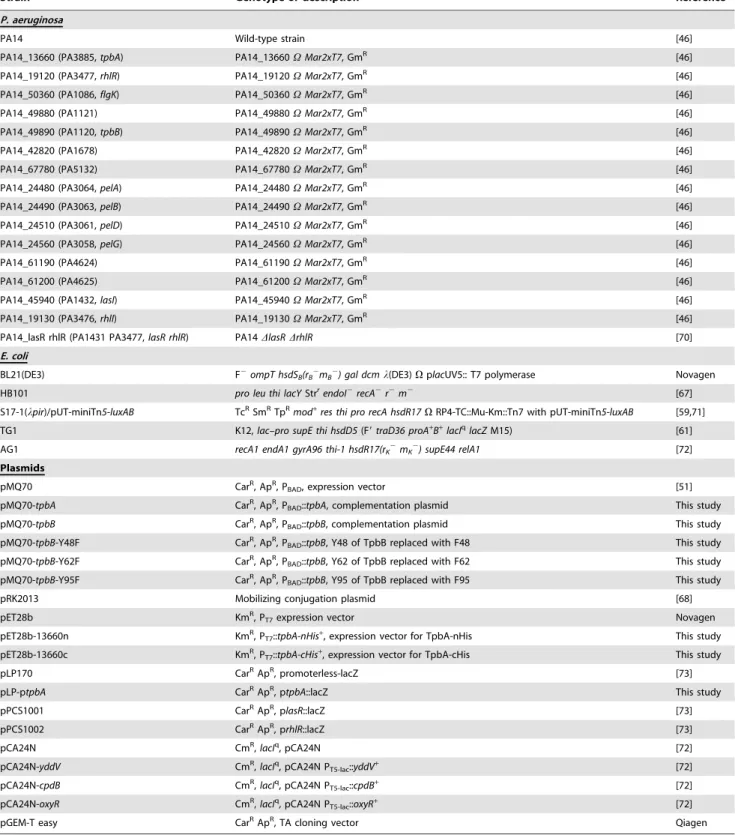
Bacterial strains and growth conditions
Strains used in this study are listed in Table 2.P. aeruginosaPA14 wild-type and its isogenic mutants were obtained from the Harvard Medical School [46]. Transposon insertion of thetpbA
Table 2.Strains used in this study.
Strain Genotype or description Reference
P. aeruginosa
PA14 Wild-type strain [46]
PA14_13660 (PA3885,tpbA) PA14_13660VMar2xT7, GmR [46]
PA14_19120 (PA3477,rhlR) PA14_19120VMar2xT7, GmR [46]
PA14_50360 (PA1086,flgK) PA14_50360VMar2xT7, GmR [46]
PA14_49880 (PA1121) PA14_49880VMar2xT7, GmR [46]
PA14_49890 (PA1120,tpbB) PA14_49890VMar2xT7, GmR [46]
PA14_42820 (PA1678) PA14_42820VMar2xT7, GmR [46]
PA14_67780 (PA5132) PA14_67780VMar2xT7, GmR [46]
PA14_24480 (PA3064,pelA) PA14_24480VMar2xT7, GmR [46]
PA14_24490 (PA3063,pelB) PA14_24490VMar2xT7, GmR [46]
PA14_24510 (PA3061,pelD) PA14_24510VMar2xT7, GmR [46]
PA14_24560 (PA3058,pelG) PA14_24560VMar2xT7, GmR [46]
PA14_61190 (PA4624) PA14_61190VMar2xT7, GmR [46]
PA14_61200 (PA4625) PA14_61200VMar2xT7, GmR [46]
PA14_45940 (PA1432,lasI) PA14_45940VMar2xT7, GmR [46]
PA14_19130 (PA3476,rhlI) PA14_19130VMar2xT7, GmR [46]
PA14_lasR rhlR (PA1431 PA3477,lasR rhlR) PA14DlasRDrhlR [70]
E. coli
BL21(DE3) F2ompT hsdS
B(rB2mB2) gal dcml(DE3)VplacUV5:: T7 polymerase Novagen
HB101 pro leu thi lacYStrrendoI2recA2r2m2 [67]
S17-1(lpir)/pUT-miniTn5-luxAB TcRSmRTpR
mod+
res thi pro recA hsdR17VRP4-TC::Mu-Km::Tn7 with pUT-miniTn5-luxAB [59,71] TG1 K12,lac–pro supE thi hsdD5(F9traD36 proA+
B+
lacIqlacZM15) [61]
AG1 recA1 endA1 gyrA96 thi-1 hsdR17(rK2mK2) supE44 relA1 [72]
Plasmids
pMQ70 CarR, ApR, P
BAD, expression vector [51]
pMQ70-tpbA CarR, ApR, P
BAD::tpbA, complementation plasmid This study
pMQ70-tpbB CarR, ApR, P
BAD::tpbB, complementation plasmid This study
pMQ70-tpbB-Y48F CarR, ApR, P
BAD::tpbB, Y48 of TpbB replaced with F48 This study
pMQ70-tpbB-Y62F CarR, ApR, P
BAD::tpbB, Y62 of TpbB replaced with F62 This study
pMQ70-tpbB-Y95F CarR, ApR, P
BAD::tpbB, Y95 of TpbB replaced with F95 This study
pRK2013 Mobilizing conjugation plasmid [68]
pET28b KmR, P
T7expression vector Novagen
pET28b-13660n KmR, P
T7::tpbA-nHis+, expression vector for TpbA-nHis This study
pET28b-13660c KmR, P
T7::tpbA-cHis+, expression vector for TpbA-cHis This study
pLP170 CarRApR, promoterless-lacZ [73]
pLP-ptpbA CarRApR, p
tpbA::lacZ This study
pPCS1001 CarRApR, plasR::lacZ [73]
pPCS1002 CarRApR, prhlR::lacZ [73]
pCA24N CmR,
lacIq, pCA24N [72]
pCA24N-yddV CmR,lacIq, pCA24N P
T5-lac::yddV+ [72]
pCA24N-cpdB CmR,lacIq, pCA24N P
T5-lac::cpdB+ [72]
pCA24N-oxyR CmR,lacIq, pCA24N P
T5-lac::oxyR+ [72]
pGEM-T easy CarRApR, TA cloning vector Qiagen
GmR, TcR, KmR, CarR, CmR, and ApRindicate gentamicin, tetracycline, kanamycin, carbenicillin, chloramphenicol, and ampicillin resistance, respectively.
used for growth of the P. aeruginosa transposon mutants, carbenicillin (300mg/mL) was used to maintain P. aeruginosa
plasmids, and kanamycin (50mg/mL) and chloramphenicol (50mg/mL) were used to maintainE. coliplasmids (Table 2).
Complementation ofP. aeruginosamutants
For complementation of thetpbAandtpbBmutations,tpbAand
tpbBwere expressed under the control of the pBAD promoter in pMQ70 [51]. tpbA and tpbB were amplified using a Pfu DNA polymerase with primers F1-NheI and PA14_13660-R-cHis-HindIII and PA14_49890-F1-NheI and PA14_49890-R-cHis-HindIII, respectively (Table S3). PCR products were cloned into the NheI and HindIII sites of pMQ70. The resulting plasmids, pMQ70-tpbA and pMQ70-tpbB, were transformed into PA14 and the mutants by conjugation. Briefly, 1 mL of overnight culture of the recipient strain (PA14 or the mutant), helper strain (HB101/pRK2013), and donor strain (TG1/pMQ70, TG1/ pMQ70-tpbA, or pMQ70-tpbB) was washed with 1 mL of fresh LB medium. The mixture of three strains was incubated on LB plates at 37uC overnight. PA14 strains with pMQ70-based plasmid were selected on LB plates with 100mg/mL rifampicin (to kill the donor and helper), 300mg/mL carbenicillin (to kill P. aeruginosa without pMQ70-based plasmids), and 15mg/mL gentamicin (if a recipient was a PA14 mutant constructed using a transposon insertion with the GmRgene). If indicated, 0.05% arabinose was added to induce gene expression.
Biofilm formation
Biofilm formation was examined in 96-well polystyrene plates using crystal violet staining [52]. Overnight cultures ofP. aeruginosa
were diluted to a turbidity of 0.05 at 600 nm with fresh LB medium, and then 150mL of diluted bacterial culture was incubated in 96-well polystyrene plates for 2, 4, 8, 24, and 50 h. Ten wells were used for each strain and three independent cultures were used for each experiment. Trisodium orthovanadate, a tyrosine phosphatase-specific inhibitor, was added to LB medium at 10 mM.
Colony morphology
To observe colony morphology, overnight cultures were diluted to a turbidity of 0.005 at 600 nm with T-broth (10 g/L tryptone), and 2mL of diluted cultures were spotted on Congo-red plates (10 g/L tryptone, 40mg/mL Congo-red, and 20mg/mL Coo-massie brilliant blue) [3]. Plates were incubated at 37uC or room temperature for 3 to 7 days.
Motility assays
Swimming motility was examined with cells grown to a turbidity of 1 at 600 nm using 0.3% agar plates with 1% tryptone and 0.25% NaCl [53] and swarming motility was examined with BM-2 plates (62 mM potassium phosphate, 2 mM MgSO4, 10mM FeSO4, 0.1% casamino acid, 0.4% glucose, and 0.5% Bacto agar) [54]. Motility was measured after 24 h. Five plates were tested for each culture, and two independent cultures were used. The flgK
[22] and rhlR [23] mutants were used as negative controls for swimming and swarming, respectively.
Aggregation assay
Aggregation was examined by diluting overnight cultures with fresh LB medium in 5 mL screw-capped tubes from 0% (no added fresh LB medium) to 100% (pure fresh LB medium). Cells were inverted gently several times and placed at room temperature for 15 min.
Pellicle formation
Overnight cultures of PA14, the tpbA mutant, and the pelA
mutant were diluted to a turbidity of 0.005 at 600 nm in 4 mL T-broth, and the bacterial cultures were placed in a polycarbonate glass tube at 37uC or room temperature [3].
EPS assay
Pel-dependent EPS production was quantified as described previously [6] based on the amount of Congo red that binds to the EPS. Briefly, 1 mL of overnight culture was washed with 1 mL T-broth. Due to aggregative phenotype of the tpbA mutant, cell pellets of the tpbA mutant, wild-type, andpelAmutant (negative control) were sonicated three times at 3W for 10 sec. Bacterial suspensions in T-broth (500mL) were incubated with 40mg/mL Congo-red at 37uC or room temperature with vigorous shaking. After 2 h, the absorbance of the supernatants of the each suspension was measured at 490 nm using a spectrophotometer. T-broth with 40mg/mL Congo-red was used as a blank.
Rhamnolipids assay
Production of rhamnolipids was determined as described previously [55]. Overnight cultures were diluted to a turbidity of 0.05 at 600 nm in 25 mL LB medium and were re-grown at 250 rpm for 24 h to eliminate the effect of antibiotics. The supernatants of the bacterial cultures were used to determine the relative concentrations of rhamnolipids using orcinol/sulfuric acid. Rhamnose (Fisher Scientific, Pittsburgh, PA) was used as a standard.
Whole-transcriptome analysis
The P. aeruginosagenome array (Affymetrix, P/N 510596) was used to investigate differential gene expression in biofilm cells between PA14 and thetpbAmutant. Biofilm cells were harvested from 10 g of glass wool [56] after incubation for 4 h and 7 h in LB with shaking at 250 rpm, and RNA was extracted with a RNeasy Mini Kit (Qiagen) [57]; note the RNase inhibitor RNAlater
(Applied Biosystems, Austin, TX) was used for the 4 h and second 7 h set of microarrays. Global scaling was applied so the average signal intensity was 500. The probe array images were inspected for any image artifact. Background values, noise values, and scaling factors of both arrays were examined and were comparable. The intensities of polyadenosine RNA controls were used to monitor the labeling process. If the gene with the larger transcription rate did not have a consistent transcription rate based on the 13 probe pairs (p-value less than 0.05), these genes were discarded. A gene was considered differentially expressed when the
p-value for comparing two chips was lower than 0.05 (to assure that the change in gene expression was statistically significant and that false positives arise less than 5%) and when the expression ratio was higher than the standard deviation for the whole microarrays [58], 1.4 for 4 h, 1.7 for the first 7 h replicate, and 2.2 for second 7 h replicate. All three sets of whole-transcriptome data were deposited at the Gene Expression Omnibus (GSE13871).
qRT-PCR
qRT-PCR was performed using the StepOnePlusTMReal-Time PCR System (Applied Biosystems, Foster City, CA). Expression of
Genetic screening
To isolate the suppressive loci for TpbA functions, a double mutant library was generated using the Tn5-luxABtransposon with the background of thetpbAmutation as described previously [59]. Briefly, 1 mL of overnight culture of theP. aeruginosa tpbAmutant andE. coliS17-1 (lpir) with Tn5-luxABwere grown on LB plates together overnight. Cells were harvested from the plate and resuspended in 10 mL of LB medium. Screening of cells with mutations in addition to tpbA was performed in three steps. Suppression of the highly-aggregative phenotype of the tpbA
mutant was used first; the cell mixture (P. aeruginosa single and double mutants along with E. coli S17-1) was placed at room temperature for 15 min and the supernatant was used for secondary screening (cells with the tpbA mutant aggregative phenotype were therefore discarded). Supernatant cells were spread on Congo-red plates with 50mg/mL gentamicin (to killE. coli), and 75mg/mL tetracycline (to kill the tpbA single mutant), and incubated for 3–4 days. P. aeruginosa double mutants with smooth surfaces were picked (the tpbA mutant was red and wrinkled). The crystal violet biofilm assay was used for the third screening, and mutants showing decreased biofilm formation in comparison to that of thetpbAmutant were chosen as phenotype reversal mutants. The insertion position of Tn5-luxABtransposon was determined by two-step PCR as described previously [59] with primers LuxAB inside and Arb1 for the first round of PCR and LuxAB outside and Arb2 for the second round of PCR (Table S3). The PCR product was ligated into pGEM-T easy (Promega, Madison, MI) and sequenced using a BigDye Terminator Cycle Sequencing Kit (Applied Biosystems, Foster City, CA).
Quantification of c-di-GMP
c-di-GMP was isolated as described previously [60].P. aeruginosa
was grown in 1 L of LB medium for 16 h at 250 rpm, and formaldehyde (final concentration of 0.18%) was added to inactivate degradation of c-di-GMP. Cells were harvested by centrifugation at 8,000g for 10 min at 4uC. Nucleotide extract was prepared as described previously [60]. Cell pellets were washed with 40 mL of phosphate buffered saline (pH 7) [61] with 0.18% formaldehyde and centrifuged at 8,000gfor 10 min at 4uC. The cell pellets were dissolved in H2O and boiled for 10 min. After cooling the samples on ice for 10 min, nucleotides were extracted in 65% ethanol. Supernatants were transferred, and the extraction was repeated. Pooled supernatants were lyophilized, and pellets were dissolved in 1 mL of 0.15 M triethyl ammonium acetate (TEAA, pH 5.0). The samples were filtered using a PVDF filter (0.22mm), and 20mL of each sample was fractionated using HPLC (Waters 515 with photodiode array detector, Milford, MA) with a reverse-phase column (Nova-PakH C18 column; Waters, 15063.9 cm, 4mm). Separations were conducted in 0.15 M TEAA at a 1 mL/min flow rate using gradient elution with acetonitrile (0% to 15% concentration). Synthetic c-di-GMP (BIOLOG Life Science Institute, Bremen, Germany) was used as a standard. The peak corresponding to c-di-GMP from the extract of thetpbAmutant was verified by co-elution with standard c-di-GMP.E. coliAG1/pCA24N-yddVthat has an elevated c-di-GMP concentration [62] was also used as a positive control.
Plasmid construction of pET28b-13660c and purification of recombinant TpbA-cHis
To determine if TpbA is a phosphatase,tpbAwas amplified with a Pfu DNA polymerase using primers PA14_13660-F-XbaI and PA14_13660-R-XhoI (Table S3). The PCR product was digested with XbaI and XhoI and was ligated in-frame to the polyhistidine
tag sequence of the pET28b vector. The resulting plasmid, pET28b-13660c has thetpbAgene fused to a 66His tag at the C-terminus (TpbA-cHis) and under control of the T7 promoter. The pET28b-13660c plasmid was confirmed by DNA sequencing with the T7 promoter and T7 terminator primers (Table S3). Production of TpbA-cHis was induced inE. coliBL21(DE3) cells with 1 mM IPTG at room temperature overnight. TpbA-cHis was purified using a Ni-NTA resin (Qiagen, Valencia, CA) as described in a manufacturer’s protocol. Purified TpbA-cHis was dialyzed against buffer (50 mM Tris-HCl, 100 mM NaCl, 10% glycerol, 0.01% Triton X-100, pH 7.5) at 4uC overnight.
Phosphatase assay
Thep-nitrophenyl phosphate assay (pNPP) was used to examine TpbA-cHis phosphatase activity [63]. Purified TpbA-cHis protein was incubated in 100mL of reaction buffer (50 mM Tris-acetate, 10 mM MgCl2, 10 mM pNPP, 5 mM DTT, pH 5.5) at 37uC for 1 h. The reaction was quenched by adding 900mL of 1 M NaOH. Trisodium orthovanadate, a specific inhibitor for tyrosine phosphatase [30], was used at 10 mM. p-nitrophenol was measured at an absorbance of 405 nm. An extinction coefficient of 1.786104M21cm21was used to calculate the concentration of
p-nitrophenol.
To examine if TpbA is a tyrosine specific phosphatase, a tyrosine phosphatase assay was performed using the Tyrosine Phosphatase Assay System (Qiagen). Eight micrograms of TpbA-cHis were incubated with either 50mM phosphotyrosine peptide type I (END(pY)INASL) or peptide type II (DADE(pY)LIPQQG) in a reaction buffer (50 mM Tris-Acetate, 10 mM MgCl2, pH 5.5) at 37uC for 3 h. The reaction was quenched using a molybdate dye solution and incubated for 30 min at room temperature. Released phosphate was quantified by measuring the absorbance at 630 nm.
Determination of the subcellular localization of TpbA TpbA-cHis protein was expressed in BL21(DE3) cells with 1 mM IPTG for 4 h at 37uC. Periplasmic proteins were purified using a PeriPreps Periplasting Kit (Epicentre Technologies, Madison, WI) as well as cytoplasmic and membrane proteins.
Escherichia coli CpdB [64] was used as a control of periplasmic protein and theE. coli OxyR [65] was used for the cytoplasmic control. Fractionated proteins as well as TpbB were analyzed by 12% SDS-PAGE.
Site-directed mutagenesis of the TpbB periplasmic tyrosine residues
Site-directed mutagenesis of the predicted periplasmic tyrosine residues of TpbB was performed to convert them to phenylalanine (Y48F, Y62F, and Y95F); it was reasoned that phenylalanine would provide a similar bulky side chain but remove the hydroxyl moiety needed for phosphorylation [66]. The mutations were introduced into pMQ70-tpbB using Pfu DNA polymerase and QuikChange Site-Directed Mutagenesis Kit (Stratagene, La Jolla, CA), and the primers are listed in Table S3. The resulting plasmids, pMQ70-tpbB-Y48F, pMQ70-tpbB-Y62F, and
pMQ70-tpbB-Y95F were transformed into thetpbBmutant by conjugation and aggregation was assayed. DNA sequencing was used to confirm the tyrosine mutations and that no other mutations were introduced into the promoter or protein-coding sequences.
b-galactosidase activity
amplified using Pfu DNA polymerase with primers pPA14_13660-F-HindIII and pPA14_13660-R-BamHI (Table S3). The PCR product (430 bp) was cloned into the HindIII/BamHI sites of pLP170 to produce pLP-ptpbA, and it was conjugated into PA14 and the QS-related mutants using helper strain HB101/pRK2013 [67,68]. Transformants were grown overnight in LB medium with 300mg/mL carbenicillin, reinoculated at a turbidity of 0.05 at 600 nm, and grown for another 6 h. Biofilm cells were harvested from 4 g of glass wool after incubation for 6 h in LB at 37uC with shaking at 250 rpm.b-galactosidase activity was measured using suspension cells and biofilm cells as described previously [69]. Similarly, b-galactosidase activity of plasR::lacZ (pPCS1001) and prhlR::lacZ (pPCS1002) was examined in PA14 and the tpbA
mutant.
Supporting Information
Figure S1 Complementation of the tpbA mutant using biofilm formation and aggregation. Total biofilm formation (A) and bottom biofilm formation on polystyrene plates (B) byP. aeruginosa
PA14 and thetpbAmutant with either pMQ70 or pMQ70-tpbAin LB with 300mg/mL carbenicillin and 0.05% arabinose after 24 h at 37uC. Six wells were used for each culture. Data show the average of the two to four independent experiments6s.d. Cell aggregation of PA14 and thetpbAmutant with either pMQ70 or pMQ70-tpbA (C). Overnight cultures (1 mL) were mixed with 3 mL of fresh LB medium, and the tubes were placed at room temperature for 15 min.
Found at: doi:10.1371/journal.ppat.1000483.s001 (2.54 MB TIF)
Figure S2 Complementation of thetpbBmutant using aggrega-tion. Bacterial cultures were grown at 37uC at 250 rpm overnight. Found at: doi:10.1371/journal.ppat.1000483.s002 (6.07 MB TIF)
Figure S3 Quantification of cellular c-di-GMP concentrations by HPLC from 30 mg of cells. 10mM synthetic c-di-GMP (A), nucleotide extract from PA14 (B), nucleotide extract from thetpbA
mutant (C), and nucleotide extract from the tpbAmutant spiked with 10mM c-di-GMP (D). Spectra of synthetic c-di-GMP (E) and nucleotide extract from thetpbAmutant (F).
Found at: doi:10.1371/journal.ppat.1000483.s003 (1.71 MB TIF)
Figure S4 Normalized biofilm formation of PA14 at 37uC in LB after 24 h with and without tyrosine phosphatase inhibitor Na3VO4 (10 mM). Six wells were used for each culture. Data show the average of the two independent experiments6s.d. Found at: doi:10.1371/journal.ppat.1000483.s004 (0.49 MB TIF)
Figure S5 Aggregation is reduced by site-directed mutagenesis at Y48 and Y62 of TpbB. ThetpbBmutant was grown in 25 mL of LB medium supplemented with carbenicillin (300mg/mL) and gentamicin (15mg/mL) with pMQ70 (negative control) and pMQ70-tpbB (positive control) (A), pMQ70-tpbB-Y48F (B), pMQ70-tpbB-Y62F (C) and pMQ70-tpbB-Y95F (data not shown). Percentage of cultures with each phenotype (aggregation or no aggregation are indicated). A total of 46 and 37 independent cultures was tested for tpbB/pMQ70-tpbB-Y48F and tpbB/ pMQ70-tpbB-Y62F, respectively, and representative images are shown. Note that aggregates were always formed with tpbB/ pMQ70-tpbB (positive control) andtpbB/pMQ70-tpbB-Y95F, but not withtpbB/pMQ70 (negative control).
Found at: doi:10.1371/journal.ppat.1000483.s005 (9.95 MB TIF)
Table S1 Partial list of induced genes in biofilm cells in LB medium after 4 and 7 h at 37uC for thetpbAmutant versus wild-type PA14 using three sets of DNA microarrays (note RNAlater
was used for the 4 h set and the second set at 7 h).
Found at: doi:10.1371/journal.ppat.1000483.s006 (0.07 MB DOC)
Table S2 Partial list of repressed genes in biofilm cells in LB medium after 4 and 7 h at 37uC for thetpbAmutant versus wild-type PA14 using three sets of DNA microarrays (note RNAlater
was used for the 4 h set and the second set at 7 h).
Found at: doi:10.1371/journal.ppat.1000483.s007 (0.10 MB DOC)
Table S3 Primers used in this study.
Found at: doi:10.1371/journal.ppat.1000483.s008 (0.05 MB DOC)
Acknowledgments
We are grateful to Professor Frederick Ausubel for the PA14 transposon mutant library, Professor Barbara Iglewski for the pLP170 plasmid, Professor Marvin Whiteley for Tn5-luxAB, Professor You-Hee Cho for the PA14lasR rhlR mutant, and the National Institute of Genetics for the ASKA clones.
Author Contributions
Conceived and designed the experiments: AU TKW. Performed the experiments: AU. Analyzed the data: AU TKW. Wrote the paper: AU TKW.
References
1. Ryder C, Byrd M, Wozniak DJ (2007) Role of polysaccharides inPseudomonas aeruginosabiofilm development. Curr Opin Microbiol 10: 644–648.
2. Jackson KD, Starkey M, Kremer S, Parsek MR, Wozniak DJ (2004) Identification ofpsl, a locus encoding a potential exopolysaccharide that is essential forPseudomonas aeruginosaPAO1 biofilm formation. J Bacteriol 186: 4466–4475.
3. Friedman L, Kolter R (2004) Genes involved in matrix formation inPseudomonas aeruginosaPA14 biofilms. Mol Microbiol 51: 675–690.
4. Kulasakara H, Lee V, Brencic A, Liberati N, Urbach J, et al. (2006) Analysis of
Pseudomonas aeruginosadiguanylate cyclases and phosphodiesterases reveals a role for bis-(39–59)-cyclic-GMP in virulence. Proc Natl Acad Sci U S A 103: 2839–2844.
5. Ross P, Mayer R, Weinhouse H, Amikam D, Huggirat Y, et al. (1990) The cyclic diguanylic acid regulatory system of cellulose synthesis inAcetobacter xylinum. Chemical synthesis and biological activity of cyclic nucleotide dimer, trimer, and phosphothioate derivatives. J Biol Chem 265: 18933–18943.
6. Lee VT, Matewish JM, Kessler JL, Hyodo M, Hayakawa Y, et al. (2007) A cyclic-di-GMP receptor required for bacterial exopolysaccharide production. Mol Microbiol 65: 1474–1484.
7. Sakuragi Y, Kolter R (2007) Quorum-sensing regulation of the biofilm matrix genes (pel) ofPseudomonas aeruginosa. J Bacteriol 189: 5383–5386.
8. Passador L, Cook JM, Gambello MJ, Rust L, Iglewski BH (1993) Expression of
Pseudomonas aeruginosa virulence genes requires cell-to-cell communication. Science 260: 1127–1130.
9. Ochsner UA, Reiser J (1995) Autoinducer-mediated regulation of rhamnolipid biosurfactant synthesis inPseudomonas aeruginosa. Proc Natl Acad Sci U S A 92: 6424–6428.
10. Diggle SP, Cornelis P, Williams P, Ca´mara M (2006) 4-quinolone signalling in
Pseudomonas aeruginosa: old molecules, new perspectives. Int J Med Microbiol 296: 83–91.
11. Wagner VE, Bushnell D, Passador L, Brooks AI, Iglewski BH (2003) Microarray analysis of Pseudomonas aeruginosaquorum-sensing regulons: effects of growth phase and environment. J Bacteriol 185: 2080–2095.
12. Davies DG, Parsek MR, Pearson JP, Iglewski BH, Costerton JW, et al. (1998) The involvement of cell-to-cell signals in the development of a bacterial biofilm. Science 280: 295–298.
13. Davey ME, Caiazza NC, O’Toole GA (2003) Rhamnolipid surfactant production affects biofilm architecture in Pseudomonas aeruginosa PAO1. J Bacteriol 185: 1027–1036.