Role of
in the Regulation of
albicans
Biofilm Formation
Hsueh-Fen Chen1, Chung-Yu Lan1,2*
1Institute of Molecular and Cellular Biology, National Tsing Hua University, Hsinchu, 30013, Taiwan, R.O.C,
2Department of Life Science, National Tsing Hua University, Hsinchu, 30013, Taiwan, R.O.C
*cylan@life.nthu.edu.tw
Abstract
Candida albicansis a major human fungal pathogen. One of the important features ofC.
albicanspathogenicity is the ability to form biofilms on mucosal surfaces and indwelling medical devices. Biofilm formation involves complex processes inC.albicans, including cell adhesion, filamentous growth, extracellular matrix secretion and cell dispersion. In this work, we characterized the role of the transcription factor Sfp1, particularly with respect to its function in the regulation of biofilm formation. The deletion of theSFP1gene enhanced cell adhesion and biofilm formation in comparison to the wild-type strain. Interestingly, the
sfp1-deleted mutant also exhibited an increase in the expression of theALS1,ALS3and
HWP1genes, which encode adhesin proteins. In addition, Sfp1 was demonstrated to func-tion downstream of the Rhb1-TOR signaling pathway. Bcr1 and Efg1 are transcripfunc-tion fac-tors that are critical for controlling biofilm formation, and Efg1 is also required for hyphal growth. Deleting either theBCR1orEFG1gene in thesfp1-null background led to reduced adhesin gene expression. As a result, thebcr1/sfp1orefg1/sfp1double deletion mutants exhibited dramatically reduced biofilm formation. The results indicated that Sfp1 negatively regulates theALS1,ALS3andHWP1adhesin genes and that the repression of these genes is mediated by the inhibition of Bcr1 and Efg1.
Introduction
Candida albicansis a part of the normal microbial flora and typically inhabits the skin, the mu-cosal surfaces of the oral cavity, and the gastrointestinal and genitourinary tracts. However, this organism is also an opportunistic pathogen that can cause invasive and life-threatening in-fections, particularly in immunocompromised patients [1]. A critical feature that is closely re-lated to the ability ofC.albicansto cause infections is its ability to form surface-associated microbial communities called biofilms [2–4].C.albicanscan form biofilms on biotic or inert surfaces, such as the mucosal epithelia and a variety of indwelling medical devices. The in-creased use of implanted medical devices has enhanced the risk ofC.albicansbiofilm-related infections; thus,C.albicanshas emerged as a major causative agent of nosocomial infections [2,4–6]. For example, biofilm formation on intravascular catheters is a key step in the develop-ment of hematogenously disseminated candidiasis [7]. Moreover,C.albicansis ranked third
a11111
OPEN ACCESS
Citation:Chen H-F, Lan C-Y (2015) Role ofSFP1in the Regulation ofCandida albicansBiofilm Formation. PLoS ONE 10(6): e0129903. doi:10.1371/ journal.pone.0129903
Academic Editor:Alix Therese Coste, Institute of Microbiology, SWITZERLAND
Received:February 25, 2015
Accepted:May 14, 2015
Published:June 18, 2015
Copyright:© 2015 Chen, Lan. This is an open access article distributed under the terms of the
Creative Commons Attribution License, which permits unrestricted use, distribution, and reproduction in any medium, provided the original author and source are credited.
Data Availability Statement:All relevant data are within the paper and its Supporting Information file.
Funding:This work was supported by grants NSC101-2311-B-007-010-MY3 and NSC100-2627-B-007-002 (to CYL) from the National Science Council (Taiwan). The funders had no role in study design, data collection and analysis, decision to publish, or preparation of the manuscript.
among the leading causes of intravascular catheter-related infections with high levels of mortal-ity [5,8–11].
Complex processes for biofilm formation inC.albicanshave been proposed. Biofilm forma-tion begins with the adhesion of yeast cells to a supporting surface, which is followed by cell proliferation that forms a basal layer along the surface [12]. Filamentous growth is then in-duced, and the biofilm is enclosed by secreted extracellular matrix materials, forming a com-plex three-dimensional architecture [11–13]. Finally, cells disperse from the mature biofilm into the surroundings. Biofilms protectC.albicansfrom antifungals and may also help cells es-cape from the host immune system [14–17]. The antifungal resistance ofC.albicansremains a great threat in clinical settings. Therefore, to understandC.albicanspathogenesis and develop novel antifungal strategies, it is necessary to elucidate different aspects of biofilm formation.
In addition,C.albicansbiofilm formation involves complex signaling and gene regulation networks [11,18]. The identification and study of transcription factors related to biofilm forma-tion should provide important insights into the molecular mechanisms that control biofilm formation. For example, the deletion of theBCR1gene or theMSS11gene, which both encode transcription factors, affects biofilm formation inC.albicans[19,20]. Moreover, using a computational approach, we constructed the gene regulatory networks of biofilm and plank-tonic cells [21]. By comparing the network structure and performing statistical analysis, we re-vealed the differences between the networks and identified several potential transcription factors related to biofilm formation [21]. Among those candidates, orf19.5953 exhibited the most statistical significance and was considered to be a transcription factor closely related to biofilm formation. In this study, we characterized the function ofC.albicansorf19.5953. We demonstrated thatC.albicansorf19.5953 is a functional homolog ofSaccharomyces cerevisiae
Sfp1. Moreover, the deletion ofC.albicans SFP1promotes biofilm formation in comparison to the wild-type (WT) strain. TheSFP1-deleted mutant also exhibited higher expression of the
ALS1,ALS3andHWP1genes, which encode cell wall adhesion proteins [22–24]. Finally, Sfp1 functions downstream of the Rhb1-Tor1 signaling pathway and may coordinate with the tran-scriptional factors Bcr1 and Efg1. Our findings highlight a previously unknown mechanism for signaling and transcription regulation duringC.albicansbiofilm formation.
Materials and Methods
Yeast strains and growth conditions
All of theC.albicansstrains used in this study are listed in Table A inS1 File. The cells were routinely grown in YPD medium (2% glucose, 1% yeast extract and 2% peptone). Plates were prepared with 1.5% agar. For the assay of cell adhesion and biofilm formation, synthetic com-plete (SC) medium (0.67% yeast nitrogen base [YNB] with ammonium sulfate, 2.0% glucose, and 0.079% complete supplement mixture) was used.
DNA manipulation and strain construction
integration into the chromosome between the 5’and 3’flanking sequences ofSFP1via homolo-gous recombination. The transformants were selected for nourseothricin resistance and verified by PCR. To remove the integratedSAT1-flipper cassette from theSFP1locus, the cells were grown in YPM medium (2% maltose, 1% yeast extract and 2% peptone) to induce theMAL2
promoter-regulated recombinase forSAT1-FLIP excision. The heterozygoussfp1-deleted mu-tants (sfp1Δ/SFP1) were used for a second round of deletion cassette integration and excision to knock out the second allele ofSFP1. Two independently generated heterozygous and homozy-goussfp1-deleted mutants (sfp1Δ/sfp1Δ) were used for further study.
To construct theSFP1-reintegrated strain, the DNA fragment composed of theSFP1 pro-moter along with the full-lengthSFP1coding sequence was amplified from the SC5314 genome using the primer pair CaSFP1uF-KpnI and CaSFP1-2R-XhoI. This fragment was cloned into pSFS2AdSFP1 upstream of theSAT1-flipper cassette to replace the originalKpnI-XhoI frag-ment, generating pSFP1R. The DNA fragment carrying the full-lengthSFP1gene, theSAT1 -flipper cassette and the 5’and 3’flanking regions ofSFP1was excised from pSFP1R viaKpnI/
SacI digestion, purified and transformed into the homozygousSFP1-deleted strains. Nourseo-thricin selection and removal of theSAT1-cassette were performed as described above. The strains carrying the integration in the first allele ofSFP1were used to integrateSFP1into the second allele. Two independentSFP1-reintegrated strains were used in different experiments.
Similar procedures were used to delete and reintegrateEFG1andBCR1in the homozygous
sfp1-deleted background, generating theefg1Δ/efg1Δ/sfp1Δ/sfp1Δandbcr1Δ/bcr1Δ/sfp1Δ/sfp1Δ
double mutant strains. The primer pair EFG1-UR-F-KpnI and EFG1-UR-R-XhoI was used for the 5’flanking region of theEFG1gene, and the primer pair EFG1-DR-F-SacII and
EFG1-DR-R-SacI was used for the 3’flanking region of theEFG1gene. In addition, the primer pair BCR1 UR F1-KpnI and BCR1 UR R2-XhoI was used for the 5’flanking region ofBCR1, and the primer pair BCR1 DR F1-SacII and BCR1 DR R2-SacI was used for the 3’flanking re-gion ofBCR1.
(2) Overexpression of theC. albicans SFP1gene. To construct a strain capable of tetracy-cline-inducedSFP1overexpression, theGFPgene was excised from the pNIM1 plasmid via
BglII/SalI digestion [26] and replaced with the full-lengthSFP1sequence, generating pNIM1-SFP1. A DNA fragment containing the tetracycline-inducible promoter (Ptet)-SFP1
-SAT1cassette was obtained from pNIM1-SFP1 viaSacII/KpnI digestion. This linear DNA frag-ment was purified and transformed into the SC5314 genome for integration into one allele of theADH1locus.
One-hybrid assay
To generate the strains used in the one-hybrid assay, theSFP1gene was PCR-amplified from the SC5314 genome using the primer pair SFP1-one-hybrid-1 and SFP1-one-hybrid-2 (Table B inS1 File). The PCR product was cloned into the pCIp-lexA-F1 plasmid [27,28] between the
MluI andSphI restriction enzyme sites to generate pCIp-LexA-F-SFP1. The DNA fragment containing thelexA-SFP1fusion was obtained from pCIp-LexA-F-SFP1 viaStuI digestion, purified and introduced into theRPS1locus of the COP1 and CCR1 strains [28] to yield COP-LexASFP1 and CCR-LexASFP1, respectively (Table A inS1 File). The COP1-derived strains use a LexA operator and anADH1basal promoter to drive thelacZreporter, whereas the CCR1-derived strains lack the LexA operator.
chloroform solution and air-dried the plates for 10 min. The plates were overlaid with X-Gal—
agarose (0.25 or 0.5 mg/ml of X-Gal, 0.1 M sodium phosphate buffer [pH 7.0] and 1% agarose). After the gel solidified, the plates were incubated overnight at 37°C until the blue color devel-oped. For the liquidβ-galactosidase assays, the cells were grown overnight in YPD at 30°C, sub-cultured into fresh YPD and grown to mid-log phase (~5 h). The cell pellets were collected, washed with sterile double-distilled water (ddH2O), and stored at -80°C until use. The cells were resuspended in 300μl of Z buffer (60 mM Na2HPO4, 40 mM NaH2PO4, 10 mM KCl, 1 mM MgSO47H2O, and 4μl/ml of 98% mercaptoethanol, pH 7.0). The cell suspension was
di-vided equally into three tubes, and 900μl of Z buffer were added to each tube. The cells were
lysed by adding 15μl of 0.1% SDS and 30μl of chloroform and vortexing the samples for 15
sec. Then, 0.2 ml of 4 mg/ml o-nitrophenyl-β-D-galactopyranoside (ONPG) in potassium phosphate buffer (pH 7.0) was added and the mixture was incubated at 37°C for 30 min. When the mixture turned yellow, the reaction was stopped via the addition of 400μl of 1 M Na2CO3. The cell debris was removed by centrifugation, and the absorbance of the supernatants was measured at 420 and 550 nm. Theβ-galactosidase activity was calculated as follows: 1 Miller Unit = 1000 × [(OD420)–(1.75 × OD550)] / [(t) × (v) × (OD600)], where OD420is the absorbance derived from ONPG,tis the duration of the reaction (in minutes),vis the volume of the super-natant used in the assay (in milliliters), OD600is the cell density at the beginning of the reac-tion, and OD550is the light scattering from cell debris. The assays were performed in triplicate, with at least three independent experiments for each tested strain.
RNA preparation and reverse transcription (RT) real-time quantitative
PCR (qPCR)
The biofilm cells were scraped from the microplates and washed with ddH2O. The cell pellets were collected via centrifugation and stored at -80°C until use. Total RNA extraction and cDNA synthesis were performed as described previously [27]. ThePMA1transcripts were used as an internal control for the RNA input [29].
Real-time qPCR was performed using the StepOne Plus real-time PCR system (Applied Bio-systems, Framingham, MA, USA). The primers used are listed in Table B inS1 File. Briefly, each 20μl reaction mixture contained 80 ng of cDNA, 300 nM of each forward and reverse
primer, and 10μl of the Power SYBR green PCR master mixture (Applied Biosystems). The
re-actions were performed with 1 cycle at 95°C for 10 min, followed by 40 repeated cycles at 95°C for 15 sec and 60°C for 1 min. ThePMA1transcripts were used as an endogenous control for qPCR [29]. All experiments were repeated independently at least two times, and the average CT values were determined. The relative fold change in the expression of each gene was calculated using the 2–ΔΔCTmethod [30].
Cell susceptibility to rapamycin and hygromycin B
Cells from overnight cultures were harvested via centrifugation, washed and diluted to 3 × 10⁷ cells per ml in sterile ddH2O. Five microliters of 10-fold serial dilutions were spotted onto YPD agar plates containing 25 ng/ml of rapamycin (catalog no. 553210; Merck KGaK, Darmstadt, Germany). The rapamycin stock solution (100μg/ml) was prepared in methanol. In addition,
cells were grown on YPD plates that contained the same volume of methanol (without rapamy-cin) and used as controls. For the assay of cell susceptibility to hygromycin B, YPD agar plates with or without 400μg/ml of hygromycin B (catalog no. H7772-250MG; SIGMA) were used.
Measurement of cell adhesion and biofilm formation
The WT,SFP1-deleted andSFP1-reintegrated strains ofC.albicanswere grown overnight in YPD broth at 30°C. The overnight culture was subcultured into fresh SC medium and grown for 5 h. For the cell adhesion assay, 107cells were suspended in 0.25 ml of SC medium/per well of a 24-well flat polystyrene microplate (Cat number 4430300, Orange Scientific, Braine-l’ Al-leud, Belgium) and incubated for 1 h at 37°C with 5% CO2. The plate was washed twice with sterile phosphate-buffered saline (PBS). Adhered cells were evaluated by assessing the reduc-tion of 2,3-bis (2-methoxy-4-nitro-5-sulfophenyl)-2H-tetrazolium-5-carboxanilide (XTT) and by measuring the optical density at 600 nm (OD600). For the XTT reduction assay, XTT (1 mg/ ml) and menadione (14.28μM) were added to each well and the samples were incubated at
37°C for 20 minutes. The reaction was measured at 490 nm using a microplate reader [31]. For OD600measurement, adherent cells were scraped from each well and resuspended in PBS and the OD600was measured [32].
For the assay of biofilm formation, 3×10⁵cells were suspended in 1 ml of SC medium and incubated in each well of a 24-well polystyrene microplate at 37°C with 5% CO2. After 24 h, the wells were washed twice with sterile PBS. The degree of biofilm formation was determined using the XTT assay, as described above, except 0.125 mg/ml of XTT was used, and no menadi-one was added.
Biofilm evaluation using scanning electron microscopy (SEM) and
confocal scanning laser microscopy (CSLM)
The examination ofC.albicansbiofilm structure using SEM was performed as described previ-ously [20]. Briefly, 3×10⁵cells were grown on polystyrene coverslips (Thermanox plastic cover-slip 174950, Thermo Scientific), which were placed in each well of a 24-well microplate containing 1 ml of SC medium. After biofilm formation, the coverslip was washed twice with PBS and fixed with 3.75% formaldehyde (in PBS) for 40 minutes. The post-fixed coverslip was treated with 1% osmium tetroxide for 5 minutes. After fixation, the samples were dehydrated with serial ethanol solutions (35% for 5 min; 50% for 5 min; 70% for 5 min; 80% for 5 min; 95% for 5 min; and 100% for 10 min twice), followed by overnight treatment with hexamethyl-disilazane. The samples were then dried in a 60°C oven. Finally, the biofilms were examined and micrographs were collected using a SEM (Hitachi, S-4700, Type II).
To further examine the morphology and architecture of the biofilms, CSLM was used. The cells were grown on a polystyrene coverslip (Thermanox plastic coverslip 174950) in a 24-well microplate. After biofilm formation, the coverslip was washed with PBS and the cells were stained with 0.2 mg/ml of calcofluor (Sigma F-3543) for 1 h. The coverslip with the biofilm cells was placed on a regular microscope slide and covered with a regular microscope cover glass. A reinforcing ring was glued between the coverslip and the regular microscope cover glass to prevent the biofilm from becoming flattened. The morphology and architecture of the biofilms were examined using a Carl Zeiss LSM 510 confocal microscope with a 405 nm diode excitation laser.
Results
C
.
albicans
orf19.5953 is a homolog of
S
.
cerevisiae
Sfp1
In this work, to study the functions of Orf19.5953, we first aligned the amino acid sequences ofC.albicansorf19.5953 andS.cerevisiaeSfp1 using Clustal W2 (Fig A inS1 File). The se-quences of these two proteins share ~40% similarity and ~31% identity. In addition, both of these proteins contain two C2H2-type zinc finger domains in their C-terminal regions, which may be responsible for DNA binding (Fig A inS1 File).
S.cerevisiaeSfp1 acts as an activator to regulate ribosome protein gene expression [33]. To in-vestigate the role ofC.albicansSfp1 in transcriptional regulation, the one-hybrid assay [28] was performed. In the one-hybrid assay, both the X-gal overlay and the liquidβ-galactosidase assay were performed. Strains that express LexA-Gcn4 and LexA-Nrg1 were used as controls for the activator and repressor, respectively. As shown inFig 1, LexA-Sfp1 can strongly activate the ex-pression of thelacZreporter to the same extent as LexA-Gcn4. Moreover, in the liquidβ-gal assay, LexA-Sfp1 activated thelacZreporter gene even more strongly than LexA-Gcn4 (Fig 1). These results indicate thatC.albicansSfp1 is a transcriptional activator. Therefore, based on the sequence comparison and one-hybrid analysis, we refer to orf19.5953 asC.albicansSfp1.
C
.
albicans
Sfp1 is involved in the regulation of ribosomal gene
expression and is related to the TOR signaling pathway
To further reveal the functions ofC.albicansSfp1, we generated thesfp1-deleted andSFP1 -re-integrated strains (Fig A inS1 File). The successful construction of these strains was verified by Southern blot analysis and real-time PCR (Fig A inS1 File). The growth rates of the WT,
sfp1Δ/sfp1ΔandSFP1-reintegrated strains were then determined. As shown in Fig B inS1 File,
C.albicans sfp1Δ/sfp1Δmutants exhibited slower growth than the WT andSFP1-reintegrated strains, in both YPD and SC broth. A similar delay in cell growth was also observed afterSFP1
deletion inS.cerevisiae[34].
In addition,S.cerevisiaeSfp1p functions downstream of the target of rapamycin (TOR) ki-nase to regulate ribosomal protein (RP) and ribosome biogenesis (Ribi) gene transcription [33–36]. Moreover, theS.cerevisiae sfp1-deleted mutant is sensitive to hygromycin B, which is a drug that inhibits translation [37]. We hypothesize thatC.albicansSfp1 functions in a similar
Fig 1.C.albicansSfp1 functions as a transcriptional activator, as demonstrated by a one-hybrid analysis.Sfp1 was fused to LexA and regulated by theACT1promoter. LexA-Sfp1 binds to the LexA operator (“OP”) upstream of thelacZreporter gene. LacZ activity was measured using the X-Gal overlay assay (A) and the liquidβ-galactosidase assay (B). A strain expressing only the LexA protein and a strain without the LexA operator (“basal”) upstream of the
lacZreporter gene were used as controls. The known activator Gcn4 and the repressor Nrg1 fused with LexA were used as positive and negative controls, respectively.
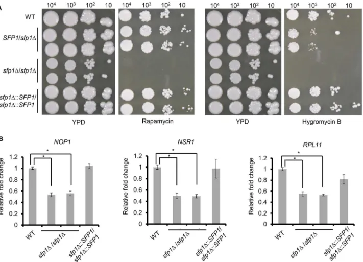
manner. To test this hypothesis, the WT,sfp1Δ/SFP1,sfp1Δ/sfp1ΔandSFP1-reintegrated strains were spotted onto agar plates with or without rapamycin. This drug is an inhibitor of the Tor1 kinase, which is the key enzyme of theC.albicansTOR signaling pathway. InFig 2A, thesfp1Δ/sfp1Δmutants were more sensitive to rapamycin than the WT,sfp1Δ/SFP1and
SFP1-reintegrated strains, suggesting thatC.albicansSfp1 is related to the TOR signaling path-way. Moreover, the WT,sfp1Δ/SFP1,sfp1Δ/sfp1ΔandSFP1-reintegrated strains were spotted onto agar plates with or without hygromycin B. Similar to the findings obtained forS. cerevi-siae, our results demonstrated that thesfp1Δ/sfp1Δstrains were much more sensitive to hygro-mycin B than the WT,sfp1Δ/SFP1andSFP1-reintegrated strains (Fig 2A).
To determine the role ofC.albicansSfp1 in the regulation of the RP and Ribi genes, RNAs were isolated from exponential phase cells and used for RT real-time qPCR. The expression of the RP (RPL11) and Ribi (NOP1andNSR1) genes was detected. As shown inFig 2B, the ex-pression ofNOP1,NSR1andRPL11decreased in thesfp1Δ/sfp1Δmutants compared to the WT andSFP1-reintegrated strains.
Fig 2.C.albicansSfp1 regulates ribosomal gene expression and is related to the TOR signaling pathway.(A) Deletion ofSFP1increases
susceptibility to rapamycin and hygromycin B. The cells were ten-fold serially diluted and spotted onto YPD agar plates with or without rapamycin (25 ng/ml) and hygromycin B (400μg/ml). The plates were incubated at 30°C for 5 days. (B) Deletion ofSFP1affects ribosomal gene expression. The cells were grown in YPD medium overnight at 30°C, subcultured into fresh YPD medium and incubated until log phase (OD600= 2). RNAs were isolated, cDNAs were generated and quantitative real-time PCR was performed. The expression levels of each gene are calculated as the mean±standard deviation (SD) for three
independent experiments. For each gene, the relative fold changes were displayed as the expression levels of an individual strain normalized to the WT strain (as 1).*, p<0.05.
Sfp1 affects cell adhesion and biofilm formation
Using a computational approach, we hypothesized that Sfp1 is involved in biofilm formation [21]. Cell adhesion is the first step of biofilm formation. Therefore, the ability of WT,sfp1Δ/
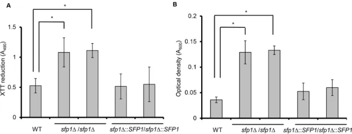
sfp1ΔandSFP1-reintegrated cells to adhere to a 24-well polystyrene microplate was com-pared using the XTT reduction assay. In metabolically active cells, the yellow tetrazolium salt XTT was reduced to a water-soluble orange-colored product whose absorbance can be mea-sured at 490 nm [20]. InFig 3A, the adhesion ability of thesfp1Δ/sfp1Δstrains was much higher than that of the WT andSFP1-reintegrated strains after 1 h incubation in SC medium. To further verify this finding, the adherent cells were scraped from the polystyrene surface and the optical density was measured at 600 nm. The results indicated that density of the ad-herent cells of thesfp1Δ/sfp1Δstrain was higher than that of the WT andSFP1-reintegrated strains (Fig 3B). Similar results were also obtained when cell adhesion was tested in YPD me-dium (Fig C inS1 File).
To further determine whethersfp1Δ/sfp1Δstrains can affect biofilm formation, cells were grown in SC medium in a 24-well polystyrene microplate for 24 h, and the mature biofilms in the plate were photographed and assayed using the XTT reduction method. Thesfp1Δ/
sfp1Δstrain exhibited much stronger biofilm formation than the WT andSFP1-reintegrated strains (Fig4Aand4B). To further examine the structure of the formed biofilms, SEM was also used. The WT andSFP1-reintegrated strains exhibited a similar biofilm structure; the cells formed only a single layer of predominantly blastopore cells, which lacked a complex three-dimensional structure (Fig 4C). However, the structure of thesfp1Δ/sfp1Δmutants differed considerably; the cells formed complex multilayered filamentous networks and in-ternally embedded blastopore cells (Fig 4C).C.albicansbiofilm formation was related to the growth medium used in a previous study [38]. Our finding that the deletion ofSFP1can lead to increased biofilm formation was also observed in YPD medium (Fig C inS1 File). Collectively, our results indicate that Sfp1 negatively affects cell adhesion and biofilm inC.
albicans.
Fig 3. Deletion ofSFP1enhancesC.albicansadhesion.Cell adhesion was assessed in a 24-well polystyrene microplate in SC medium at 37°C with 5% CO2for 1 h. The adherent cells were washed twice with PBS buffer. (A) Cell adhesion was determined using the XTT reduction assay. The results are presented as the mean±SD from three independent experiments.*p<0.05 forsfp1Δ/sfp1Δvs. WT cells. (B) Cell adhesion was evaluated by measuring the density of the adherent cells. After washing, the adherent cells were scraped from the well, collected, and quantified by measuring the cell density (OD600). The results are presented as the mean±SD from three independent experiments.*p<0.05.
Fig 4. Deletion ofSFP1enhancesC.albicansbiofilm formation.(A) Biofilms were formed on the surface of a 24-well polystyrene microplate in SC medium at 37°C with 5% CO2for 24 h. (B) Biofilm formation was assessed using the XTT reduction assay. The results are presented as the mean±SD from
three independent experiments.*p<0.05 forsfp1Δ/sfp1Δvs. WT cells. (C) Biofilm structure was examined using scanning electron microscopy with a 1,000× magnification. Biofilm formation was carried out as described above, except the cells were grown on polystyrene coverslips for 24 h.
Sfp1 controls adhesin gene expression downstream of the Rhb1-TOR
signaling pathway
The cell wall adhesin proteins Als1, Als3 and Hwp1 play critical roles in promotingC.albicans
biofilm development [11,39]. The Tor1 kinase is involved in the regulation of adhesin gene ex-pression [40]. Because thesfp1Δ/sfp1Δmutants enhanced cell adhesion (Fig 3), we suspected that Sfp1 is involved in the regulation of theALS1,ALS3andHWP1adhesin genes. RT real-time qPCR was performed, and dynamic patterns of gene expression were observed. For the
ALS1gene, very low levels of expression were detected in both the WT andsfp1Δ/sfp1Δstrains at time 0, when the cells are planktonic and nonadherent (Fig 5A). During the cell adherence stage (approximately 0.5 to 1 h), theALS1gene began to be induced in thesfp1Δ/sfp1Δmutant compared to the WT strain (Fig 5A). Moreover, the expression of theALS1gene was enhanced in thesfp1Δ/sfp1Δmutant during the stages of biofilm development and maturation (approxi-mately 2 to 24 h). A similar enhancement ofALS3andHWP1gene expression was also de-tected in thesfp1Δ/sfp1Δmutants compared to the WT strain during the development and maturation ofC.albicansbiofilms (Fig5Band5C).
The association of Sfp1 with the TOR signaling pathway was proposed based onFig 2. Inter-estingly, our previous studies linked the small GTPase Rhb1 to the Tor1 kinase in the control of filamentation and the secreted aspartyl protease 2 (Sap2), which are both important viru-lence factors ofC.albicans[41,42]. Therefore, this finding suggests a possible connection be-tween Sfp1 and Rhb1. To test this hypothesis, biofilm formation was compared among the
sfp1Δ/sfp1Δ,rhb1Δ/rhb1Δandsfp1Δ/sfp1/Δrhb1Δ/rhb1Δstrains. Fig D inS1 Fileshowed that all thesfp1Δ/sfp1Δ,rhb1Δ/rhb1Δandsfp1Δ/sfp1/Δrhb1Δ/rhb1Δformed a robust biofilm in SC medium compared to the controls (WT,SFP1-reintegrated andRHB1-reintegrated strains). Therefore, these results were not helpful to detect epistatic relationship betweenSFP1and
RHB1genes. Alternatively, we compared the strains withSFP1overexpression in thesfp1Δ/
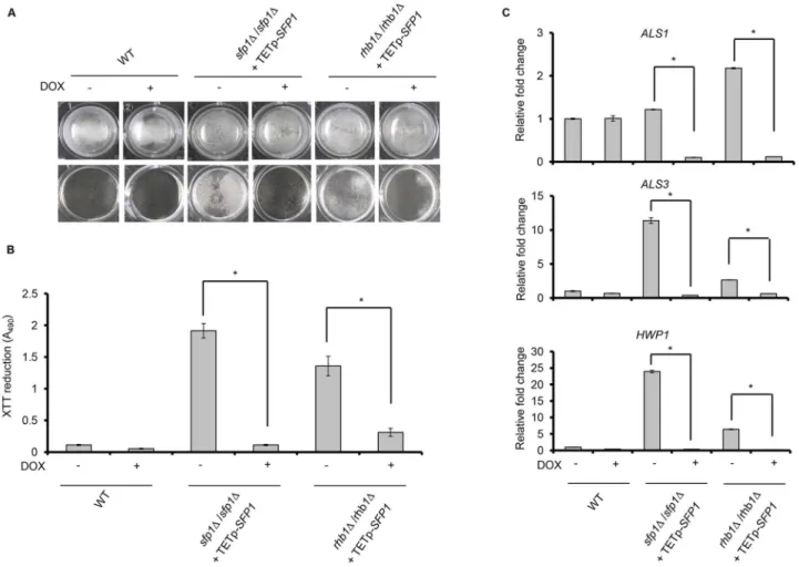
sfp1Δorrhb1Δ/rhb1Δbackground. In the presence of the tetracycline derivative doxycycline a gene can be overexpressed under the control of a tetracycline-inducible promoter [26]. As shown in Fig6Aand6B, the biofilm formation ability of the WT strain was not affected in the
Fig 5. Deletion ofSFP1enhances adhesin gene expression.The expression of adhesin genes was compared between the WT andsfp1Δ/sfp1Δstrains during biofilm formation in SC medium at 37°C with 5% CO2. Real-time qPCR was used to detect the expression of adhesin genes.C.albicans PMA1 transcripts were used as an endogenous control. The results are presented as the mean±SD from at least three independent experiments. ForHWP1gene expression, the fold change was represented on a log2 scale.
presence or absence of doxycycline. Thesfp1Δ/sfp1Δmutant exhibited enhanced biofilm for-mation, whereas the overexpression ofSFP1in thesfp1Δ/sfp1Δbackground significantly de-creased biofilm formation. Similar to thesfp1Δ/sfp1Δmutant, enhanced biofilm formation was observed for therhb1Δ/rhb1Δstrain without doxycycline, but doxycycline-inducedSFP1 over-expression in therhb1Δ/rhb1Δbackground significantly suppressed biofilm formation. Finally, reduced expression of adhesin genes was also detected afterSFP1overexpression in thesfp1Δ/
sfp1Δandrhb1Δ/rhb1Δstrains (Fig 6C). Based on the results of this study and other studies [40], Sfp1 appears to function downstream of the Rhb1-Tor1 signaling pathway.
Sfp1 may regulate adhesin genes and biofilm formation through Bcr1
and Efg1
In addition to Tor1, the transcription factors Efg1 and Bcr1 are also known to control the
ALS1,ALS3andHWP1genes and play a role in the activation ofC.albicansbiofilm formation
Fig 6. Rhb1 is involved in Sfp1-mediated regulation of biofilm formation.Biofilm formation was compared between thesfp1Δ/sfp1Δstrain containing TETp-SFP1and therhb1Δ/rhb1Δstrain containing TETp-SFP1. Biofilms were formed for 24 h in SC medium at 37°C with 5% CO2, with or without 50μg/ml of doxycycline (DOX). Doxycycline induces the overexpression of theSFP1gene, which is under the control of a tetracycline-inducible promoter (TETp). (A) Biofilms were formed on the surface of a 24-well polystyrene microplate in SC medium at 37°C with 5% CO2for 24 h. Biofilm cells are shown before washing (top) and after washing (bottom). Representative images from three independent experiments with similar results are shown. (B) Biofilm formation was measured using the XTT reduction assay. The results are presented as the mean±SD from three independent experiments.*p<0.05 for cells without DOX
treatment vs. DOX-treated cells. (C) The expression of adhesin genes was compared in the presence and absence ofSFP1overexpression in theSFP1- or
RHB1-deleted background. Real-time qPCR was performed, andC.albicans PMA1transcripts were used as an endogenous control. The results are presented as the mean±SD from at least three independent experiments.*p<0.05 for cells without DOX treatment vs. cells with DOX treatment.
[19,43–45]. Because Sfp1 can regulate the expression of theALS1,ALS3andHWP1genes (Fig 5), the relationship between Sfp1, Efg1 and Bcr1 in adhesin gene regulation and biofilm formation is of interest. To address this question, we constructedbcr1Δ/bcr1Δ/sfp1Δ/sfp1Δand
efg1Δ/efg1Δ/sfp1Δ/sfp1Δdouble mutants. Biofilm formation determined by the XTT reduction method showed that thesfp1Δ/sfp1Δmutant significantly enhanced biofilm formation com-pared to the controls (WT,bcr1Δ/bcr1Δandefg1Δ/efg1Δstrains). Interestingly, both thebcr1Δ/
bcr1Δ/sfp1Δ/sfp1Δandefg1Δ/efg1Δ/sfp1Δ/sfp1Δdouble deletion mutants exhibited poor bio-film-forming abilities similar to that of the controls (Fig 7A). Moreover, SEM examination demonstrated that thesfp1Δ/sfp1Δmutant formed a complex multilayered biofilm, while both thebcr1Δ/bcr1Δandefg1Δ/efg1Δmutants formed only a single layer of cells similar to that of
Fig 7. Sfp1 regulates biofilm formation through Bcr1 and Efg1.(A) Biofilm formation was assessed using the XTT reduction assay. The results are presented as the mean±SD from three independent experiments.*p<0.05 forsfp1Δ/sfp1Δvs. WT,bcr1Δ/bcr1Δ/sfp1Δ/sfp1Δorefg1Δ/efg1Δ/sfp1Δ/sfp1Δ mutant cells. (B) The biofilm structure was examined using scanning electron microscopy. Pictures were taken at a 1,000× magnification. The cells were grown on polystyrene coverslips for 24 h to form biofilms. (C) The biofilm structure was examined using confocal scanning laser microscopy (CSLM). Biofilms were formed on the polystyrene coverslips in SC medium at 37°C with 5% CO2for 24 h. After washing, the cells were stained with 0.2 mg/ml of calcofluor (Sigma F-3543,“Fluorescent brightener 28”) for CSLM visualization. Pictures were taken at a 400× magnification. (D) The expression of adhesin genes in biofilm cells was detected using RT real-time qPCR.PMA1transcripts were used as an endogenous control. The results are presented as the mean±SD
from at least two independent experiments.*p<0.05 forsfp1Δ/sfp1Δvs. WT,bcr1Δ/bcr1Δ/sfp1Δ/sfp1Δorefg1Δ/efg1Δ/sfp1Δ/sfp1Δmutant cells.
WT (Fig 7B). However, thebcr1Δ/bcr1Δ/sfp1Δ/sfp1Δandefg1Δ/efg1Δ/sfp1Δ/sfp1Δdouble dele-tion mutants also formed a single layer of structure containing predominantly blastopore cells (Fig 7B). The biofilm structures were also examined using CSLM, and the biofilm of thesfp1Δ/
sfp1Δmutant exhibited a complex multilayered structure with a thickness of ~80μm. However,
thebcr1Δ/bcr1Δ/sfp1Δ/sfp1Δandefg1Δ/efg1Δ/sfp1Δ/sfp1Δdouble mutants exhibited reduced biofilm formation and only formed a very thin layer (~10μm) of cell structure (Fig 7C).
Be-cause both Bcr1 and Efg1 are known to promote biofilm formation [44,46], our results suggest that Sfp1 suppresses biofilm formation via the negative regulation of Bcr1 and Efg1.
To further reveal the relationships among Bcr1, Efg1 and Sfp1, the expression of adhesin genes was also examined using RT real-time qPCR. TheALS1,ALS3andHWP1genes were high-ly expressed in thesfp1Δ/sfp1Δmutant, in agreement with the real-time qPCR analysis inFig 5. However, the deletion ofBCR1orEFG1in thesfp1Δ/sfp1Δbackground significantly reduced the expression levels of all three adhesin genes (Fig 7D). Based on the biofilm structure and adhesin gene expression, Bcr1 and Efg1 appear to function as downstream effectors of Sfp1.
Discussion
The use of indwelling medical devices has become routine in the clinical setting.C.albicans
easily adheres to and forms a biofilm on indwelling medical devices. Such biofilms require sub-sequent surgical removal and replacement of the infective devices.C.albicanscells can also ad-here to and form a biofilm on mucosal surfaces, leading to resistance to antifungal agents and the initiation of infections. Therefore,C.albicansbiofilms are a serious public health problem. Many recent studies focused on the regulation of biofilm formation. One study that was per-formed by Nobileet al. identified a transcriptional network that controls biofilm development [18]. This study combined‘‘classical”genetics, genome-wide approaches, and RNA deep se-quencing technology to comprehensively map the transcriptional circuitry that controls bio-film formation inC.albicans. In addition, this study also described a master circuit of six transcription regulators, including Bcr1, Tec1, Efg1, Ndt80, Rob1, and Brg1, that controls ap-proximately 1,000 target genes and biofilm formation in vitro and in two animal models [18]. This master circuit led to many new predictions about the genes involved in biofilm formation, and some of those predicted genes have been confirmed to play roles in biofilm development. Another study that was performed by Wanget al. computationally screened for potential tran-scription factors that could regulateC.albicansbiofilm [21]. In that study, gene expression pro-files and ChIP-chip were used to establish biofilm and planktonic gene regulatory networks. The two networks were subsequently compared, and the relevance value was calculated to identify potential transcription factors related to biofilm formation. Among the identified can-didates, the relevance value of Sfp1 (Orf19.5943) was the highest and that gene was considered the most relevant for biofilm formation. The aim of this study was to examine the functions of Sfp1, particularly the role of Sfp1 in biofilm formation.
Previous studies demonstrated that the growth medium may affectC.albicansbiofilm for-mation under laboratory conditions [32,38,47,48]. In this study, we tested several growth media for biofilm formation, including Lee’s, Spider, SC and YPD media (Fig 4, Fig C inS1 File
and Fig F inS1 File). Among the tested media, although the cells showed a growth delay (Fig B inS1 File), both the WT andsfp1Δ/sfp1Δstrains formed biofilms in the Lee’s and Spider media (Fig F inS1 File). However, the WT andsfp1Δ/sfp1Δstrains exhibited significant differences in biofilm formation in SC and YPD media (Fig 4and Fig C inS1 File). Particularly, the WT and
SFP1-reintegrated strains produced only a rudimentary biofilm in the SC medium, but the
biofilm that is not as stable as a normal mature biofilm that contains both cell morphologies [49]. Thesfp1Δ/sfp1Δmutants exhibited highly enhanced biofilm formation compared to the WT strain, suggesting that Sfp1 negatively regulates biofilm formation under these experimen-tal conditions. To reveal the mechanisms via which Sfp1 affects biofilm formation, we thus used the SC medium for most of our experiments.
Adhesion is the first key step for biofilm formation. Cell adhesion may be mediated by non-specific factors, including hydrophobicity and electrostatic forces of the cell surface, or by spe-cific adhesins on the surface ofC.albicans.Fig 3demonstrated that thesfp1Δ/sfp1Δmutants exhibit significantly enhanced cell adhesion in polystyrene microplates, indicating that Sfp1 suppresses cell adhesion. TheSFP1deletion may somehow alter theC.albicanscell wall archi-tecture and composition, leading to changes in the non-specific properties of the cell surface. Coincidentally, thesfp1Δ/sfp1Δmutants were resistant to the cell wall disrupting agents Congo red and calcofluor white and hyper-resistant to zymolyase, which has strong lytic activity against cell wallβ-1,3-glucan (data not shown). These results implied that theSFP1gene dele-tion alters cell wall structure and composidele-tion, leading to changes in adhesion-related cell surface properties.
However, other evidence suggests that Sfp1 can also regulate cell adhesion by regulating the expression of cell wall adhesin genes. The deletion ofSFP1increasedALS1andHWP1adhesin gene expression 0.5 h and 1 h after the cells adhered to the substrate (Fig 5). The easy detection of theALS1gene during the early adhesion stage ofC.albicansyeast cells was reported previ-ously [50]. Hwp1 is also required for covalent attachment to host epithelial cells and virulence [23]. Strains that lack eitherALS1orHWP1can lose their abilities to attach to an abiotic sur-face and form a biofilm [18,51]. Moreover,ALS3andHWP1are expressed primarily in hyphal cells [50,52]. InFig 5, the expression ofALS3andHWP1in thesfp1Δ/sfp1Δstrain began to be induced after cell attachment for 2 h, which is when the cells began to form germ tubes (Fig E inS1 File). Moreover, the Als1, Als3 and Hwp1 adhesins also play a complementary role in bio-film formation. A previous study demonstrated that both thehwp1Δ/hwp1Δmutant and the
als1Δ/als1Δ/als3Δ/als3Δdouble mutant strain are defective in biofilm formation; however, a mixture of these two strains can form robust biofilms both in vitro and in vivo [39]. In addi-tion, thehwp1Δ/hwp1Δmutant produced a biofilm with significantly less biomass than the WT strain [39].C.albicansmutants that lack Als3 produce scarce, defective biofilms on cathe-ter macathe-terial in vitro [46]. Although the functions of Als1 and Als3 in biofilm formation are somewhat overlapping, Als1 may not play as pivotal a role as Als3 [39]. Anals1Δ/als1Δmutant had only a partial defect in biofilm formation [18], whereas theals3mutant displayed a severe defect in biofilm formation [46]. Following the early adhesion stage, the expression of all three adhesin genes increased gradually in thesfp1Δ/sfp1Δstrain (Fig 5). Together, these results sug-gest thatSFP1deletion can enhance biofilm development, possibly through the derepression of adhesin gene expression.
In addition to Sfp1, the transcription factors Bcr1 and Efg1 can also regulate adhesin gene expression [19,43,46,53,54] in a manner dependent on the Tor1 kinase [40]. Moreover, the
among Sfp1, Efg1 and Bcr1. Thebcr1Δ/bcr1Δ/sfp1Δ/sfp1Δdouble mutant exhibited dramatical-ly reduced adhesin gene expression in comparison to thesfp1Δ/sfp1Δmutant (Fig 7D). More importantly, the biofilm of thebcr1Δ/bcr1Δ/sfp1Δ/sfp1Δdouble mutant contained fewer adher-ent cells after a 24 h incubation than the biofilm of thesfp1Δ/sfp1Δmutant, which contained a large number of attached cells (Fig7A–7C). In addition, thebcr1Δ/bcr1Δ/sfp1Δ/sfp1Δmutant had nearly no filamentous cells in the biofilm, in contrast to thesfp1Δ/sfp1Δmutant (Fig 7B). Although Bcr1 is not required for hyphal growth, this transcription factor is required for the activation of several hyphal-specific genes, includingALS3andHWP1. Our result is consistent with a previous biofilm assay, in which the WT strain formed a biofilm that consisted of abun-dant hyphal cells but thebcr1Δ/bcr1Δstrain generated a thin rudimentary biofilm that was comprised largely of yeast form cells [46]. Thus, our results suggested that Bcr1 appeared to in-duce adhesin expression in the absence of theSFP1gene during biofilm formation in SC medi-um. In addition, thesfp1Δ/sfp1Δ/efg1Δ/efg1Δexhibited reduced biofilm formation and was defective in filamentous growth compared with thesfp1Δ/sfp1Δstrain (Fig 7B). Moreover, the
efg1Δ/efg1Δ/sfp1Δ/sfp1Δdouble mutant also exhibited dramatically reduced adhesin gene ex-pression in comparison to thesfp1Δ/sfp1Δmutant (Fig 7D).
Taken together, these findings suggest a simple model for the functions ofC.albicansSfp1, as presented inFig 8. Similar toS.cerevisiaeSfp1,C.albicansSfp1 functions as an activator to regulate ribosomal gene expression. However, we further expand the function of Sfp1 to in-clude a role inC.albicansbiofilm formation, downstream of the Rhb1-Tor1 signaling pathway. Moreover, the functions of Sfp1 appear to be mediated by the negative regulation of the tran-scription factors Bcr1 and Efg1, which activate adhesin genes and promote biofilm formation (Fig 8). Although this study provides some new insights into the regulation ofC.albicans bio-film formation, many questions must still be addressed. For example, the relationships among Sfp1, Ndt80 and Rob1 during biofilm formation remain unclear. Moreover, most of the tran-scription factors that have been identified to date are related to the activation of biofilm forma-tion. In this study, we demonstrate that Sfp1 plays a negative role in biofilm formaforma-tion. The
Fig 8. A simple model of the role of Sfp1 in the regulation of ribosomal gene expression and biofilm formation.Sfp1 is related to the Tor1 signaling pathway and plays a role in the induction of RP and Ribi gene expression. Moreover, during the regulation of adhesin gene expression and biofilm formation, the small GTPase Rhb1 coordinates with Tor1 to function upstream of Sfp1, while the transcription factors Bcr1 and Efg1 function downstream of Sfp1.
identification of additional transcriptional repressors of biofilm formation is important. These studies will allow us to understand the complex interplay of negative and positive regulation in the context of biofilm formation.
Supporting Information
S1 File. Supplementary Tables and Figures.
(DOCX)
Acknowledgments
We are grateful to A. J. P. Brown (University of Aberdeen, UK) and Joachim Morschhäuser (Universität Würzburg, Germany) for generously providing strains and plasmids.
Author Contributions
Conceived and designed the experiments: HFC CYL. Performed the experiments: HFC. Ana-lyzed the data: HFC. Contributed reagents/materials/analysis tools: HFC CYL. Wrote the paper: HFC CYL.
References
1. Pfaller MA, Diekema DJ. Epidemiology of invasive candidiasis: a persistent public health problem. Clin Microbiol Rev. 2007; 20: 133–163. PMID:17223626
2. Douglas LJ.Candidabiofilms and their role in infection. Trends Microbiol. 2003; 11: 30–36. PMID: 12526852
3. Nobile CJ, Mitchell AP. Genetics and genomics ofCandida albicansbiofilm formation. Cell Microbiol. 2006; 8: 1382–1391. PMID:16848788
4. Ramage G, Saville SP, Thomas DP, Lopez-Ribot JL.Candidabiofilms: an update. Eukaryot Cell 2005; 4: 633–638. PMID:15821123
5. Kojic EM, Darouiche RO.Candidainfections of medical devices. Clin Microbiol Rev. 2004; 17: 255–
267. PMID:15084500
6. Douglas LJ. Medical importance of biofilms inCandidainfections. Rev Iberoam Micol. 2002; 19: 139–
143. PMID:12825991
7. Hajjeh RA, Sofair AN, Harrison LH, Lyon GM, Arthington-Skaggs BA, Mirza SA, et al. Incidence of bloodstream infections due toCandidaspecies and in vitro susceptibilities of isolates collected from 1998 to 2000 in a population-based active surveillance program. J Clin Microbiol. 2004; 42: 1519–
1527. PMID:15070998
8. Crump JA, Collignon PJ. Intravascular catheter-associated infections. Eur J Clin Microbiol Infect Dis. 2000; 19: 1–8. PMID:10706172
9. Viudes A, Peman J, Canton E, Ubeda P, Lopez-Ribot JL, Gobernado M. Candidemia at a tertiary-care hospital: epidemiology, treatment, clinical outcome and risk factors for death. Eur J Clin Microbiol Infect Dis. 2002; 21: 767–774. PMID:12461585
10. Ramage G, Mowat E, Jones B, Williams C, Lopez-Ribot J. Our current understanding of fungal biofilms. Crit Rev Microbiol. 2009; 35: 340–355. doi:10.3109/10408410903241436PMID:19863383
11. Finkel JS, Mitchell AP. Genetic control ofCandida albicansbiofilm development. Nat Rev Microbiol. 2011; 9: 109–118. doi:10.1038/nrmicro2475PMID:21189476
12. Blankenship JR, Mitchell AP. How to build a biofilm: a fungal perspective. Curr Opin Microbiol. 2006; 9: 588–594. PMID:17055772
13. Nett J, Andes D.Candida albicansbiofilm development, modeling a host-pathogen interaction. Curr Opin Microbiol. 2006; 9: 340–345. PMID:16815078
14. Nett J, Lincoln L, Marchillo K, Massey R, Holoyda K, Hoff B, et al. Putative role of beta-1,3 glucans in
Candida albicansbiofilm resistance. Antimicrob Agents Chemother. 2007; 51: 510–520. PMID: 17130296
16. Chandra J, Kuhn DM, Mukherjee PK, Hoyer LL, McCormick T, Ghannoum MA. Biofilm formation by the fungal pathogenCandida albicans: development, architecture, and drug resistance. J Bacteriol. 2001; 183: 5385–5394. PMID:11514524
17. Andes D, Nett J, Oschel P, Albrecht R, Marchillo K, Pitula A. Development and characterization of an in vivo central venous catheterCandida albicansbiofilm model. Infect Immun. 2004; 72: 6023–6031. PMID:15385506
18. Nobile CJ, Fox EP, Nett JE, Sorrells TR, Mitrovich QM, Hernday AD, et al. A recently evolved transcrip-tional network controls biofilm development inCandida albicans. Cell 2012; 148: 126–138. doi:10. 1016/j.cell.2011.10.048PMID:22265407
19. Nobile CJ, Mitchell AP. Regulation of cell-surface genes and biofilm formation by theC.albicans tran-scription factor Bcr1p. Curr Biol. 2005; 15: 1150–1155. PMID:15964282
20. Tsai PW, Chen YT, Yang CY, Chen HF, Tan TS, Lin TW, et al. The role of Mss11 inCandida albicans
biofilm formation. Mol Genet Genomics 2014; 289: 807–819. doi:10.1007/s00438-014-0846-0PMID: 24752399
21. Wang YC, Lan CY, Hsieh WP, Murillo LA, Agabian N, Chen BS. Global screening of potentialCandida albicansbiofilm-related transcription factors via network comparison. BMC Bioinformatics 2010; 11: 53–63 doi:10.1186/1471-2105-11-53PMID:20102611
22. Loza L, Fu Y, Ibrahim AS, Sheppard DC, Filler SG, Edwards JE Jr. Functional analysis of theCandida albicansALS1 gene product. Yeast 2004; 21: 473–482. PMID:15116430
23. Staab JF, Bradway SD, Fidel PL, Sundstrom P. Adhesive and mammalian transglutaminase substrate properties ofCandida albicansHwp1. Science 1999; 283: 1535–1538. PMID:10066176
24. Zhao X, Daniels KJ, Oh SH, Green CB, Yeater KM, Soll DR, et al.Candida albicansAls3p is required for wild-type biofilm formation on silicone elastomer surfaces. Microbiology 2006; 152: 2287–2299. PMID:16849795
25. Reuss O, Vik A, Kolter R, Morschhauser J. The SAT1 flipper, an optimized tool for gene disruption in
Candida albicans. Gene 2004; 341: 119–127. PMID:15474295
26. Park YN, Morschhauser J. Tetracycline-inducible gene expression and gene deletion inCandida albi-cans. Eukaryot Cell 2005; 4: 1328–1342. PMID:16087738
27. Hsu PC, Yang CY, Lan CY.Candida albicansHap43 is a repressor induced under low-iron conditions and is essential for iron-responsive transcriptional regulation and virulence. Eukaryot Cell 2011; 10: 207–225. doi:10.1128/EC.00158-10PMID:21131439
28. Russell CL, Brown AJ. Expression of one-hybrid fusions withStaphylococcus aureuslexA inCandida albicansconfirms that Nrg1 is a transcriptional repressor and that Gcn4 is a transcriptional activator. Fungal Genet Biol. 2005; 42: 676–683. PMID:15946869
29. Nailis H, Coenye T, Van Nieuwerburgh F, Deforce D, Nelis HJ. Development and evaluation of different normalization strategies for gene expression studies inCandida albicansbiofilms by real-time PCR. BMC Mol Biol. 2006; 7: 25–33. PMID:16889665
30. Hsu PC, Chao CC, Yang CY, Ye YL, Liu FC, Chuang YJ, et al. Diverse Hap43-independent functions of theCandida albicansCCAAT-binding complex. Eukaryot Cell 2013; 12: 804–815. doi:10.1128/EC. 00014-13PMID:23543673
31. Ramage G, Vandewalle K, Wickes BL, Lopez-Ribot JL. Characteristics of biofilm formation byCandida albicans. Rev Iberoam Micol. 2001; 18: 163–170. PMID:15496122
32. Lin CH, Kabrawala S, Fox EP, Nobile CJ, Johnson AD, Bennett R J. Genetic Control of Conventional and Pheromone-Stimulated Biofilm Formation inCandida albicans. PLoS Pathog. 2013; 9: e1003305. doi:10.1371/journal.ppat.1003305PMID:23637598
33. Marion RM, Regev A, Segal E, Barash Y, Koller D, Friedman N, et al. Sfp1 is a stress- and nutrient-sen-sitive regulator of ribosomal protein gene expression. Proc Natl Acad Sci U S A 2004; 101: 14315–
14322. PMID:15353587
34. Jorgensen P, Nishikawa JL, Breitkreutz BJ, Tyers M. Systematic identification of pathways that couple cell growth and division in yeast. Science 2002; 297: 395–400. PMID:12089449
35. Jorgensen P, Rupes I, Sharom JR, Schneper L, Broach JR, Tyers M. A dynamic transcriptional network communicates growth potential to ribosome synthesis and critical cell size. Genes Dev. 2004; 18: 2491–2505. PMID:15466158
36. Lempiainen H, Uotila A, Urban J, Dohnal I, Ammerer G, Loewith R, et al. Sfp1 interaction with TORC1 and Mrs6 reveals feedback regulation on TOR signaling. Mol Cell 2009; 33: 704–716. doi:10.1016/j. molcel.2009.01.034PMID:19328065
38. Kucharikova S, Tournu H, Lagrou K, Van Dijck P, Bujdakova H. Detailed comparison ofCandida albi-cansandCandida glabratabiofilms under different conditions and their susceptibility to caspofungin and anidulafungin. J Med Microbiol. 2011; 60: 1261–1269. doi:10.1099/jmm.0.032037-0PMID: 21566087
39. Nobile CJ, Schneider HA, Nett JE, Sheppard DC, Filler SG, Andes DR, et al. Complementary adhesin function inC.albicansbiofilm formation. Curr Biol. 2008; 18: 1017–1024. doi:10.1016/j.cub.2008.06. 034PMID:18635358
40. Bastidas RJ, Heitman J, Cardenas ME. The protein kinase Tor1 regulates adhesin gene expression in
Candida albicans. PLoS Pathog. 2009; 5: e1000294. doi:10.1371/journal.ppat.1000294PMID: 19197361
41. Tsao CC, Chen YT, Lan CY. A small G protein Rhb1 and a GTPase-activating protein Tsc2 involved in nitrogen starvation-induced morphogenesis and cell wall integrity ofCandida albicans. Fungal Genet Biol. 2009; 46: 126–136. doi:10.1016/j.fgb.2008.11.008PMID:19095072
42. Chen YT, Lin CY, Tsai PW, Yang CY, Hsieh WP, Lan CY. Rhb1 regulates the expression of secreted aspartic protease 2 through the TOR signaling pathway inCandida albicans. Eukaryot Cell 2012; 11: 168–182. doi:10.1128/EC.05200-11PMID:22194462
43. Argimon S, Wishart JA, Leng R, Macaskill S, Mavor A, Alexandris T, et al. Developmental regulation of an adhesin gene during cellular morphogenesis in the fungal pathogenCandida albicans. Eukaryot Cell 2007; 6: 682–692. PMID:17277173
44. Ramage G, VandeWalle K, Lopez-Ribot JL, Wickes BL. The filamentation pathway controlled by the Efg1 regulator protein is required for normal biofilm formation and development inCandida albicans. FEMS Microbiol Lett. 2002; 214: 95–100. PMID:12204378
45. Stichternoth C, Ernst JF. Hypoxic adaptation by Efg1 regulates biofilm formation byCandida albicans. Appl Environ Microbiol. 2009; 75: 3663–3672. doi:10.1128/AEM.00098-09PMID:19346360
46. Nobile CJ, Andes DR, Nett JE, Smith FJ, Yue F, Phan QT, et al. Critical role of Bcr1-dependent adhe-sins inC.albicansbiofilm formation in vitro and in vivo. PLoS Pathog. 2006; 2: e63. PMID:16839200
47. Tumbarello M, Posteraro B, Trecarichi EM, Fiori B, Rossi M, Porta R, et al. Biofilm production by Candi-daspecies and inadequate antifungal therapy as predictors of mortality for patients with candidemia. J Clin Microbiol. 2007; 45: 1843–1850. PMID:17460052
48. Daniels KJ, Park YN, Srikantha T, Pujol C, Soll DR. Impact of environmental conditions on the form and function ofCandida albicansbiofilms. Eukaryot Cell 2013; 12: 1389–1402. doi:10.1128/EC.00127-13 PMID:23954841
49. Baillie GS, Douglas LJ. Role of dimorphism in the development ofCandida albicansbiofilms. J Med Microbiol. 1999; 48: 671–679. PMID:10403418
50. Green CB, Zhao X, Hoyer LL. Use of green fluorescent protein and reverse transcription-PCR to moni-torCandida albicansagglutinin-like sequence gene expression in a murine model of disseminated can-didiasis. Infect Immun. 2005; 73: 1852–1855. PMID:15731087
51. Nobile CJ, Nett JE, Andes DR, Mitchell AP. Function ofCandida albicansadhesin Hwp1 in biofilm for-mation. Eukaryot Cell 2006; 5: 1604–1610. PMID:17030992
52. Staab JF, Ferrer CA, Sundstrom P. Developmental expression of a tandemly repeated, proline-and glu-tamine-rich amino acid motif on hyphal surfaces onCandida albicans. J Biol Chem. 1996; 271: 6298–
6305. PMID:8626424
53. Fan Y, He H, Dong Y, Pan H. Hyphae-specific genes HGC1, ALS3, HWP1, and ECE1 and relevant sig-naling pathways inCandida albicans. Mycopathologia 2013; 176: 329–335. doi: 10.1007/s11046-013-9684-6PMID:24002103
54. Fu Y, Ibrahim AS, Sheppard DC, Chen YC, French SW, Cutler JE, et al.Candida albicansAls1p: an adhesin that is a downstream effector of the EFG1 filamentation pathway. Mol Microbiol. 2002; 44: 61–
72. PMID:11967069