LEVEL IN STREPTOCOCCUS MUTANS BIOFILM CELLS
*Arezoo Tahmourespour¹, Rasoul Salehi², Rooha Kasra Kermanshahi³
¹Assistant Professor of Microbiology, Islamic Azad University-Khorasgan branch, Isfahan, Iran; ²Associated Professor of
Genetics, Medical School, Isfahan University of Medical Sciences, Isfahan, Iran; ³Professor of Microbiology, Alzahra University,
Tehran, Iran.
Submitted: January 28, 2010; Returned to authors for corrections: April 23, 2010; Approved: June 21, 2010.
ABSTRACT
Streptococcus mutans (S. mutans), harboring biofilm formation, considered as a main aetiological factor of
dental caries. Gtf genes play an important role in S. mutans biofilm formation. The purpose of this study was
to investigate the effect of Lactobacillus acidophilus–derived biosurfactant on S. mutans biofilm formation
and gtfB/C expression level (S. mutans standard strain ATCC35668 and isolated S. mutans strain (22) from
dental plaque).
The Lactobacillus acidophilus (L. acidophilus) DSM 20079 was selected as a probiotic strain to produce
biosurfactant. The FTIR analysis of its biosurfactant showed that it appears to have a protein-like
component. Due to the release of such biosurfactants, L. acidophilus was able to interfere in the adhesion
and biofilm formation of the S. mutans to glass slide. It also could make streptococcal chains shorter. Using
realtime RT-PCR quantitation method made it clear that gtfB and gtfC gene expression were decreased in
the presence of L. acidophilus–derived biosurfactant fraction.
Several properties of S. mutans cells (the surface properties, biofilm formation, adhesion ability and gene
expression) were changed after L. acidophilus- derived biosurfactant treatment. It is also concluded that
biosurfacant treatment can provide an optional way to control biofilm development. On the basis of our
findings, we can suggest that the prepared biosurfactant may interfere with adhesion processes of S. mutans
to teeth surfaces, provided additional evaluation produce satisfactory results.
Key words: Biofilm formation, biosurfactant, gtfB/C, Lactobacillus acidophilus, real-time RT PCR,
Streptococcus mutans
INTRODUCTION
Streptococcus mutans (S. mutans), harbouring the dental
biofilm, is one of the etiological factors of dental caries (16). Its
ability to adhere to the teeth surface is vital for the initiation
and progression of dental caries (3, 6, 31). The S. mutans
adhesion mechanism is related to synthesis of both extracellular
enzymes, glucosyl transferase (GTF) and fructosyl transferase
(FTF) (7, 17, 26). These enzymes are responsible for the
synthesis of extra cellular polysaccharides such as glucans and
fructans. These polymers mediate adherence of S. mutans to the
tooth surfaces and surfaces of bacteria. They also play an
important role in the formation of plaque, the precursor of
dental caries (13, 27).
-(1-3) and -(1-6)-linked glucan polymers are encoded by
the genes gtfB, gtfC and gtfD. Recently, in vitro studies
indicated that gtfB and gtfC are essential for the
sucrose-dependent attachment of S. mutans cells to hard surfaces (2) but
gtfD is dispensable (8, 43). Therefore, these genes have become
a potential target for protection against dental caries (7).
Lactobacilli, as probiotic agents, are believed to interfere
with pathogens by different mechanisms (22, 30). One of its
mechanisms is biosurfactant production.
Biosurfactants, a structurally diverse group of surface
active molecules synthesized by microorganisms, have attracted
attentions in recent years. Because the reason, they had several
advantages on synthetic surfactants, such as low toxicity,
inherent good biodegradability and ecological acceptability.
Biosurfactants include unique amphipathic properties derived
from their complex structures, which include a hydrophilic
moiety and a hydrophobic portion (37). The use of
biosurfactants from probiotic bacteria as antimicrobial and/or
anti-adhesive agents has been studied before and their ability to
inhibit adhesion of various microorganisms isolated from
explanted voice prostheses has been demonstrated (20).
The present research focused on the influence of
Lactobacillus acidophilus (L. acidophilus)–derived
biosurfactant on the gtfB and gtfC genes expression level in S.
mutans biofilm cells by real time RT PCR for the first time.
MATERIALS AND METHODS
Bacterial strains and culture conditions
The S. mutans strains used in this study were S. mutans
ATCC35668 and dental plaque isolated S. mutans 22, with high
ability of biofilm forming.
S. mutans strains were cultured on blood agar and mitis
salivarius agar media and were incubated in a CO2 enriched, 37
°C atmosphere. The identification of strains was done by usual
biochemical tests and rapid identification kit of Streptococci
(Rap ID STR kit) and by PCR. L. acidophilus DSM 20079 as a
probiotic source was cultured in MRS broth or Agar.
Biosurfactant production
15 mL of L. acidophilus cultured overnight was inoculated
to 600-ml of MRS broth and incubated for 24 hours. The cells
were harvested by centrifugation at 10,000 g for 5 min at 10
°C, washed twice in demineralized water, and were
resuspended in 100 ml of PBS. The lactobacilli were incubated
at room temperature for 2 hours with gentle stirring for
biosurfactant production.
Subsequently, the bacteria were removed by centrifugation,
and the remaining supernatant liquid was filtered through a
0.22 mm-pore-size filter (Millipore). A 10-ml portion of the
supernatant was used immediately in the adhesion assay, and
the remainder was dialyzed against demineralized water at 4 °C
in a Spectrapor membrane tube (molecular weight cutoff, 6'000
to 8'000; Spectrum Medical Industries, Inc.), and was
freeze-dried as in method of Velraed et al (38).
Drop-collapse method
In order to test whether produced biosurfactant was able to
decrease the surface tension between water and hydrophobic
surfaces, the ability to collapse a droplet of water was tested as
follows: 25 L of extracted biosurfactant was pipetted as a
droplet onto parafilm; the flattening of the droplet and the
spreading of the droplet on the parafilm surface was followed
over seconds or minutes.
Subsequently, methylene blue (which had no influence on
the shape of the droplets) was added to the water stain and
supernatants for photographic purposes. The droplet was
allowed to dry and the diameter of the dried droplet was
recorded (14, 34).
Fourier transform infrared spectroscopy
Freeze-dried biosurfactants (2 mg) were ground with 100 mg
KBr and compressed at 7'500 kg for 3 min to obtain translucent
with a model (Nicolet Impact 400) instruments (the spectral
resolution and wave number accuracy were 4 and 0.01 cm-1,
respectively). KBr pellet was used as the background reference.
Quantity of a spectral region of interest was determined by
normalizing the area under the absorption bands regarding to the
area of the CH absorption band around 2'932 cm-1 (25, 38).
Biofilm formation assay
Glass slide method: In order to generate S. mutans biofilm
on the glass slide, 1 mL of S. mutans over night cultured (108
CFU/ml) was inoculated into a flask containing 100 mL of
sterile BHI broth supplemented with 1% sucrose and two slides
with and without L. acidophilus–derived biosurfactant. The
glass slides were washed in detergent solution, rinsed in
distilled water twice, then air dried and autoclaved at 121 °C
for 15 minutes before use. The flasks were incubated in an
orbital incubator (100 rpm) at 35-7 °C for 18-20 h. Then, the
glass slides were removed from the flasks and rinsed twice with
10 ml of PBS solution in order to remove unattached cells.
Removed glass slides were stained with 2% crystal violet for 5
minutes, washed, air dried and photographed under an optical
microscope with digital camera (Nicon, Eclipse, E200,
Japan)(5).
Microtiter plate method: In order to generate biofilm on
microtiter plate wells, 20 L of overnight cultured S. mutans
was placed in each wells of a 24 wells polystyrene multidishes
and was cultivated with 2 mL of BHI broth supplemented with
1% sucrose. The plates were incubated at 37 °C in an
atmosphere enriched with 5% CO2. After 18 hours of
incubation, the spent medium was aspirated and wells were
washed with PBS solution in order to remove unattached cells.
The biofilm was incubated again in fresh BHI with 1% sucrose;
after another 18-hour incubation, the spent medium was
aspirated again. The cells were washed and the biofilm was
incubated again in fresh BHI broth with 1% sucrose
supplemented with and without 2.5 mg/ml of freeze-dried
biosurfactant. After 4 hours of incubation, the cells of the
biofilms were dislodged into tubes containing 2 mL PBS
solution and vortexed (31).
Extraction of total RNA
The prepared biofilm cells on microtiter plates (S. mutans
ATCC 35668 & S. mutans 22 with and without biosurfactant, in
3 replicate) were used for RNA extraction. Cells were disrupted
using ribolyser instrument (Hybaid, UK.) and the supplied kit
according to the manufacturer's instruction in which
RNA-containing supernatant from ribolyser tube was transferred to
new RNase free microtube, centrifuged and treated with 300 L
of chloroform–isoamyl alcohol, vortexed and centrifuged. Then
total RNA was recovered by precipitation with isopropanol and
dried under appropriate sterile conditions. Quantitative and
qualitative evaluations performed on the extracted RNA by
spectrophoto-metrically (biophotometer, Eppendorff, Rs 232-C,
Germany) and Agarose-gel electrophoresis.
Reverse transcription
A reverse transcription (RT) reaction mixture (20 L)
containing 50 ng of random hexamers, 2 g of total RNA
sample and up to 12 L DEPC-treated water was incubated at
70 °C for 5 min to remove any secondary structure and was
placed on ice. Then 5X RT buffer (4 L), 20 U/ L
Ribonuclease Inhibitor (1 L) and 10 mM dNTPs mix
(Cinagen) were added to each reaction mixture, after 5 minutes
incubation in 37 °C, 1 L reverse transcriptase was added.
Then the mixture was incubated at 42 °C for 60 minutes. The
reaction was terminated by heating the mixture at 70 °C for 10
minutes, and the cDNA samples were stored at -20 °C to be
used later.
Real-time quantitative RT-PCR
PCR amplification of synthesized cDNAs were first
optimized by conventional PCR. Real time quantitative RT-
PCR was performed using the ABI-step I (Applied Biosystems,
USA) instrument and SYBR Green PCR Master Mix
(Qiagene).
The relative quantitation of gtfB/C genes were made
against 16s rRNA as a reference gene. All primers and their
The reaction mixture (20 L) contained 1X SYBR Green
PCR Master Mix (Qiagene), the appropriate forward, reverse
PCR primers (1 M) and 1 L of the cDNA sample. PCR
program consisted of an initial denaturation at 95 °C for 5
minutes, then 40 cycles of amplification applied as follow:
denaturation at 95 °C for 15 second and annealing and
extension at 60 °C for 1 minute. Appropriate negative and
positive controls were included. Using the two-step protocol
described above, all primer pairs were checked for primer–
dimer formation without the addition of template. As an
additional control for each primer pair and each RNA sample,
the cDNA synthesis reaction was carried out without reverse
transcriptase in order to identify whether the RNA samples
were contaminated by residual genomic DNA. The critical
threshold cycle (Ct) was defined as the cycle in which
fluorescence becomes detectable above the background
fluorescence and was inversely proportional to the logarithm of
the initial number of template molecules.
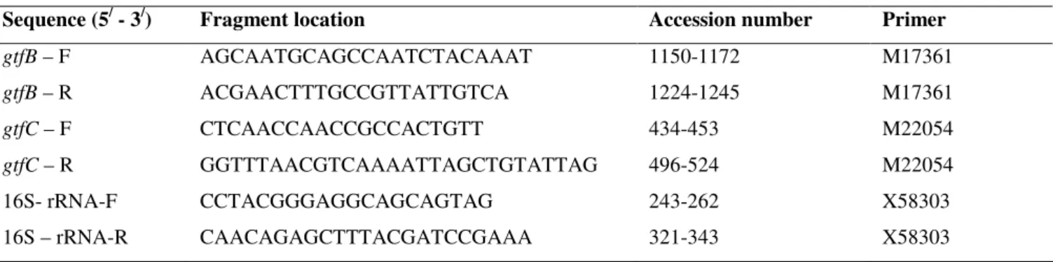
Table 1. Nucleotide sequences of primers
Sequence (5/ - 3/) Fragment location Accession number Primer
gtfB – F AGCAATGCAGCCAATCTACAAAT 1150-1172 M17361
gtfB – R ACGAACTTTGCCGTTATTGTCA 1224-1245 M17361
gtfC – F CTCAACCAACCGCCACTGTT 434-453 M22054
gtfC – R GGTTTAACGTCAAAATTAGCTGTATTAG 496-524 M22054
16S- rRNA-F CCTACGGGAGGCAGCAGTAG 243-262 X58303
16S – rRNA-R CAACAGAGCTTTACGATCCGAAA 321-343 X58303
RESULTS
Drop collapse assay
In drop collapse assay, no activity was detected for
distilled water as predicted. The biosurfactant droplets do
result in a collapsed droplet (Figure 1), indicating their effects
on reduction of surface tension.
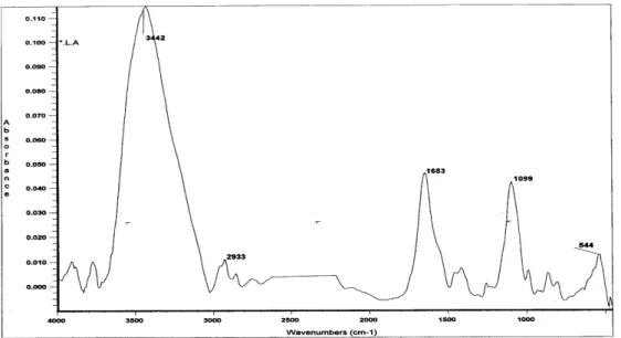
Fourier transform infrared spectroscopy
The molecular composition of the biosurfactant used in this
study was analysed by Fourier transform infrared spectroscopy
(Figure 2). The most important bands were located at 2'933 cm-1
(CH band: CH2- CH3 stretching), 1'653 cm-1 (AmI band: CAO
stretching in proteins), 1'480 cm-1 (AmII band: NOH bending in
proteins), 1'248 cm-1 (PI band: phosphates), and 1'099 cm-1 (PII
band: polysaccharides). Therefore one of the components of L.
acidophilus DSM 20079 biosurfactants appeared to be protein
(Figure 2).
Figure 1. Drop collapse assay. Collapsed droplets (1, 2 & 3)
Figure 2. The spectrum of the freeze-dried biosurfactant released from Lactobacillus acidophilus
Biofilm formation
As it is shown in Figure 3A/B,the presence of 2.5mg/ml L.
acidophilus derived biosurfactant, dramatically reduced the
process of attachment and biofilm production .It could also
make chain shortening.
The attached bacterial cells were collected from the wells
of microtiter plates (Figure 4A) and used for RNA extraction
by mentioned method (Figure 4B).
The effect of biosurfactant on gtfB and gtfC expression Real-time RT-PCR was used to quantify the effect of L.
acidophilus–derived biosurfactant on gtfB and gtfC gene
expression in biofilm of S. mutans ATCC35668 and S. mutans
22 (Figures 5A and B). As an internal reference 16srRNA gene
was used.
In the biofilm environment, tested biosurfactant
significantly reduced gtfB and gtfC gene expression (p < 0.05).
However, the effect of biosurfactant on two genes was not
identical with more pronounced gtfB gene expression
suppression. Regarding S. mutans 22, the extent of reduction of
gtfB expression level was more than the standard strain
(p,0.05), but it's effect on gtfC expression level was the same as
the standard strain.
Figure 3.S.mutans biofilm biofilm formation on glass slide. A:control group (in the absence of biosurfactant). B: Experimental group(in the
Figure 4. A: Attached cells to PVC microtiter plate well. B: total RNA extracted RNA from biofilm producing cells on PVC
micrtiter plate. (lane1,2: Total RNA from S.mutans 22 in the abscence and presence of biosurfactant, respectively. lane 3,4 Total
RNA from standard strain in the abscence and presence of biosurfactant, respectively.)
Figure 5. The effect of Lactobacillus acidophilus-derived biosurfactant, on gtfB/C in biofilm immobilized. A: S. mutans ATCC
35668; B: S. mutans 22 (isolated from dental plaque).
The mRNA expression levels were calibrated relative to the control group (in the absence of biosurfactant). The results were
expressed as the means and standard errors of dublicate experiments using primers specific for gtfB/C and 16S rRNA (normalizing
gene).
DISCUSSION
Oral infection constitute some of the most prevalent and
expensive forms of human infection. The disease is widespread
in all age groups with least success to control the actual
infection. Dental caries is a multi factorial disease tightly
related with the presence of cariogenic bacteria, Streptococcus
mutans, which are embedded in the dental plaque biofilm.
As S. mutans is involved in dental caries, it must be
inhibited. Increasing problems of resistance to synthetic
antimicrobials have encouraged the researchers to research on
their products.
Some of microorganisms such as lactic acid bacteria were
found to be biosurfactant producing strains (21, 25, 35, 38).
One of the major roles known for biosurfactants is their
negative effect on other microbial species.
In this study, 24-h culture of L. acidophilus was used to
release biosurfactant, because Velraed et al. (38) and
Rodrigues et al. (21) showed that the 24-h cultures exhibited
the largest and most rapid decreases in liquid surface tension.
The biosurfactant activity showed that we had used real
surfactant preparations, since the force or interfacial tension
between the drop containing the surfactant and the parafilm
surface was reduced and resulted in the spread of the drop.
Rodrigues et al. (25), Kuiper et al. (14) and Walencka et al.
(39) also showed that surface tension was reduced by
biosurfactants.
The dialyzed (molecular weight cutoff 6,000 to 8,000) and
freeze-dried biosurfactant derived from L. acidophilus
DSM20079 was examined by Fourier transform infrared
spectroscopy and was compared with the reference compounds
albumin, salivary glycoprotein, dextran, lipoteichoic acid and
other FTIR spectra of biosurfactants (38).
On the basis of the results, it was concluded that the
biosurfactant from L. acidophilus had more protein than
polysaccharide and phosphate. According to figure 3-A and 3-B,
it is also clear that, the biosurfactant could reduce the adhesion
of S. mutans to glass slide. Velraeds et al. (38) demonstrated
that, the biosurfactants from L. acidophilus RC14 and L.
fermentum B54 were richer in protein and also contained less
polysaccharide and phosphate than the biosurfactants from L.
casei subsp. rhamnosus 36 and ATCC7469.
Due to the release of such biosurfactant, L. acidophilus
could interfere in the adhesion and biofilm formation of the S.
mutans to glass slide. It also could make streptococcal chains
shorter (Figure 3b).
Van Hoogmoed et al. (10, 11) reported that, the release of
biosurfactant from S. mitis BMS could interfere in the adhesion
of the cariogenic S. mutans to glass in the presence and absence
of a salivary conditioning film. Others also confirmed that
biosurfactants had inhibitory effect on bacterial adhesion and
also biofilm formation (9, 22-25, 29).
However, the precise mechanisms of such effects have not yet
been explained. It seems to be highly dependent on
biosurfactant type and the properties of the target bacteria. The
simplest way to explain biosurfactant antiadhesion and
anti-biofilm activities would be their direct antimicrobial action.
However, the antimicrobial activity of biosurfactants have not
been observed in all cases (25, 39).
Thus, Walencka et al. (39) reported that the way in which
surfactants influenced bacterial surface interactions appeared to
be more closely related to the changes in surface tension and
bacterial cell-wall charge. These factors are very important in
overcoming the initial electrostatic repulsion barrier between
the microorganism cell surface and its substrate. Surfactants
may affect both cell-to-cell and cell-to-surface interactions.
Their results support the idea that lactobacilli-derived agents
remarkably have an effect on these interactions (39).
In this study, we made an effort to investigate the effect of
L. acidophilus–derived biosurfactant on gtfB and gtfC genes
expression level. The expression of these genes and the
production of insoluble extracellular glucans mediate the
attachment of S. mutans not only to the surfaces but also to
other active form of bacteria which are favorable to the
organisms for the persistent colonization of tooth surfaces (28).
Additionally, gtf genes are known virulence factors
associated with the pathogenesis of dental caries and high
content of insoluble glucans in dental plaque which is related to
elevated risk of biofilms cariogenicity in humans (19).
Vacca-Smith et al. (36) mentioned that GtfB levels in saliva were
correlated with presence of caries in humans.
Several environmental factors can influence on expression
and activity of the GTF enzymes. The existence of various
enzymes in the process of carbohydrate metabolism and
transport, glucan synthesis and secretion, and degradation in
the oral streptococci, in addition to factors that involve
post-translational modifications of the GTF enzymes, have
traditionally complicated the understanding of regulatory
Multiple regulatory networks that incorporate external
signals, such as two-component regulatory systems, are
necessary for biofilm formation. S. mutans also possesses a
LuxS-mediated signaling pathway that affects biofilm
formation. LuxS is the enzyme that catalyzes the reactions
leading to the production of the AI-2 signal molecule (40, 42).
In addition, a number of other gene products, such as BrpA (a
cell surface-associated biofilm regulatory protein), have also
been shown to play critical roles in environmental stress
responses and biofilm development by S. mutans (40, 41).
While much effort has been dedicated to understanding the
molecular mechanisms of adherence, biofilm development and
virulence gene expression by S. mutans in pure cultures, there
are large gaps in our knowledge of how this cariogenic
bacterium acts in response to inter-generic interactions with
bacteria normally found in the supragingival plaque (41).
Our results showed that the L. acidophilus-derived
biosurfactant, or a putative signaling molecule in the extract,
down regulate the expression level of genes playing important
role in the process of S. mutans attachment and biofilm
formation.
Clearly, most studies have focused on the production and
gene regulation of virulence factors such as GTFs, which play
an important role in biofilm formation by S. mutans for the
purpose of controlling dental caries (12, 32).
The ability of S. mutans to produce extracellular
polysaccharides from dietary carbohydrates has been
demonstrated to significantly enhance its cariogenicity. Thus,
the less these extracellular polysaccharides produced, the
cariogenicity of S. mutans can be less.
Tomita et al (33) demonstrated that chemical surfactants
exerted different effects on the synthesis of
glucosyltransferases in S. mutans. So that the presence of
Tween 80 in media significantly increased in
glucosyltransferases (GTFs), while Triton X-100 decreased it.
These results indicated that several properties of bacterial
cells (the surface properties, gene expression, etc) were also
changed after biosurfactant treatment. The situation in natural
systems is obviously more complex and requires the
consideration of many additional parameters (15, 39).
In general, biosurfacant treatment can provide an optional
way for controlling biofilm development and also influence the
adhesion ability of bacterial pathogens (18).
Currently, further investigations are being done in our
laboratory on the effect of other probiotic biosurfactant such as
L. fermentum and L. rhamnosus on gtfB, gtfC and ftf expression
in S. mutans biofilm.
REFERENCES
1. Allakera, R.P.; Douglasb, C.W.I. (2009). Novel anti-microbial therapies for dental plaque-related diseases. Int. J. Antimicrob. Agents. 33, 8-13. 2. Aoki, H.; Shiroza, T.; Hayakawa, M.; Sato, S.; Kuramitsu, H. K. (1986).
Cloning of a Streptococcus mutans glucosyltransferase gene coding for insoluble glucan synthesis.Infect. Immun. 53, 587-594.
3. Barbieri, V.A. D.; Vicente, V.A.; Fraiz2, F.C.; Lavoranti, O.J.; Svidzinski, T.I.E.; Pinheiro, R.L.(2007). Analysis of the In Vitro adherence of Streptococcus mutans and Candida albicans. Braz. J. Microbiol. 38, 624-631.
4. Banas, J.A.; Vickerman, M.M. (2003). Glucan binding proteins of the oral streptococci. Cri.t Rev. Oral Biol. Med.14(2):89-99.
5. Bos, R.; Mei, H.C.; Busscher, H.J. (1999). Physico – chemistry of initial microbial adhesion interactions – its mechanisms and methods for study. FEMS Microbiol. Rev. 23, 179 – 230.
6. Caglar, E.; Kargul, B.; Tanbogaet, I. (2005). Bacteriotherapy and probiotics role on Oral health. Oral Dis. 11, 1-7.
7. Chia, J.S.H.; Teng, T.Y.; Chen, L.J.; Hahn, J.Y.; Yang, L.J. (1991). Glucosyltransferase Gene Polymorphism among Streptococcus mutans Strains. Infect. Immun. 59 (5), 1656-1660.
8. Hanada, N.; Kuramitsu, H. K.(1988). Isolation and characterization of the Streptococcus mutans gtfC gene, coding for synthesis of both soluble and insoluble glucans.Infect. Immun. 56, 1999-2005.
9. Heinemann, C.; Hylckama, J.; Janssen, D.B.; Busscher, H.J. (2000). Purification and characterization of a surface binding protein from Lactobacillus fermentum RC-14 that inhibits adhesion of Enterococcus faecalis 1131. FEMS Microbiol. Lett. 190(1), 177.
10. Hoogmoed, C.G. (2000). Inhibition of S. mutans NS adhesion to glass with and without a salivary conditioning film by biosurfactant releasing S.mitis Strains.Appl. Environ. Microbiol. 66(2), 659-63.
11. Hoogmoed, C.G.; Mei, H.C.; Busscher, H.J. (2004). The influence of biosurfactants released by S.mitis BMS on the adhesion of pioneer Strains and cariogenic bacteria. Biofoul. 26(6), 261-67.
doi:10.1016/j.archoralbio.2008.07.007.
13. Jae-Gyu, J.; Klein, M.I. ; Xiao, J.; Gregoire, S.; Rosalen, P.L.; Koo, H. (2009). Influences of naturally occurring agents in combination with fluoride on gene expression and structural organization of Streptococcus mutans in biofilms. BMC Microbiol. 9, 228-38.
14. Kuiper, I.; Lagendijk, E.L.; Pickford, R.; Derrick, J.P.; Lamers, G.E.M.; Thomas-Oates, J.E.; Lugtenberg, B.J.J.; Bloemberg, G.V. (2004). Characterization of two Pseudomonas putida lipopeptide biosurfactants, putisolvin I and II,which inhibit biofilm formation and break down existing biofilms. Mol. Microbiol. 51(1), 97-113.
15. Mack, D.; Becker, P.; Chatterje, I.; Dobinsky, S.; Knobloch, J.K.M.; Peter, G.; Rohde, H.; Herrmann, M. (2004). Mechanisms of biofilm formation in Staphylococus epidermidis and Staphylococcus aureus: functional molecules, regulatory circuits, and adaptive responses.Inter. J. Med. Microbiol. 294, 203-212.
16. Marsh, P. (2003). Are dental diseases examples of ecological catastrophes? Microbiology. 149, 279-94.
17. Moreira, M,; Vicente, V.A.; Glienke, Ch.(2007) Genetic variability of Streptococcus Mutans isolated from low – income families , as shown by
RAPD markers. Braz. J. Microbiol. 38:729-735.
18. Ofek, I.; Hasty, D.L.; Sharon, N. (2003). Anti-adhesion therapy of bacterial diseases: prospects and problems. FEMS Immunol. Med. Microbiol. 38,181–191.
19. Paes Leme, A.F.; Koo, H.; Bellato, C.M.; Bedi, G.; Cury, J.A. (2006). The role of sucrose in cariogenic dental biofilm formation--new insight.J. Dent. Res. 85, 878-87.
20. Rodrigues, L.; Mei, H.C.; Teixeira, J.; Oliveira, R. (2004). Influence of Biosurfactants from Probiotic Bacteria on Formation of Biofilms on Voice Prostheses. Appl. Environ. Microbiol. 70(7), 4408-10.
21. Rodrigues, L.; Moldes, A.; Teixeira, J. A.; Oliveira, R. (2005). Modeling of biosurfactant production by Lactobacillus Strains. http://hdl.handle.net/1822/3463 Universidade do Minho (Portugal) Conference paper.
22. Rodrigues, L.; Banat, I.M.; Teixeira, J.; Oliveria, R. (2006a). Biosurfactants: Potential application in medicine. J. Antimicrob. Chemother. 57, 609-618.
23. Rodrigues, L.; Mei, H.; Banat, I.M.; Teixeria, J.; Oliveria, R. (2006b). Inhibition of microbial adhesion to silicon rubber treated with biosurfactant from Streptococcus thermophilus. FEMS Immunol. Med. Microbiol. 46(1), 107-112.
24. Rodrigues, L.; Teixeira, J.A.; Mei, H.C.; Oliveira, R. (2006c). Physicochemical and functional characterization of a biosurfactant produced by Lactococcus lactis 53. Colloids Surf. B Biointerfaces. 15(49(1)), 79-86.
25. Rodrigous, L.; Teixeira, J.A.; Mei, H.C.; Oliveria, R. (2006d). Isolation and partial characterization of a biosurfactant produced by Streptococcus thermophilus A.Colloid surf. B Biointerfaces. 53, 105-112.
26. Rozen, R.; Bachrach, G.; Bronshteyn, M. (2001). The role of fructans on dental biofilm formation by Streptococcus sobrinus, Streptococcus
mutans, Streptococcus gordonii and Actinomyces viscosus. FEMS Microbiol. Lett. 195, 205-10.
27. Schilling, K.M.; Bowen, W.H. (1988). The activity of glucosyltransferase adsorbed onto salivacoated hydroxyapatite.J. Dent. Res. 67, 2-8. 28. Schilling, K.M.; Bowen, W.H. (1992). Glucans synthesized in situ in
experimental salivary pellicle function as specific binding sites for Streptococcus mutans.Infect. Immun. 60, 284-95.
29. Schooling, S.R.; Charaf, U.K.; Allison, D.G.; Gilbert, P. (2004). A role for rhamnolipial in biofilm dispertion. Biofilms. 1, 91-99.
30. Santos, A.L.; Jorge, A.O.C.; Santos, S.S.F.; Silva, C.R.G.; Leão, M.V.P. (2009). Influence of probiotics on Candidapresence and IgA anti-Candida in the oral cavity. Braz. J. Microbiol. 40, 960-964.
31. Tam, A.; Shemesh, M.; Wormser, U.; Sintov, A.; Steinberg, D. (2006). Effect of different iodine formulations on the expression and activity of Streptococcus mutans glucosyltransferase and fructosyltransferase in biofilm and planktonic environments. J. Antimicrob. Chemother. 57, 865-871.
32. Tamwsada, M.; Kawabata, S. (2004).Synergistic effects of streptococcal glucosyltransferase on adhesive biofilm formation.J. Dent. Res. 83, 874-9.
33. Tomitab, Y.; Watanabea, T.; Takeuchi,T.; Nanbua,A.; Shinozaki, N.; kemi, T.; Fukushima, K. (1998). Effects of surfactants on glucosyltransferase production and in vitro sucrose-dependent colonization by Streptococcus mutans. Arch. Oral Bio. 43, 735-40. 34. Tugrul, T.; Cansunar, E. (2005). Detecting surfactant – producing
microorganisms by the drop collapse test. World J. Microbiol. Biotechnol. 21, 851-853.
35. Uehara, S.; Mondena, K.; Nomotob, K.; Seno, Y.; Kariyama, R.; Kumon, H. (2006). A pilot study evaluating the safety and effectiveness of Lactobacillus vaginal suppositories in patients with recurrent urinary tract infection. Inter. J. Antimicrob. Agents. 28S, S30-34.
36. Vacca-Smith A.M.; Whelehan, M.T.; Berkowitz, R.J.; Feng, C.; Bowen, W.H. (2007). Salivary glucosyltransferase B as a possible marker for caries activity.Caries Res. 41, 445-50.
37. Vater, J.; Kablits, B.; Wild, Ch.; Franke, P.; Mehta, N.; Cameotra, S.S. (2002). Matrix-Assisted Laser Desorption Ionization–Time of Flight MassSpectrometry of Lipopeptide Biosurfactants in Whole Cells and Culture filtrates of Bacillus subtilis C-1 Isolated from Petroleum Sludge. Appl. Environ. Microbiol. 68(12), 6210-19.
38. Velraeds, M.C.; Mei, H.C.; Reid, G.; Busscher, H.J. (1996). Inhibition of Initial adhesion of uropathogenic Enterococcus faecalis by biosurfactants from Lactobacillus Isolates. Appl. Environ. Microbiol. 1996. 62(6), 1958-63.
39. Walencka, E.; Rozalska, S.; Sadowska, B.; Rozalska, B. (2008). The Influence of Lactobacillus acidophilus-Derived Surfactants on Staphylococcal Adhesion and Biofilm Formation.Folia Microbiol. 53(1), 61-66.
stress tolerance and biofilm formation. J Bacteriol 186(9):2682-2691. 41. Wen, Z.T.; Yates, D.; Ahn,S.J.; Burne, R.A. (2010). Biofilm formation
and virulence expression by Streptococcus mutans are altered when grown in dual-species model. BMC Microbiol. 10: 111, doi:10.1186/1471-2180-10-111.
42. Yoshida, A.; Ansai, T.; Takehara, T.; Kuramitsu, H.K. (2005).
LuxS-based signaling affects Streptococcus mutans biofilm formation. 71 Appl. Environ. Microbiol. (5): 2372-2380.