Hexavalent Chromium Reduction and Its Distribution in the Cell
and Medium by Chromium Resistant
Fusarium solani
Mousumi Sen
1,*, Manisha Ghosh Dastidar
2and Pradip K.Roychoudhury
31
Department of Applied Chemistry, Amity School of Engineering & Technology, India
2
Centre for Energy Studies
3
Department of Biochemical Engineering and Biotechnology, Indian Institute of Technology, India Received 10 October 2012; received in revised form 13 November 2012; accepted 04 December 2012
Abstract
In the present work, batch biosorption of Cr(VI) was studied using the fungal strain isolated from soil. The
fungal strain was characterized as Fusarium solani. The total Cr distribution in the biomass (fungus) and in the media obtained from the experiment conducted at 500 mg l-1 initial Cr(VI) concentration and pH 5.0. The results indicated
both intracellular and extracellular accumulation and enzymatic reduction of Cr(VI) and this was supported by the Transmission Electron Microscopic (TEM) observation at the same Cr(VI) concentration and pH value. Chromium
elution from Fusarium solani containing Cr was then tried out using a number of chromium eluting reagents and a maximum Cr could be eluted using 0.5N sodium hydroxide solution without destructing the biomass structure. The
total Cr was recovered by pH adjustment from both biomass and media was found to be 44% of the initial Cr(VI) concentration (500 mg l-1).
Keywords: batch biosorption, Cr (VI), fusarium solani, growing cells
1.
Introduction
Chromium is one of the toxic heavy metals, which exists in nature as stable hexavalent and trivalent forms. The
hexavalent form of chromium is more toxic than trivalent chromium and is often present in wastewater as chromate (CrO42-) and
dichromate (Cr2O72-). This is of serious environmental concern as Cr(VI) persists indefinitely in the environment and
complicating its remediation. The persistent nature makes it accumulate in the food chain which with time reaches harmful levels in living beings and resulting in serious health hazards such as irritation in lungs and stomach, cancer in digestive tract,
low growth rates in plants, death of animals etc. Therefore, removal of Cr(VI) from waste water prior to its discharge into natural water systems, adjoining landmasses, sewer systems, etc. requires serious and immediate attention.
The conventional physico-chemical techniques used for the removal of Cr(VI) include chemical reduction which followed by precipitation with caustic soda. This process requires a large excess of chemicals and produces voluminous sludges,
disposal of which again create secondary pollution. Other available Cr(VI) removal treatments include ion-exchange, electrolysis and reverse osmosis etc. which are not only expensive and high energy processes, but also are ineffective when
metal ions are present at lower concentration in a large volume of waste waters [1]. Bioremediation processes which lead to the
*Corresponding author. E-mail address: [email protected]
production of harmless products need to be developed to clean up the environment. Bioremediation involves potential
application of microorganisms in removal of heavy metals and has been recognized as a potential alternative to the conventional methods for treatment of contaminated wastewaters [2].
The growing, resting and non-living cells of microorganisms are reported to remove Cr(VI) from aqueous solutions [3-13]. However, most of the work to remove Cr(VI) have been carried out by using non-living fungal cells [14-15] and a very
little information is available on use of growing and resting cells [6, 16-18]. The use of non-living cells has advantages over growing and resting cells due to the absence of both toxicity limitations and requirements of growth media and nutrients. Both
growing and resting cells can be maintained biochemically active. However, growing systems have the advantage over the non-living and resting cells that the simultaneous removal of metal is obtained during growth of the organism and separate
biomass production processes such as cultivation, harvesting, drying, processing and storage can be avoided. However, the major limitation of using growing systems for biosorption of metals is that cell growth is inhibited when the metal concentration is high. This problem can be overcome by the use of metal tolerant organism. The tolerance and removal capacities are the
essential characteristics of growing biomass used in a metal ion removal process.
In the present study, the fungal strain used for the removal of Cr(VI) was isolated from soil and characterized as
Fusarium solani. Studies were also conducted in Transmission Electron Microscope in order to have an insight mechanism of distribution of Cr in the biomass and in the media at 500 mg l-1 initial Cr(VI) concentration and at pH 5.0. Batch studies were
further conducted on chromium elution from Fusarium solani by using several chromium eluting reagents. 0.5N sodium hydroxide solution was found to be the best eluting reagent. Recovery of chromium from chromium loaded biomass and also
from liquid medium was done and a maximum 44 % chromium was recovered. This is assumes great importance from the point of resource recovery as the metals are non-renewable.
2.
Materials and Method
2.1. Microorganism
The fungal strain used in the present study was isolated from soil near an electroplating industry.
2.1.1 Microorganism acclimatization
To have substantial amount of biomass, the isolated organism was grown in a sterile media at 30o C and 180 rpm. The
organism was then cultured in Potato Dextrose Agar (PDA) plates and stored at 40 C. To maintain the potency and metabolic activity of the organism, the stock culture was sub cultured every 1-2 weeks.
2.1.2 Microorganism characterization
The fungal strain used in the present study was characterized (morphology) as Fusarium solani at Indian Type Culture
Collection (ITCC) at Indian Agricultural Research Institute (IARI), New Delhi, India. The fungal strain grown in the media in shake flasks was microscopically examined by using the phase transfer microscope (model: Nikon: HB-10101AF).
2.2. Microorganism and inoculum preparation
Fusarium solani isolated from soil was grown in 250 ml Erlenmeyer flask in a shaking incubator at 30o C and 180 rpm by using media of the following composition (g l-1): Glucose, 10.0; K2HP04, 0.5; NaCl, 1.0; MgS04, 0.1; NH4N03, 0.5 and Yeast
extract, 5.0. The pH of the media was 5.0. An inoculum of 10 % (v/v) of a 36 h old culture was used for the Cr(VI) removal
2.3. Batch studies
To find out the potential of the fungus for removal of Cr(VI), batch studies were carried out by using growing cells of Fusarium solani. In the batch experiments, sterile synthetic media (100 ml) containing Cr(VI) was inoculated and incubated in Erlenmeyer flask (250 ml). A 10 % (v/v) inoculum was added and the flask was incubated at 30o C and 180 rpm. The pH was adjusted by using sulphuric acid. The process was monitored with time and analyzed for residual Cr(VI) concentration and
residual sugar concentration and pH. The biomass collected by centrifugation was washed, dried at 80 0 C and the dry weight of biomass was estimated gravimetrically.
The distribution of total chromium in the biomass as well as in the media was studied by using Fusarium solani at 500 mg l-1 initial Cr(VI) concentration and at pH 5.0.The chromium loaded biomass was separated from the media by centrifuging at
5000 rpm for 30 mins. The residual Cr(VI) concentration and total chromium concentration were determined separately in the media by using UV-VISIBLE Spectrophotometer and Atomic Absorption Spectrophotometer (AAS), respectively. The
biomass separated from the media was mechanically destructed by suspending in 100 ml water and grinding at 5000 rpm for 30 seconds. The cell wall debris was then separated from the liquid phase (cytoplasmic fluid contained in water) by centrifuging at
5000 rpm for 30 mins. The Cr(VI) concentration and total chromium were determined in the liquid phase. The cell debris was acid digested and the total chromium was determined using Atomic Absorption Spectrophotometer (AAS).
A sample of Fusarium solani grown in the absence of Cr(VI) and another sample of Fusarium solani grown in the presence of 500 mg l-1 initial Cr(VI) concentration were analysed by using a Transmission Electron Microscope (CM 10 TEM PHILIPS). The samples were kept in 0.1 M phosphate buffer overnight at 40C and then cut to the appropriate size and fixed in
1% OSO4 solution. The samples were then dehydrated by using absolute alcohol and then embedded in Epon 812 resin followed
by polymerization in an oven at 600C overnight. The ultra thin section of 60-90 nm were cut with an ultramicrotom and located
on grid and analyzed by using Transmission Electron Microscope (TEM).
Batch studies were further conducted on chromium elution from Fusarium solani. Fusarium solani (4.5 g l-1 on dry wt.
basis) containing Cr was harvested from the batch experiments carried out at 500 mg l-1 initial Cr(VI) concentration by using Fusarium solani under growing conditions. The harvested biomass was then suspended in 100 ml of different concentrations of chromium eluting reagents taken in 250 ml Erlenmeyer flasks. The media was agitated in a shaker at 150 rpm for 2 h. The following chromium eluting reagents were used in the present study:
Distilled water
Sodium chloride solution---- (0.01, 0.1, 0.5 N)
EDTA---- (0.01, 0.1, 0.5 N)
Sodium hydroxide solution--- (0.01, 0.1, 0.5, 1.0 N)
Hydrochloric acid--- (0.01, 0.1, 0.5 m)
Sulphuric acid--- (0.01, 0.1, 0.5 m)
The liquid samples were periodically withdrawn, centrifuged and analysed for total chromium using AAS. In Erlenmeyer
flask the biomass (4.5 g l-1 on dry wt. basis) was suspended in 100 ml of the eluant which was found to give maximum elution of chromium from the biomass.
The recovery of metal was tried from the metal concentrated eluant by pH adjustment in the alkaline range. The precipitate obtained was separated from the media by centrifugation and the total chromium content in the media was analysed
by AAS. The total chromium recovery was calculated on the basis of chromium recovered from the media and the chromium recovered from Fusarium solani (biomass) containing Cr.
3.
Assay Techniques
The residual Cr(VI) concentrations in the medium was determined spectrophotometrically (Sytronics UV-VIS spectrophotometer 117) at 540 nm using di-phenyl carbazide (DPC) as the complexing agent [19]. The sugar concentration in the medium was analysed by di-nitro salicylic acid (DNS) method at 540 nm [20]. Total chromium was determined using
Atomic Absorption Spectrophotometer (AAS).
4.
Results and Discussions
Earlier batch studies by using growing cells of the fungus in sterile synthetic media indicated that the fungus was able to
grow in presence of Cr(VI) and was tolerant to very high concentrations of Cr(VI) (up to 2000 mg l-1), beyond which no growth was observed. Batch studies also indicated that the growth and Cr(VI) removal by Fusarium solani were dependent on initial
Cr(VI) concentration. The maximum specific Cr(VI) removal was found to be 71 mg g-1 at 500 mg l-1 initial Cr(VI) concentration and pH 5.0 [17].
In order to have an insight into the process of Cr(VI) removal by Fusarium solani during its growth, an attempt was made to estimate the distribution of total chromium (at 500 mg l-1 initial Cr(VI) concentration and pH 5.0) between the biomass and the media. The results are shown in Fig.3.
Table 1 Characteristics of Fusarium solani
Characteristics Figures Remarks
Growth of mycelium (Vegetative cell) 1a
The length and the number of mycelia were increased as the organism was grown and this resulted in increase in size and density
Formation of
sporulative cell 1b
At the end of the exponential phase, depletion of nutrients in the growth media resulted in cessation of vegetative growth and starting of sporulation
Formation of
Macrocinidias 1c Long and slightly curved in nature
(a) Growth of the mycelium of Fusarium solani (b) Sporulative cell of Fusarium solani
(c) Formation of Macroconidias of Fusarium solani (d) Septate mycelium during growth of Fusarium solani
Fig. 1 Microscope Nikon phot, Magnification 400 X
A visual observation of the media after the growth when glucose was completely utilized is in Fig. 2.
Fig. 2 A visual observation of the growth media (a) without inoculation, (b) immediately after inoculation and (c) after 36 h of growth when glucose was completely utilized
From Fig. 3, it appears that the total chromium content in the media was 374 mg, whereas the total chromium content in the biomass (63 mg in cytoplasmic fluid and 48 mg in cell debris) was 111 mg. The Fusarium solani during its growth has followed two different pathways, i.e., accumulation of Cr(VI) in biomass and conversion of Cr(VI) into Cr(III). Further, out of the total chromium content in the biomass, 43.2% (48 mg) was present in cell debris and about 56.8% (63 mg) was present in
The chromium content in the cell debris suggests an extracellular accumulation of Cr on the cell surface. Absence of any
Cr in the washings given with water to the cell debris indicates strong binding between the Cr and the functional groups present on the cell surface. Presence of Cr in the liquid phase separated from cell debris is due to the intracellular accumulation which
might have occurred by diffusion of Cr through the cell membrane. The figure shows biologically reduced Cr(III) along with the chemically reduced Cr(III) in the media. The biological conversion of Cr(VI) to Cr(III) is either due to the extracellular
enzymatic reduction in the biomass followed by diffusion of Cr(III) from the biomass into the media. Again, the possibility of diffusion of Cr(III) from the media into the biomass cannot be ruled out.
Fig. 3 Distribution of chromium between biomass and media at 500 mg l-1 Cr(VI) concentration and pH 5.0
In the literature, various mechanisms have been suggested for detoxification of Cr(VI). Under actively growing condition the organism can remove Cr(VI) by extracellular accumulation/precipitation, complexation between intracellular constituents
and intracellularly accumulated metals, etc. Cr(VI) removal is also possible by intracellular reduction of Cr(VI) to Cr(III), in which Cr(VI) enters in the cell via a non-specific anion carrier, the permease system [21,22]. Removal of Cr(VI) is also possible
by the presence of functionally active vacuoles [23] and certain enzymes in the cell membrane. Therefore, to elucidate the exact mechanism for Cr(VI) removal, in the present study, more in-depth investigations are required.
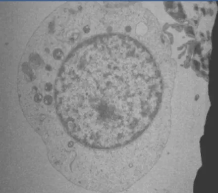
Figs. 4 a & b show the micrographs of Transmission Electron Microscopic analysis of Fusarium solani grown in the absence of Cr(VI) and in the presence of Cr(VI) of 500 mg l-1 concentration. A nearly round shaped cell with a well defined cell
wall was observed for the cell grown in the absence of Cr(VI) (Fig. 4a). The shape of the cell seemed to be distorted when the organism was grown in presence of Cr(VI) (Fig. 4b).
Both intracellular accumulation of Cr within the cell and extracellular accumulation of Cr on the cell surface are evident from the Fig 4b. The change in shape of the cell in presence of Cr(VI) could be due to the damage of the cell wall component
(glucan) caused by extracellular accumulation of Cr. The Transmission Electron Microscopic analysis of a facultative Gram-negative bacterium Shewanella oneidensis MR-1 in the presence of 150 µM of Cr(VI) has been reported [24]. The study showed that the reduced chromium precipitates Cr(III) were present both extracellularly on the cell surface and intracellularly
(a) Fusarium solani grown in absence of Cr(VI) (b) Fusarium solani grown in presence of Cr(VI) Fig. 4 magnification 8, 400 x; 500 x
In order to explore the feasibility of recovering Cr contained in biomass (4.5 g l-1, dry wt. basis) produced in the batch experiment conducted at 500 mg l-1 initial Cr(VI) concentration, the biomass was treated with different eluting reagents as
shown in Table 2.
Table 2 Elution of Chromium from Fusarium solani containing Cr with time
Elution Reagent Chromium eluted (mg)
5 mins 30 mins 60 mins 120 mins
Distilled water Nil Nil Nil Nil
Sodium chloride
0.01 N, 0.1N,0.5N 4,12,14 6,14,14 8,14,14 8,14,14
Sodium hydroxide 0.01N,0.1N,0.5 N,1N
0.4,2,19.5,19.5 1.5,15,19.5,19.5 1.5,15,19.5,19.5 1.5,15,19.5,19.5
EDTA
0.01N,0.1N,0.5 N 0.2,0.5,0.5 0.5,0.5,0.5 0.5,1,1 0.5,1,1 Hydrochloric acid
0.01m, 0.1m,0.5 m 0.2,0.5,0.5 0.5,0.5,0.5 0.5,1,1 0.5,1,1 Sulphuric acid
0.01 m,0.1m,0.5 m 1.5,0.6,1 3,1.6,3.5 3,1.6,3.5 3,1.6,3.5 Nitric acid
0.01m,0.1m,0.5 m Nil,0.5,1 2,1,1.5 2,1,1.5 2,1,1.5
Recovery of Cr assumes great importance from resource conservation point of view along with an added benefit of reusing the eluted biomass for Cr(VI) removal. The table shows maximum elution of chromium (19.5 mg) using 0.5 N sodium
hydroxide solution within 5 mins time period. This was 18% of total chromium (111 mg) removed by the Fusarium solani. Significant elution of 14 mg of Cr was also observed in 5 mins using 0.5 N sodium chloride solution. However, no significant elution was obtained by using other eluents. The above results indicate that the Cr elution from the biomass was favoured by the
alkaline eluting reagent. It was expected that in the presence of alkaline solution the elution of anionic dichromate from the biomass would follow an ion-exchange mechanism. The Cr recovered from the Cr concentrated eluent through pH adjustment
The chromium recovery was also attempted from the media from which the biomass was harvested (Fig. 5). About 55%
(i.e., 205.7 mg) of the total chromium (374 mg) present in the media could be recovered through pH adjustment. Therefore, the total chromium recovery from the media and the biomass was possible to the extent of 219.3 mg, which was found to be 44% of
the initial Cr(VI), i.e., 500 mg.
5.
Conclusions
The fungal strain isolated from soil and used in the present study was characterized as Fusarium solani. The Cr distribution in the biomass and in the media obtained from the experiment conducted at 500 mg l-1 initial Cr(VI) concentration and at pH 5.0 indicated both intra cellular and extracellular accumulation of Cr and this was supported by the Transmission Electron Microscopic observation under similar conditions. However, it is difficult to elucidate the exact mechanism by which
the metal has been taken up by the growing cells of Fusarium solani. Therefore, more in-depth studies are required in this direction.
Fig. 5 Recovery of chromium from biomass and media
Studies conducted on elution of chromium from Fusarium solani containing Cr, indicated that a maximum of 18% of Cr
could be eluted using 0.5 N sodium hydroxide solution without destructing the biomass structure. The recovery of Cr from the Cr concentrated eluant through pH adjustment in the alkaline range was found to be 12.25% of total Cr (111 mg) removed by the
biomass. However, the Cr recovery from the media was found to be 55% of the total Cr (205.7 mg) present in the media.Therefore, the total Cr recovered from both biomass and media was found to be 44% of the initial Cr(VI)
concentration (500 mg l-1).
References
[1] B. R. Sudha, and E. Abraham, “Biosorption of Cr(VI) from aqueous solution by Rhizopus nigrificans,” Bioresource Technology, vol. 79, pp. 73-81, 2001.
[2] B. Volesky, Biosorption and biosorbents in Biosorption of Heavy Metals, Boca Raton: CRC press Inc, 1990.
[3] O. Muter, A. Patmalnieks, and A. Rapoport, “Interrelations of the yeast Candida utilis and Cr (VI): metal reduction and its
distribution in the cell and medium,” Process Biochemistry, vol. 36, pp. 963-970, 2001.
[5] T. Srinath, T. Verma, P.W. Ramteka, S.K. Garg, “Chromium (VI) biosorption and bioaccumulation by chromate resistant
bacteria,” Chemophere, vol. 48, pp. 427- 435, 2001.
[6] M. Sen, “A Comparative Study on Biosorption of Cr(VI) by Fusarium solani under Different Growth Conditions,” Open Journal of Applied Sciences, vol. 2, pp. 146-152, 2012.
[7] M. Nourbaksh, Y. Sag, Z. Ozer, Z. Aksu, T. Kustal, and A. Caglar, “A comparative study of Various Biosorbent for Removal of Chromium (VI) Ions from Industrial Waste Waters,” Process Biochemistry, vol. 29, pp. 1-5, 1994.
[8] P. Pattanapipitpaisal, N.L.Brown, and L.E. Macaskie, “Chromate reduction by Microbacterium liquefaciens immobilized in polyvinyl alcohol,” Biotech Letter, vol. 23, pp. 41-43, 2001.
[9] B. Barkhordar, and M. Ghiasseddin, “Comparision of Langmuir and Freundlich Equilibriums in Cr, Cu and Ni Adsorption by Sargassum,” Iranian J Env Health Sci Eng, vol. 1, pp. 58-64, 2004.
[10] K.S. Selvaraj, S. Manonmai, and S. Pattabhi, “Removal of hexavalent chromium using distillery sludge,” Bioresource Technology, vol. 89, pp. 207-211, 2003.
[11] A.Y. Dursan, G. Ulsu, Y. Cuci, and Z.Aksu, “Bioaccumulation of cupper (II), lead (II) and chromium (VI) by growing Aspergillus niger,” Process Biochemistry, vol. 38, pp. 1647-1651, 2003.
[12] J.M. Panacastro, F.M.-Jeronimo, E.-Garcia, and F.R.O. Canizares-Villanueva, “Heavy metals removal by the micro algae Sceneunus in crassatulus in continuous cultures,” Bioresource Technology, vol. 94, pp. 219-222, 2004.
[13] A. Khanafari, S. Eshghdoost, and A. Mashinchian, “Removal of lead and chromium from aqueous solution by bacillus
circulans,” Iran. J. Environ. Health. Sci. Eng., vol. 5, pp. 195-200, 2008.
[14] M. Sen, M.G. Dastidar, and P.K. Roychoudhury, “Biosorption of Cr(VI) using fungal strain of Fusarium sp.,” American Society of Civil Engineers (ASCE), Prac. Period. Hazard.,Toxic Radioact Waste Manag., vol. 9, pp. 147-151, 2005. [15] M. Sen, and M.G. Dastidar, “Adsorption- Desorption Studies on Cr(VI) Using Non-Living Fungal Biomass,” Asian Journal
of Chemistry, vol. 22, pp. 2331-2338, 2010.
[16] M.Sen, and M.G. Dastidar, “Biosorption of Cr(VI) by resting cells of Aspergillus sp.,” Iran. J. Environ.Health. Sci. Eng., vol. 4, pp. 9-12, 2006.
[17] M. Sen, M.G. Dastidar, and P. K. Roychoudhury, “Comparative studies of batch and continuous biological treatment of Cr(VI) using Fusarium solani,” Enzyme and Microbial Technology, vol. 41, pp 51-56, 2007.
[18] M.Sen, and M.G. Dastidar,”Biosorption of Cr(VI) by resting cells of Fusarium Solani,” Iran. J. Environ. Health. Sci. Eng., vol. 8, pp. 153-158, 2011.