microRNA Expression in Relapsing Remitting Multiple
Sclerosis
Anne Waschbisch1*, Monika Atiya1, Ralf A. Linker1, Sergej Potapov2, Stefan Schwab1, Tobias Derfuss1,3
1Department of Neurology, Friedrich-Alexander University, Erlangen-Nu¨rnberg, Germany,2Institute of Medical Informatics, Biometry and Epidemiology, Friedrich-Alexander University, Erlangen-Nu¨rnberg, Germany,3Department of Neurology, University Hospital Basel, Basel, Switzerland
Abstract
The expression of selected microRNAs (miRNAs) known to be involved in the regulation of immune responses was analyzed in 74 patients with relapsing remitting multiple sclerosis (RRMS) and 32 healthy controls. Four miRNAs (miR-326, miR-155, miR-146a, miR-142-3p) were aberrantly expressed in peripheral blood mononuclear cells from RRMS patients compared to controls. Although expression of these selected miRNAs did not differ between treatment-naı¨ve (n = 36) and interferon-beta treated RRMS patients (n = 18), expression of miR-146a and miR-142-3p was significantly lower in glatiramer acetate (GA) treated RRMS patients (n = 20) suggesting that GA, at least in part, restores the expression of deregulated miRNAs in MS.
Citation:Waschbisch A, Atiya M, Linker RA, Potapov S, Schwab S, et al. (2011) Glatiramer Acetate Treatment Normalizes Deregulated microRNA Expression in Relapsing Remitting Multiple Sclerosis. PLoS ONE 6(9): e24604. doi:10.1371/journal.pone.0024604
Editor:Christoph Kleinschnitz, Julius-Maximilians-Universita¨t Wu¨rzburg, Germany
ReceivedJune 4, 2011;AcceptedAugust 14, 2011;PublishedSeptember 16, 2011
Copyright:ß2011 Waschbisch et al. This is an open-access article distributed under the terms of the Creative Commons Attribution License, which permits unrestricted use, distribution, and reproduction in any medium, provided the original author and source are credited.
Funding:This study was funded by a research grant of the Interdisciplinary Centre for Clinical Research (IZKF, project J6) of the Friedrich Alexander University Erlangen to AW. The funders had no role in study design, data collection and analysis, decision to publish, or preparation of the manuscript.
Competing Interests:AW has received funding for travel or speaker honoraria from Bayer Schering Pharma, Biogen Idec/Elan Corporation, Merck Serono, and Teva Pharmaceuticals. RL has received funding for travel and speaker honoraria from Bayer Schering Pharma, Biogen Idec/Elan Corporation, Merck Serono, Novartis, Sanofi-Aventis, and Teva Pharmaceuticals as well as research support from Biogen Idec, Merck Serono, Novartis, and Teva Pharmaceuticals. TD has received funding for travel and speaker honoraria from Bayer Schering Pharma, Biogen Idec/Elan Corporation, Novartis, Merck Serono, and Teva Pharmaceuticals as well as research support from Merck Serono and Novartis. This does not alter the authors’ adherence to all the PLoS ONE policies on sharing data and materials. The commercial companies mentioned above did not fund this particular study.
* E-mail: [email protected]
Introduction
microRNAs (miRNAs) have recently emerged as potential biomarkers of disease in different autoimmune disorders including rheumatoid arthritis [1], systemic lupus erythematosus [2] or Sjo¨gren’s syndrome [3]. miRNAs are short (,22 nucleotides) non-coding RNA molecules that regulate gene expression at the post-transcriptional level. One miRNA may target several hundreds of messenger RNAs and it has been proposed that up to 50% of mammalian genes underlay miRNA fine-tuning [4]. miRNAs are critically involved in the regulation of a wide array of cellular and developmental processes including the differentiation of immune cells and the outcome of immune responses [5].
Recent data indicate that miRNA dysregulation may also contribute to the pathogenesis of multiple sclerosis (MS): Overexpression of miRNAs that target CD47 was found to contribute to macrophage-mediated damage in active MS lesions and miRNA expression profiling in whole blood or leukocytes derived from MS patients suggests an aberrant expression of various miRNAs in the peripheral immune compartment [6,7]. Here we studied the expression of five selected immunologically relevant miRNAs in peripheral blood mononuclear cells (PBMC) derived from patients with relapsing-remitting multiple sclerosis (RRMS) and healthy controls and addressed the impact of immunomodulatory therapy on miRNA expression by comparing treatment-naı¨ve to glatiramer acetate (GA) or interferon-beta (IFN-beta) treated patients.
Results
Overexpression of miR-142-3p, miR-155, miR-146a and miR-326 in PBMC of RRMS Patients
Aberrant Expression of miR-142-3p and miR-146a is Restored in GA Treated MS Patients
In parallel we analyzed the expression of miRNA in a group of glatiramer acetate (n = 20) and interferon-beta (n = 18) treated RRMS patients to detect a potential impact of immunomodulatory therapy on deregulated miRNA. The expression of miR-142-3p, miR-146a, miR-155, miR-326 and miR-20b did not differ between treatment-naı¨ve and IFN-beta treated RRMS patients. In contrast, the expression of miR-142-3p and miR-146a was significantly decreased in GA treated RRMS patients to values that resembled the level of normal (p = 0,003; p = 0,028). miR-155 and miR-326 did not differ between untreated and the glatiramer acetate treated group (Figure 1). In vitro stimulation of PBMC derived from
untreated MS patients with GA for up to 72 hours (40mg/ml) did
not result in the downregulation of the miRNAs under investigation suggesting that the effects of miRNA modulation by GA are of more complex nature requiring long-term GA exposure or interaction with resident cells of secondary lymphoid organs (Figure S1).
Discussion
analyze the expression of selected, immune relevant miRNAs by quantitative PCR in a cohort of treatment-naive RRMS patients and healthy controls.
Two of the miRNAs found to be overexpressed in MS, miR-326 and miR-155 have recently been identified as crucial regulators of T cell development and Th17 differentiation [10,14] and were found to promote CNS inflammation in experimental autoim-mune encephalomyelitis (EAE). The first-time demonstration of miR-326 and miR-155 overexpression in PBMC derived from European MS patients underlines the clinical significance of these findings and adds to previous data on miR-326 dysregulation in Chinese patients with relapsing MS [10].
While the expression levels of miR-326 and miR-155 did not differ between untreated and GA or IFN-beta treated patients, GA treatment seemed to normalize miR-146a and miR-142-3p expression in MS patients. miR-142-3p expression has been linked to immune tolerance, since it was found to be repressed by FOXP3 resulting in an increased production of cyclic AMP and suppressor function in T regulatory cells [15]. Here we demonstrate upregulation of miR-142-3p in PBMC from RRMS patients which is in line with a previous report [11]. The mechanisms by which GA influences the expression of miR-142-3p remain unknown. However, it is well perceivable that an expansion of T regulatory cells by glatiramer acetate and/or changes in the composition of the T cell compartment may account for the downregulation of miR-142-3p.
miR-146a has emerged as a key player in the regulation of innate immunity and seems to be critical for the suppressor function of T regulatory cells [16]. miR-146a was found to be overexpressed at the site of inflammation in RA [17], psoriasis [18] and within active MS lesions [19]. We hypothesize that overexpression of miR-146a in PBMC of MS patients is reactive to the proinflammatory milieu in MS. In this light, downregulation of miR-146a in the glatiramer acetate treatment group may reflect
restoration of the cytokine milieu by GA dependent induction of a Th1 to Th2 shift and inhibition of monocyte reactivity [20–22].
There are certain possible confounders to this study. The mean age and disease duration was higher in GA and IFN treated patients compared to treatment-naı¨ve patients which is an obvious conse-quence of an early treatment strategy. Since miRNA expression in PBMC may change with age and immune senescence or may be influenced by a chronic inflammatory state we analysed whether expression of the studied miRNAs was biased by age or disease duration. However, we did not find significant correlations of the miRNA expression levels with age or disease duration in our study.
The patients we studied represent a rather active MS population as reflected by the high percentage of patients that experienced relapse within the last 3 months (up to 39% within the no treatment group). It has been suggested that certain miRNAs levels may increase during MS relapse [10,13]. Since recent MRI as a surrogate marker of disease activity was not available in most of the patients we cannot control a possible influence of disease activity on miRNA expression in our study which may have biased our results.
Summarizing, we report the aberrant expression of four miRNAs, previously reported to be critically involved in TH17 differentiation (i.e. miR-326, miR-155), the regulation of immune tolerance 142-3p, miR-146a) or innate immunity (miR-146a). Immunomodulatory treatment with IFN-beta did not restore the expression of deregulated miRNAs, whereas GA treatment seemed to normalize the levels of 146a and miR-142-3p but not miR-155 and miR-326 expression in RRMS patients. The biologic significance of our findings is further underlined by the fact that the overexpressed miRNA in peripheral blood leukocytes, were also among the most signifi-cantly upregulated miRNAs in active MS lesions in a seminal study by Junker et al. [19]. In conclusion, our study adds to the emerging evidence of miRNA dysregulation in MS.
Figure 2. ROC analysis.To assess the sensitivity and specificity of miRNAs to predict disease, receiver operator characteristic curves were computed based on the relative miRNA expression in treatment-naı¨ve patients and healthy controls. The ROC curves for miR-326 and miR-142-3p are exemplary depicted.
Materials and Methods
Patients and Sample Collections
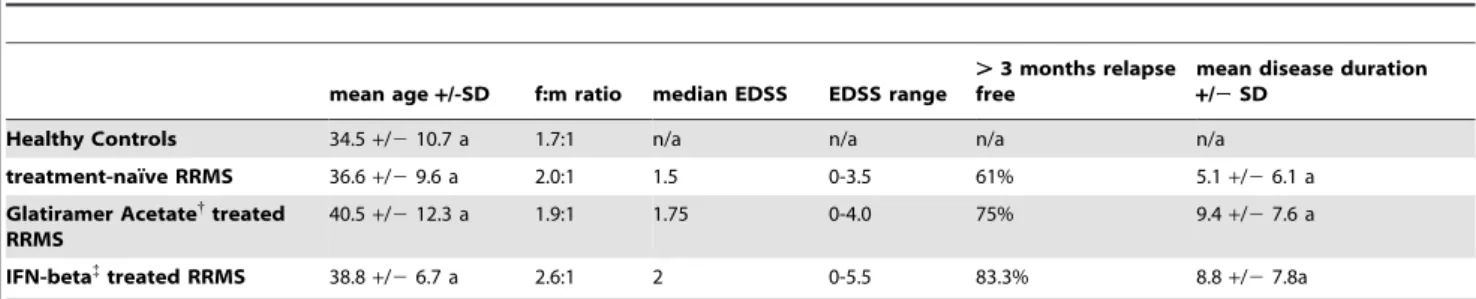
The study was approved by the ethics committee of the Friedrich-Alexander University Erlangen (No. 4203) and written informed consent was obtained from all participants. RRMS patients that had been diagnosed according to the McDonald criteria were eligible for participation in this study. Patients had to be either treatment naı¨ve, or treated with the immunomodulatory drugs glatiramer acetate (GA, CopaxoneH) or Interferon-beta (IFN-beta, AvonexHn = 5, RebifHn = 8, BetaferonHn = 5) for at least 3 months. Patients that were treated with glucocorticoids during the last four weeks before study entry were excluded from the study. .97% of patients and healthy controls were of Caucasian origin. All patients were assessed for EDSS and clinical parameters of disease activity by their treating physician. Patients that had been relapse-free for at least 3 months were considered as clinically stable. Peripheral blood was obtained by venipuncture and immediately processed for isolation of PBMCs. Demographic and clinical details are summarized in table 1.
Isolation of PBMCs and RNA Isolation
PBMCs were isolated from EDTA blood via ficoll density gradient centrifugation. 16107cells were resuspended in QiazolH
(Qiagen, Hilden, Germany) and stored at280uC. RNA enriched in miRNA was isolated using the miRNAeasyHMini and RNeasyH
MinEluteHCleanup Kit according to the manufacturer’s protocol. Total RNA concentrations were determined using a NanoDrop spectrophotometer (NanoDrop technologies, USA).
In VitroGA Stimulation of PBMC
PBMC were cultured in RPMI 1640 (Gibco Invitrogen GmbH, Karlsruhe, Germany) supplemented with 10% of FCS (PAA, Pasching, Austria), glutamine and antibiotics. Cells were grown at a densitiy of 26106ml in 6-well plates (Gibco Invitrogen GmbH, Karlsruhe, Germany) under standard culture conditions (37uC, 5%CO2). GA (CopaxoneH, 40mg/ml) was added to the medium
for different periods of time. RNA was isolated as described above.
Real-Time PCR
The TaqManHMicroRNA Reverse Transcription Kit was used according to the manufacturer’s instruction for reverse transcrip-tion with target specific stem loop primers provided in TaqManH
miRNA Assays. The TaqMan Universal MasterMix was used according to the manufacturer’s protocol for amplification of the targets on an ABI PRISMH7900 HT Real Time PCR System.
The following TaqMan miRNA assays were used: hsa-miR-155, Assay ID 002623; hsa-miR-326, Assay ID 000542; hsa-miR-142-3p, Assay ID 000464; hsa-miR-146a, Assay ID 000468; has-miR-20b, Assay ID 001014. RNU6B (Assay ID 001093) that has been previously used in several studies analyzing miRNA expression in immune cells10–12was used as an endogenous control to calculate
DCT. All samples were analysed in triplicates. The averageDCT of healthy donors was used as a calibrator to calculate DDCT. Results are expressed as 2-DDCT.
Statistical Analysis
Differences in miRNA expression were determined using nonparametric tests (Mann-Whitney-U, Kruskal-Wallis-Test with Dunn’s multiple comparison). p-values ,0.05 were considered significant. Correlation of miRNA expression with age, disease duration and gender was assessed by calculating the spearman and point-biserial correlation coefficient. Receiver operator character-istic curves (ROC) were computed and the area under the curve (AUC) was calculated to assess the discriminatory power of individual miRNA. A quadratic discriminant analysis (QDA) was employed to assess the predictive power of a combination of miRNAs to differentiate between healthy controls or RRMS patients. The adapted model was evaluated by the leave-one-out cross-validation method. Statistical analysis was performed using the R-system for statistical computing [23] (version 2.12.2, R Development Core Team 2011) and GraphPad Prism (version 5.0, GraphPad Software Inc.).
Supporting Information
Figure S1 miRNA expression was analyzed in PBMC derived from RRMS patients (n = 6) after in vitro treatment with glatiramer acetate (GA, 40mg/ml) for the indicated periods of time.
(TIF)
Acknowledgments
The help of S. Cursiefen, J. Kratzer and T. Stirnweiss in the acquisition of patient samples is greatly acknowledged. The authors thank K. Bitterer for excellent technical assistance.
Author Contributions
Conceived and designed the experiments: AW MA SS TD. Performed the experiments: AW TD MA RL. Analyzed the data: AW SP TD. Contributed reagents/materials/analysis tools: MA RL SP SS TD. Wrote the paper: AW MA RL SP SS TD.
Table 1.Clinical and demographic details of patients and healthy controls.
mean age+/-SD f:m ratio median EDSS EDSS range
.3 months relapse free
mean disease duration
+/2SD
Healthy Controls 34.5+/210.7 a 1.7:1 n/a n/a n/a n/a
treatment-naı¨ve RRMS 36.6+/29.6 a 2.0:1 1.5 0-3.5 61% 5.1+/26.1 a
Glatiramer Acetate{
treated RRMS
40.5+/212.3 a 1.9:1 1.75 0-4.0 75% 9.4+/27.6 a
IFN-beta{
treated RRMS 38.8+/26.7 a 2.6:1 2 0-5.5 83.3% 8.8+/27.8a
{
CopaxoneH; {Avonex
References
1. Pauley KM, Satoh M, Chan AL, Bubb MR, Reeves WH, et al. (2008) Upregulated miR-146a expression in peripheral blood mononuclear cells from rheumatoid arthritis patients. Arthritis Res Ther 10: R101.
2. Tang Y, Luo X, Cui H, Ni X, Yuan M, et al. (2009) MicroRNA-146A contributes to abnormal activation of the type I interferon pathway in human lupus by targeting the key signaling proteins. Arthritis Rheum 60: 1065–1075. 3. Alevizos I, Alexander S, Turner RJ, Illei GG (2011) MicroRNA expression profiles as biomarkers of minor salivary gland inflammation and dysfunction in Sjogren’s syndrome. Arthritis Rheum 63: 535–544.
4. Lewis BP, Burge CB, Bartel DP (2005) Conserved seed pairing, often flanked by adenosines, indicates that thousands of human genes are microRNA targets. Cell 120: 15–20.
5. O’Connell RM, Rao DS, Chaudhuri AA, Baltimore D (2010) Physiological and pathological roles for microRNAs in the immune system. Nat Rev Immunol 10: 111–122.
6. Junker A, Hohlfeld R, Meinl E (2011) The emerging role of microRNAs in multiple sclerosis. Nat Rev Neurol 7: 56–59.
7. Tufekci KU, Oner MG, Genc S, Genc K (2011) MicroRNAs and Multiple Sclerosis. Autoimmune Dis 2011: 807426.
8. Cox MB, Cairns MJ, Gandhi KS, Carroll AP, Moscovis S, et al. (2010) MicroRNAs miR-17 and miR-20a inhibit T cell activation genes and are under-expressed in MS whole blood. PLoS One 5: e12132.
9. De Santis G, Ferracin M, Biondani A, Caniatti L, Rosaria Tola M, et al. (2010) Altered miRNA expression in T regulatory cells in course of multiple sclerosis. J Neuroimmunol 226: 165–171.
10. Du C, Liu C, Kang J, Zhao G, Ye Z, et al. (2009) MicroRNA miR-326 regulates TH-17 differentiation and is associated with the pathogenesis of multiple sclerosis. Nat Immunol 10: 1252–1259.
11. Keller A, Leidinger P, Lange J, Borries A, Schroers H, et al. (2009) Multiple sclerosis: microRNA expression profiles accurately differentiate patients with relapsing-remitting disease from healthy controls. PLoS One 4: e7440. 12. Lindberg RL, Hoffmann F, Mehling M, Kuhle J, Kappos L (2010) Altered
expression of miR-17-5p in CD4+lymphocytes of relapsing-remitting multiple sclerosis patients. Eur J Immunol 40: 888–898.
13. Otaegui D, Baranzini SE, Armananzas R, Calvo B, Munoz-Culla M, et al. (2009) Differential micro RNA expression in PBMC from multiple sclerosis patients. PLoS One 4: e6309.
14. O’Connell RM, Kahn D, Gibson WS, Round JL, Scholz RL, et al. (2010) MicroRNA-155 promotes autoimmune inflammation by enhancing inflamma-tory T cell development. Immunity 33: 607–619.
15. Huang B, Zhao J, Lei Z, Shen S, Li D, et al. (2009) miR-142-3p restricts cAMP production in CD4+CD25- T cells and CD4+CD25+TREG cells by targeting AC9 mRNA. EMBO Rep 10: 180–185.
16. Lu LF, Boldin MP, Chaudhry A, Lin LL, Taganov KD, et al. (2010) Function of miR-146a in controlling Treg cell-mediated regulation of Th1 responses. Cell 142: 914–929.
17. Nakasa T, Miyaki S, Okubo A, Hashimoto M, Nishida K, et al. (2008) Expression of microRNA-146 in rheumatoid arthritis synovial tissue. Arthritis Rheum 58: 1284–1292.
18. Sonkoly E, Wei T, Janson PC, Saaf A, Lundeberg L, et al. (2007) MicroRNAs: novel regulators involved in the pathogenesis of psoriasis? PLoS One 2: e610. 19. Junker A, Krumbholz M, Eisele S, Mohan H, Augstein F, et al. (2009)
MicroRNA profiling of multiple sclerosis lesions identifies modulators of the regulatory protein CD47. Brain 132: 3342–3352.
20. Burger D, Molnarfi N, Weber MS, Brandt KJ, Benkhoucha M, et al. (2009) Glatiramer acetate increases IL-1 receptor antagonist but decreases T cell-induced IL-1beta in human monocytes and multiple sclerosis. Proc Natl Acad Sci U S A 106: 4355–4359.
21. Racke MK, Lovett-Racke AE (2011) Glatiramer acetate treatment of multiple sclerosis: an immunological perspective. J Immunol 186: 1887–1890. 22. Weber MS, Starck M, Wagenpfeil S, Meinl E, Hohlfeld R, et al. (2004) Multiple
sclerosis: glatiramer acetate inhibits monocyte reactivity in vitro and in vivo. Brain 127: 1370–1378.