Chemokines in the cerebrospinal
fluid of patients with active and stable
relapsing-remitting multiple sclerosis
1Centro de Investigação em Esclerose Múltipla, Hospital das Clínicas,
2Laboratório de Imunofarmacologia, Departamento de Bioquímica e Imunologia,
Instituto de Ciências Biológicas, Universidade Federal de Minas Gerais, Belo Horizonte, MG, Brasil
M.A. Moreira1,
A.L.S. Souza2,
M.A. Lana-Peixoto1,
M.M. Teixeira2
and A.L. Teixeira1,2
Abstract
Multiple sclerosis (MS) is a chronic inflammatory demyelinating disease of the human central nervous system. Although its etiology is unknown, the accumulation and activation of mononuclear cells in the central nervous system are crucial to its pathogenesis. Chemokines have been proposed to play a major role in the recruitment and activation of leukocytes in inflammatory sites. They are divided into subfamilies on the basis of the location of conserved cysteine residues. We determined the levels of some CC and CXC chemokines in the cerebrospinal fluid (CSF) of 23 relapsing-remitting MS patients under interferon-ß-1a therapy and 16 control subjects using ELISA. MS patients were categorized as having active or stable disease. CXCL10 was significantly increased in the CSF of active MS patients (mean ± SEM, 369.5 ± 69.3 pg/mL) when compared with controls (178.5 ± 29.1 pg/mL, P < 0.05). CSF levels of CCL2 were significantly lower in active MS (144.7 ± 14.4 pg/mL) than in controls (237.1 ± 16.4 pg/ mL, P < 0.01). There was no difference in the concentration of CCL2 and CXCL10 between patients with stable MS and controls. CCL5 was not detectable in the CSF of most patients or controls. The qualitative and quantitative differences of chemokines in CSF during relapses of MS suggest that they may be useful as a marker of disease activity and of the mechanisms involved in the pathogenesis of the disease.
Correspondence
A.L. Teixeira
Laboratório de Imunofarmacologia Departamento de Bioquímica e Imunologia, ICB, UFMG Avenida Antônio Carlos, 6627 Bloco O4, Sala 202
31270-901 Belo Horizonte, MG Brasil
Fax: +55-31-3499-2651 E-mail: [email protected]
Received April 8, 2005 Accepted December 14, 2005
Key words
•CXCL10 •CCL2 •Inflammation •Multiple sclerosis •Leukocyte recruitment
Introduction
Multiple sclerosis (MS) is a chronic in-flammatory disease of the human central nervous system (CNS) resulting in the for-mation of plaques of demyelination. Although the cause of MS is unknown, it is generally considered to be an autoimmune disease mediated by CD4+ T cells, which produce
high levels of type 1 cytokines, such as inter-feron-γ (1,2). These Th1 cells initiate an
cytokines that mediate the migration of leu-kocytes to inflammatory sites (5,6). They are divided into subfamilies (CC, CXC, XC, CX3C) based on the location of conserved cysteine residues. Since mononuclear cell recruitment and activation in the CNS are fundamental steps in the pathogenesis of MS, chemokines have been proposed to play a major role in this autoimmune demyelinat-ing process (7). Previous studies have dem-onstrated that in MS CXCL10 and CCL5 are increased in the cerebrospinal fluid (CSF) of patients with MS compared with control subjects, while CCL2 is significantly de-creased (7-10). There is a growing body of evidence suggesting that these altered CSF chemokine levels could be used as surrogate markers of MS disease activity (9).
In the present study, we evaluated the CSF levels of CC and CXC chemokines from active and stable relapsing-remitting MS (RR-MS) patients and control subjects. This is the first study to evaluate chemo-kines in Brazilian MS patients.
Subjects and Methods
Subjects
All subjects (patients and controls) en-rolled in this study were from Belo Hori-zonte, State of Minas Gerais, Brazil, where the prevalence of MS is about 15 cases per 100,000 persons (11). Twenty-three patients diagnosed as having clinical definite RR-MS according to Poser’s criteria (12) were studied (Table 1). Twelve MS patients were clinically stable under interferon-ß-1a thera-py (mean score on the Expanded Disability Status Scale (EDSS) 3.0 ± 1.5). The remain-ing 11 patients had active disease (mean EDSS 4.9 ± 1.2). Disease activity was de-fined according to clinical criteria, i.e., pa-tients were in relapse with the presence of new neurological symptoms or deterioration of previous ones. Accordingly, the EDSS scores of patients with active RR-MS were higher than those of patients with stable RR-MS (P < 0.05). Active RR-MS patients were also under interferon-ß-1a therapy and the CSF was collected before any steroid therapy.
Sixteen individuals with no neurological disease undergoing lumbar puncture as part of an anesthetic procedure for surgical interven-tion (orthopedic and gynecologic) were re-cruited as the control group. The absence of neurological disease was established on the basis of clinical history and neurological ex-amination of the subjects. This group was matched with MS patients for age and gender. CSF samples were collected aseptically and stored at -70ºC until required. The study was approved by the Ethics Committee of Universidade Federal de Minas Gerais and all subjects gave written informed consent to par-ticipate.
Chemokine analysis
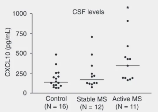
The concentration of chemokines in the CSF of MS patients and controls was meas-ured by sandwich ELISA with matched anti-Figure 1. CXCL10 levels in
cere-brospinal fluid (CSF) of patients with active multiple sclerosis (MS; N = 11), stable MS (N = 12) and control subjects (N = 16). *P < 0.05 compared to control (Newman-Keuls multiple com-parison post-test).
Table 1. Baseline demographic and clinical characteristics of the subjects studied.
MS patients Control group
(N = 16) Active MS (N = 11) Stable MS (N = 12)
Age 34.6 ± 8.9 (21-48) 35.0 ± 9.7 (20-47) 39.5 ± 10.3 (24-52)
Gender (M/F) 1/10 1/11 4/12
EDSS 4.9 ± 1.2 (2-6.5) 3.0 ± 1.5 (1-4)
body pairs for CXCL10 (formerly γ
-interfer-on inducible protein of 10 kDa; Pharmingen, San Diego, CA, USA), and with ELISA kits for CCL2 (formerly monocyte chemotactic protein-1) and CCL5 (formerly regulated upon activation of normal T cell expressed and secreted; R&D Systems, Minneapolis, MN, USA). All samples were used undi-luted and assayed in duplicate in the same plate. The intra-assay variation was less than 5%. The lower detection limits of the assays were 5 pg/mL for CCL2 and CCL5, and 10 pg/mL for CXCL10.
Statistical analysis
Differences among groups were deter-mined by analysis of variance (ANOVA) and the Newman-Keuls multiple compari-son post-test, with the level of significance set at P < 0.05. All calculations were per-formed using Instat software (GraphPad, San Diego, CA, USA).
Results
CXCL10 levels were significantly higher in CSF of active RR-MS patients (mean ± SEM, 369.5 ± 69.3 pg/mL; median (25th-75th percentile), 345.4 (190.7-521.6) pg/ mL) when compared with controls (178.5 ± 29.1 pg/mL; 137.0 (97.1-231.3) pg/mL; P < 0.05; Figure 1). Mean CXCL10 value for stable RR-MS was 241.5 ± 54.1 pg/mL and did not differ significantly from control or active RR-MS values.
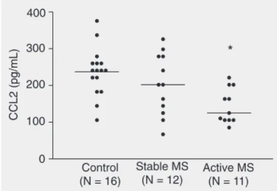
CCL2 levels were significantly lower in CSF of active RR-MS patients (mean ± SEM, 144.7 ± 14.4 pg/mL; median (25th-75th per-centile), 125.1 (105.8-202.2) pg/mL) when compared with controls (237.1 ± 16.4 pg/ mL; 240.7 (202.2-260.0) pg/mL; P < 0.01; Figure 2). The mean CCL2 value for stable RR-MS was 202.9 ± 24.0 pg/mL and was not statistically different from control or active RR-MS values.
CCL5 was detected in only one patient
with RR-MS and in one control subject, with no difference between them.
Discussion
Chemokines are involved in physiologic and pathologic leukocyte trafficking and have been investigated in several inflammatory diseases of the CNS (8,9). In the present study, we evaluated CSF levels of CXCL10, CCL2 and CCL5 in active RR-MS and stable RR-MS patients and in individuals with no neurological disorders.
The CSF levels of CXCL10 were signifi-cantly increased in active RR-MS patients compared with control subjects. In contrast, the CSF levels of CCL2 were lower in pa-tients with active RR-MS than in controls. The levels of both CCL2 and CXCL10 in stable RR-MS patients were similar to those of controls and active RR-MS patients. This is the first study to report levels of chemo-kines in CSF of MS patients in Brazil and in the Southern Hemisphere. The present re-sults demonstrate that there is a similar in-crease in the concentration of CXCL10 as well as a decrease in the concentration of CCL2 in Brazilian patients, as also reported for European/North American patients (10, 13-17). Thus, although the genetic back-ground of our population is different and the prevalence of the disease is lower than in European countries or in the United States (11,18,19), the pattern of CSF levels of CXCL10 and CCL2 is similar. This is con-sistent with a related pathogenetic
ism for the disease in these different popula-tions.
Several studies have now shown that CXCL10 may activate CXCR3 to induce the migration and activation of Th1 lympho-cytes and monolympho-cytes, cells relevant for MS pathogenesis (6). The finding of increased CXCL10 in CSF of patients with active dis-ease is consistent with current paradigms that MS is a predominantly Th1 disease (1,2). Studies of experimental autoimmune encephalomyelitis suggest a pathogenic role for CCL2 (20,21). This is reinforced by the ability of CCL2 to induce the migration of mononuclear cells into the CNS and the presence of CCL2 at sites of demyelination in patients with MS (20,21). However, CCL2 may also have a role in facilitating Th2 responses under certain circumstances (5,6). In this regard, the lower concentrations of CCL2 in CSF of patients with active MS may be due to the sequestration of the chemo-kine within MS lesions or, alternatively, may be a consequence of the altered Th1/Th2 balance observed in MS patients (20). The levels of both chemokines are consistent with their role in disease pathogenesis and in mediating leukocyte recruitment and the Th1/ Th2 cytokine balance.
In the present study, only two samples had detectable levels of CCL5. Thus, no
difference could be established between groups. There are discordant results in the literature concerning CSF CCL5 levels in MS. Some studies failed to find any differ-ence among groups (14,15), while others demonstrated increased CSF levels of CCL5 during MS relapses (10,22,23). The fact that the CSF concentration of this chemokine is very low and near the lower limit of detec-tion by ELISA may explain these discrepant results.
In the present study, all patients were using interferon-ß-1a therapy at the time the samples were collected. At least one study has demonstrated that such therapy fails to modify the expression of chemokines (13). This is an interesting finding since chemo-kines may play a role in the disease and lack of interference with the production of these mediators may be one of the reasons why this therapy only partially ameliorates pro-gression in only some of the treated patients (4).
The results of the present study support the view that CXCL10 and CCL2 are differ-ently released during relapses of MS. There appears to be a profile of secretion compat-ible with an enhanced Th1 response. More-over, it appears that levels of chemokines in CSF may be useful as markers of disease activity (24).
References
1. Lassmann H & Ransohoff RM (2004). The CD4-Th1 model for multiple sclerosis: a crucial re-appraisal. Trends in Immunology, 25: 132-137.
2. Hafler DA (2004). Multiple sclerosis. Journal of Clinical Investiga-tion, 113: 788-794.
3. Kornek B & Lassmann H (2003). Neuropathology of multiple sclero-sis. Brain Research Bulletin, 61: 321-326.
4. Noseworthy JH, Lucchinetti C, Rodriguez M et al. (2000). Multiple sclerosis. New England Journal of Medicine, 343: 938-952. 5. MacKay CR (2001). Chemokines: immunology’s high impact
fac-tors. Nature Immunology, 2: 95-101.
6. Moser B, Wolf M, Walz A et al. (2004). Chemokines: multiple levels of leukocyte migration control. Trends in Immunology, 25: 75-84. 7. Trebst C & Ransohoff RM (2001). Investigating chemokines and
chemokine receptors in patients with multiple sclerosis:
opportuni-ties and challenges. Archives of Neurology, 58: 1975-1980. 8. Ransohoff RM (2002). The chemokine system in neuroinflammation:
an update. Journal of Infectious Diseases, 186 (Suppl 2): S152-S156.
9. Sellebjerg F & Sorensen TL (2003). Chemokines and matrix metal-loproteinase-9 in leukocyte recruitment to the central nervous sys-tem. Brain Research Bulletin, 61: 347-355.
10. Sorensen TL, Tani M, Jensen J et al. (1999). Expression of specific chemokines and chemokine receptors in the central nervous system of multiple sclerosis patients. Journal of Clinical Investigation, 103: 807-815.
11. Lana-Peixoto MA, Frota E, Campos GB et al. (2002). The preva-lence of multiple sclerosis in Belo Horizonte, Brazil. Multiple Sclero-sis, 8 (Suppl 1): 38-39.
criteria for multiple sclerosis: guidelines for research protocols. An-nals of Neurology, 13: 227-231.
13. Franciotta D, Martino G, Zardini E et al. (2001). Serum and CSF levels of MCP-1 and IP-10 in multiple sclerosis patients with acute and stable disease and undergoing immunomodulatory therapies.
Journal of Neuroimmunology, 115: 192-198.
14. Sindern E, Niederkinkhaus Y, Henschel M et al. (2001). Differential release of ß-chemokines in serum and CSF of patients with relaps-ing-remitting multiple sclerosis. Acta Neurologica Scandinavica, 104: 88-91.
15. Mahad DJ, Howell SJ & Woodroofe MN (2002). Expression of chemokines in the CSF and correlation with clinical disease activity in patients with multiple sclerosis. Journal of Neurology, Neurosur-gery, and Psychiatry, 72: 498-502.
16. Scarpini E, Galimberti D, Baron P et al. (2002). IP-10 and MCP-1 levels in CSF and serum from multiple sclerosis patients with differ-ent clinical subtypes of the disease. Journal of Neurological Sci-ences, 195: 41-46.
17. Narikawa K, Misu T, Fujihara K et al. (2004). CSF chemokine levels in relapsing neuromyelitis optica and multiple sclerosis. Journal of Neuroimmunology, 149: 182-186.
18. Lana-Peixoto MA & Lana-Peixoto MI (1992). Is multiple sclerosis in
Brazil and Asia alike? Arquivos de Neuropsiquiatria, 50: 119-125. 19. Arruda WO, Scola RH, Teive HA et al. (2001). Report on 200 cases
from Curitiba, Southern Brazil and comparison with other Brazilian series. Arquivos de Neuropsiquiatria, 59: 165-170.
20. Mahad DJ & Ransohoff RM (2003). The role of MCP-1 (CCL2) and CCR2 in multiple sclerosis and experimental autoimmune encepha-lomyelitis (EAE). Seminars in Immunology, 15: 23-32.
21. Santos AC, Barsante MM, Arantes RM et al. (2005). CCL2 and CCL5 mediate leukocyte adhesion in experimental autoimmune encephalomyelitis: an intravital microscopy study. Journal of Neuro-immunology, 162: 122-129.
22. Bartosik-Psujek H, Belniak E, Mitosek-Szewczyk K et al. (2004). Interleukin-8 and RANTES levels in patients with relapsing-remitting multiple sclerosis treated with cladribine. Acta Neurologica Scandi-navica, 109: 390-392.
23. Bartosik-Psujek H & Stelmasiak Z (2005). The levels of chemokines CXCL8, CCL2 and CCL5 in multiple sclerosis patients are linked to the activity of the disease. European Journal of Neurology, 12: 49-54.