© 2012 S.K. Srivastava et al., This open access article is distributed under a Creative Commons Attribution (CC-BY) 3.0 license
doi:10.3844/ajbb.2012.63.70 Published Online 8 (2) 2012 (http://www.thescipub.com/ajbb.toc)
AJBB
MICRORNAS ARE NOT THAT SMALL TO IGNORE
1
Sanjeev Kumar Srivastava,
2Sonal Somvanshi and
2Vivek Pratap Singh
1
Department of Oncologic Sciences, Mitchell Cancer Institute, Mobile, AL, 36604,
2
Department of Pharmacology and Cell Biophysics, University of Cincinnati, College of Medicine, Cincinnati, Ohio
Received 2012-04-14; Revised 2012-05-7; Accepted 2012-05-20
ABSTRACT
MicroRNAs (miRNAs) are small, noncoding RNA molecule that negatively regulates gene expression by causing either degradation or translational inhibition of their target mRNAs. These miRNAs are key regulators of various biological processes such as differentiation, maturation and maintenance of immune homeostasis. Furthermore, deregulated expression of miRNA has now been shown to regulate several disease processes by regulating tumorigenesis, angiogenesis, apoptosis and metastasis in cancer and other diseases such as rheumatoid arthritis, diabetes and cardiovascular diseases. This review article summarizes the present status of miRNA research, with major emphasis on the roles of miRNAs in the development of various human diseases.
Keywords: MicroRNA, diabetes, cardiovascular disease, cancer
1. INTRODUCTION
MicroRNAs (miRNAs) are small non-coding RNAs (~22 nt), which are involved in the regulation of gene expression. These miRNAs negatively regulates gene expression at the posttranscriptional level by binding to the 3’UTR of target mRNAs through base pairing, resulting in either target mRNAs cleavage or causing translation inhibition (Ambros et al., 2004; Bartel, 2004). According to recent studies, in humans, more than 500 miRNAs have been identified and it is predicted that the total number may exceed up to 1000 (Bentwich et al., 2005; Griffiths-Jones et al., 2008). Despite the large number of identifying miRNAs, the target mRNAs and the biological function have been attributed to only a few of them (Bhardwaj et al., 2010). Growing evidences suggest that miRNAs play significant roles in several biological processes like differentiation, cell proliferation, embryonic development, cell proliferation and apoptosis (Esquela-Kerscher and Slack, 2006). With the continuous influx of studies on miRNAs, it is now becoming evident that these miRNAs exhibit differential expression in several human diseases, including cancer (Calin and Croce, 2006; Esquela-Kerscher and Slack, 2006) and play important roles in the development of other disease processes such as cardiovascular disease
(Latronico et al., 2007), schizophrenia (Hansen et al., 2007; Perkins, 2007), renal function disorders (Amrouche et al., 2011), fragile-X mental retardation syndrome (Provost, 2010), diabetes (Dehwah et al., 2012), hepatitis (Murakami et al., 2010) and obesity (Dehwah et al., 2012). Considering the growing number of studies relating miRNAs and various diseases this review article will focus more on newly identified miRNAs in disease (Table 1) and will discuss about their role and its significance in diagnostics and prognostics. Furthermore, this study will provide a deeper insight about the role of recently identified miRNAs.
1.1. miRNAs in Cancer
development and progression of cancer (Sotiropoulou et al., 2009). Growing studies also reveal that the aberrant expression of miRNAs is associated with the stage and metastasis of cancers (Shimono et al., 2009). These miRNA may function as either tumor promoters (oncomirs) or tumor suppressors (anti-oncomirs) (Bhardwaj et al., 2010). Thus, miRNAs may play important roles in cancer development, progression, prognosis and diagnosis. The expression profile of miRNA in multiple cancer types Including Chronic Lymphocytic Leukemia (CLL) (Moussay et al., 2011), breast (Schooneveld et al., 2012), colon (Piepoli et al., 2012), lung (Wang et al., 2012), pancreatic (Srivastava et al., 2011), prostate (Hassan et al., 2012), cancers and glioblastomas (D'Urso et al., 2012), reveal that a large number of miRNAs are aberrantly expressed in various cancers. In CLL, expression of miRNAs, miR-15a and miR-16 was downregulated (Hanlon et al., 2009). Bcl2, an anti-apoptotic protein is expressed at very high levels in CLL and is considered as an important survival factor of the cancer cells. Down regulated expression of miR-15a and miR-16-1 maintains the enhanced levels of BCL-2 and thus confers apoptosis resistance in CLL (Cimmino et al., 2005). In another study, miR-16 is shown to suppress the growth of prostate cancer cells by regulating the expression of CDK1 and CDK2, which are associated with cell cycle control and proliferation (Takeshita et al., 2010). Another study on miRNA in Lymphoma has shown that miR-17-92 cluster is upregulated in lymphomas. Their study demonstrated that the miR-17-92 cluster can enhance tumorigenesis by inhibiting apoptosis (Croce, 2008). Furthermore, several studies also show that this cluster is overexpressed in a wide array of cancers including breast, colon, lung, prostate and pancreatic endocrine. Among others oncogenic miRNAs, miR-21 has been shown to promote apoptosis through activation of caspases in human glioblastoma cells (Chan et al., 2005). In breast cancer cells, silencing of miR-21 inhibited cell growth in vitro and in vivo by causing downregulation of Bcl-2 and induction of apoptosis (Si et al., 2007). MiR-155 is overexpressed in many cancers including B-cell lymphomas, Burkitt lymphoma, Hodgkin’s lymphoma and breast, lung, colon and thyroid cancers. Its expression was significantly correlated with poor survival in pancreatic cancer patients (Greither et al., 2010). A study performed on mice also identified that mice overexpressing miR-155 are able to develop B-cell malignancy (Costinean et al., 2009). A recent study conducted by Srivastava et al. (2011) and coworkers identified a tumor suppressor miRNA in pancreatic cancer. Their study demonstrated that miR-150 exerts its tumor suppressor function by directly targeting an oncoprotein MUC4 (Srivastava et al., 2011). In lung and
colon cancer cells Let-7 family members of miRNAs are downregulated and that correlates with the shortened postoperative survival in lung cancer patients (Takamizawa et al., 2004). Studies on miR-125a and miR-125b suggest that their expression is either downregulated or lost in breast cancer (Scott et al., 2007). These miRNAs exert their function by regulating the oncogenes ERBB2 and ERBB3. Furthermore, miR- 143 and miR-145 are frequently downregulated in colorectal and breast cancers as well as B-cell malignancies suggesting their involvement in cancer (Luo et al., 2011; Akao et al., 2007). With the huge influx of data, it cannot be overruled that there may be many other miRNAs which are involved in cancers development and progression that need to be explored.
1.2. miRNAs in Cardiovascular Diseases
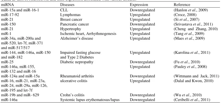
Table 1. List of MicroRNAs and their expression associated with various diseases
miRNA Diseases Expression Reference
miR-15a and miR-16-1 CLL Downregulated (Hanlon et al., 2009)
miR-17-92 Lymphomas Upregulated (Croce, 2008)
miR-21 Breast cancer Upregulated (Si et al., 2007)
miR-150 Pancreatic cancer Downregulated (Srivastava et al., 2011)
miR-21 Hypertrophy Upregulated (Cheng and Zhang, 2010)
miR-1 Ischemic heart, Arrhythmogenesis Upregulated (Tang et al., 2009)
miR-34a, miR-200a and Alzheimer’s disease Upregulated (Maes et al., 2009)
miR-520, let-7f, miR-371 and miR-517/517
miR-144, miR-146a, miR-150 Impaired fasting glucose Upregulated (Karolina et al., 2011)
and miR-182 and Type 2 Diabetes
miR-25 Diabetic nepropathy Downregulated (Fu et al., 2010)
miR-146a, miR-155, Upregulated (Pauley et al., 2008)
miR-132 and miR-16
miR-124a and miR-15a Rheumatoid arthritis Downregulated (Wittmann and Jack, 2011)
miR-16, miR-21, miR-23a, ulcerative colitis Upregulated (Dalal and Kwon, 2010)
miR-24, miR-29a, miR-126, miR-195 and let-7f
miR-19b and miR- 629 Crohn’s colitis Downregulated (Wu et al., 2010)
miR-146a Systemic lupus erythematosus/lupus Downregulated (Ceribelli et al., 2011)
In a rat model of myocardial infarction and in human hearts with coronary heart disease, miR-1 was significantly unregulated in ischemic heart tissue (Tang et al., 2009; Brattelid et al., 2011). Several studies have shown the significance of miRNAs in cardiac function and maintaining its structure. MiR-1, miR-133, miR-208, miR-21 and miR- 221/222 are associated with cardiac growth, erythropoiesis, angiogenesis, proliferation (Latronico et al., 2007; Bauersachs et al., 2007). Overexpression of mature miR-1 increases the severity of arrhythmogenesis and its downregulation suppress arrhythmias (Tang et al., 2009; Brattelid et al., 2011; Latronico et al., 2007; Latronico and Condorelli, 2009). All these evidences thus support the potential of miRNAs in the development of cardiovascular diseases.
1.3. miRNAs in Neurodegenerative Diseases
Neurodegenerative Diseases (NDs) are the diseases which result from many genetic and environmental factors. Several lines of evidence suggests the role of miRNAs in causing neurodegeneration or neuroprotection. In the current situation, with the emergence of miRNAs it is now considered that miRNAs play significant role in several mammalian brain-related biochemical pathways, including neuroplasticity and stress responses and contributes to various neurological disorders in humans, like fragile X syndrome (Caudy et al., 2002), Alzheimer’s Disease (AD) (Hebert et al., 2010). The correlation between
1.4. miRNAs in Diabetes
Diabetes is an autoimmune disorder and is often genetically inherited. So far more than 19 Insulin-Dependent Diabetes Mellitus (IDDM) susceptibility loci have been identified in humans. MiRNAs are linked to the regulation of different biological pathways associated and are now widely implicated with the diabetes. In a study, more than 27 miRNAs were observed to be located on 9 human IDDM loci and it was also revealed that some of the miRNAs may target autoimmune- and
β-cell-related genes (Zhou et al., 2008). In the diabetic rat model, an altered profile of miRNA expression in insulin target tissues was observed. A growing body of data suggests significant roles of miRNAs in insulin production, secretion and in glucose homeostasis and adipocyte differentiation. Karolina et al. (2011), performed an miRNA expression profiling in pancreas, liver, adipose and skeletal muscle and in blood samples from the T2D rat model as well as from Impaired Fasting Glucose (IFG) and T2D male patients and their investigation revealed that four miRNAs i.e. miR-144, miR-146a, miR-150 and miR-182 to be a potential signature miRNAs that distinguished IFG and T2D (Karolina et al., 2011). According to published studies, role of several miRNAs like 124a2, 195, miR-15a, miR-15b and miR-16 have been identified in regulating pancreatic development (Baroukh et al., 2007; Joglekar et al., 2007). Up-regulation or down-modulation of miR-124a2 can cause either elevated or decreased insulin mRNA levels, respectively (Baroukh et al., 2007). Notably, overexpression of miR-124a in MIN6 cell line was able to decrease glucose-stimulated insulin secretion (Lovis et al., 2008). Krek et al. (2005) have shown that miR-375 and miR-124 may share common pathways in the regulation of insulin secretion (Krek et al., 2005). Furthermore, overexpression of miR-410 elevates the level of glucose-stimulated insulin secretion in MIN6 cells (Hennessy et al., 2010). MiR-7 and miR-9 have also been identified as important regulators of insulin secretion (Bravo-Egana et al., 2008). The expression of 21, 34a and miR-146a was increased in the islets of non-obese diabetic mice during development of pre-diabetic insulitis (Roggli et al., 2010). Additionally, miR-126 was identified to be involved in the development of insulin resistance induced by mitochondrial dysfunction (Ryu et al., 2011). In diabetic nephropathy, reduced level of miR-25 was seen in the kidney from both diabetic rats and high glucose-treated mesangial cells which indicate
that miR-25 may regulate NOX4 expression and function in diabetic nepropathy (Fu et al., 2010).
1.5. miRNAs in Rheumatoid Arthritis
Rheumatoid Arthritis (RA) is a common systemic inflammatory disease. The disease is characterized by inflammation of synovial tissue, progressive cartilage destruction and bone erosion of affected joints (Srivastava et al., 2011). RA is a major cause of disability and is even correlated with increased mortality in severe cases. It is now known that miRNAs are involved in the modulation of inflammatory process in the joints. Several studies demonstrate that in Peripheral Blood Mononuclear Cells (PBMC) of RA high levels of miR-146a, miR-155, miR-132 and miR-16, were observed (Pauley et al., 2008). In a separate, study elevated expression of miR-146a in different cells and tissues of RA patients was observed, that suggest its association with the disease severity (Nakasa et al., 2011). Also, miR-146a expression in RA positively correlated with the levels of TNF-α in both Peripheral Blood (PB) and Synovial Fluid (SF) (Nakasa et al., 2011). Some groups have also provided their input and their data shows that miR-155 and miR-146a are up-regulated, while miR-124a and miR-15a are down-regulated miRNAs in the synovial membrane in RA (Wittmann and Jack, 2011). It was identified and reported that miR-124a regulates the cell cycle (Kawano and Nakamachi, 2011) and miR-155 regulates the metalloproteinases production (Leng et al., 2011) of RA synovial fibroblasts. Furthermore overexpression of miR-15a expression induces apoptosis in these cells (Nagata et al., 2009).
1.6. miRNAs in Inflammatory Bowel Disease
done on samples of colon biopsies of 5 patients with chronically active CD and 13 control individuals revealed upregulated expression of miR-23b, miR-106 and miR-191 in tissues from patients with active CD, while miR-19b and miR- 629 were downregulated in Crohn’s colitis patients (Wu et al., 2010). Studies also demonstrate that miRNA expression profiles correlates with the disease progression.
1.7. miRNAs
in
Systemic
Lupus
Erythematosus/Lupus
Systemic Lupus Erythematosus (SLE) is a chronic disease that causes inflammation, pain and swelling. Sometimes it is also called as “great imitator” because it can affect the skin, joints, kidneys, lungs, nervous system and other organs of the body. In SLE patients, expression of miR-146a expression is downregulated in PBMCs as compared to healthy controls. This decrease in miR-146a was associated with disease severity (Ceribelli et al., 2011). Working on the line to get the more insight about involvement of miRNAs, an miRNA profiles in SLE patients, Idiopathic Thrombocytopenic Purpura (ITP) patients and healthy controls was performed that identified a group of 13 miRNAs exhibiting same expression pattern in SLE and ITP whereas, miR-184, miR-198 and miR-21 were observed to have a differential expression in SLE compared to controls (Dalal and Kwon, 2010). In case of ITP, Six miRNAs were downregulated but this downregulation was not observed in patients with SLE. In kidney biopsies taken from lupus nephritis patients, miRNAs are differentially expressed compared to controls (Te et al., 2010).
1.8. Conclusion and Perspective
Growing evidences suggest that miRNAs play important role in a various developmental process including cell proliferation, cell cycle, cell differentiation, metabolism, apoptosis, developmental timing. These miRNAs are either down-regulated or overexpressed in various organs including lung, liver and kidney and exert diverse functions. Several studies have experimentally proved the links between miRNA and various disease conditions including cancer, cardiovascular diseases, arthritis, diabetes, Lupus. Additionally, identification of miRNAs in specific disease could provide impetus for the disease diagnosis, prognosis and evaluation of treatment response. Furthermore, targeting miRNAs may be a novel therapeutic approach in human diseases.
2. REFERENCES
Akao, Y., Y. Nakagawa, Y. Kitade, T. Kinoshita and T. Naoe et al., 2007. Downregulation of microRNAs-143 and -145 in B-cell malignancies. Cancer Sci., 98: 1914-1920. DOI: 10.1111/j. 1349-7006.2007.00618. x Ambros, V., 2004. The functions of animal microRNAs.
Nature, 431: 350-355. DOI: 10.1038/nature02871 Amrouche, L., R. Bonifay and D. Anglicheau, 2011.
MicroRNAs in pathophysiology of renal disease: An increasing interest. Med. Sci., 27: 398-404. DOI: 10.1051/medsci/2011274016
Baroukh, N., M.A. Ravier, M.K. Loder, E.V. Hill and A. Bounacer et al., 2007. MicroRNA-124a regulates foxa2 expression and intracellular signaling in pancreatic β-cell lines. J. Biol. Chem., 282: 19575-19588. DOI: 10.1074/jbc.M611841200
Bartel, D.P., 2004. MicroRNAs: Genomics, biogenesis, mechanism and function. Cell., 116: 281-297. PMID: 14744438
Bauersachs, J. and T. Thum, 2007. MicroRNAs in the broken heart. Eur. J. Clin. Invest., 37: 829-833. DOI: 10.1111/j.1365-2362.2007.01878.x
Bentwich, I., A. Avniel, Y. Karo, R. Aharonov and S. Gilad et al., 2005. Identification of hundreds of conserved and nonconserved human microRNAs. Nat Genet., 37: 766-770. DOI: 10.1038/ng1590 Bhardwaj, A., S. Singh and A.P. Singh, 2010.
MicroRNA-based Cancer Therapeutics: Big Hope from Small RNAs. Mol. Cell Pharmacol., 2: 213-219. DOI: 10.4255/mcpharmacol.10.27
Bonauer, A., G. Carmona, M. Iwasaki, M. Mione and M. Koyanagi et al., 2009. MicroRNA-92a controls angiogenesis and functional recovery of ischemic tissues in mice. Science, 324: 1710-1713. DOI: 10.1126/science.1174381
Brattelid, T., E.K. Aarnes, E. Helgeland, S. Guvaag and H. Eichele et al., 2011. Normalization strategy is critical for the outcome of miRNA expression analyses in the rat heart. Physiol. Genomics., 43: 604-610. DOI: 10.1152/physiolgenomics.00131.2010 Bravo-Egana, V., S. Rosero, R.D. Molano, A. Pileggi and C. Ricordi et al., 2008. Quantitative differential expression analysis reveals miR-7 as major islet microRNA. Biochem. Biophys. Res. Commun., 366: 922-926. DOI: 10.1016/j.bbrc.2007.12.052
Care, A., D. Catalucci, F. Felicetti, D. Bonci and A. Addario et al., 2007. MicroRNA-133 controls cardiac hypertrophy. Nat Med., 13: 613-618. DOI: 10.1038/nm1582
Caudy, A.A., M. Myers, G.J. Hannon and S.M. Hammond, 2002. Fragile X-related protein and VIG associate with the RNA interference machinery.
Genes. Dev., 16: 2491-2496. DOI:
10.1101/gad.1025202
Ceribelli, A., B. Yao, P.R. Dominguez-Gutierrez, M.A. Nahid and M. Satoh et al., 2011. MicroRNAs in systemic rheumatic diseases. Arthritis Res. Ther., 13: 229-229. DOI: 10.1186/ar3377
Chan, J. A., A.M. Krichevsky and K.S. Kosik, 2005. MicroRNA-21 Is an antiapoptotic factor in human glioblastoma cells. Cancer Res., 65: 6029-6033. PMID: 16024602
Cheng, Y. and C. Zhang, 2010. MicroRNA-21 in cardiovascular disease. J. Cardiovasc. Transl. Res., 3: 251-255. DOI: 10.1007/s12265-010-9169-7
Cimmino, A., G.A. Calin, M. Fabbri, M. Shimizu and S.E. Wojcik et al., 2005. MiR-15 and miR-16 induce apoptosis by targeting BCL2. Proc. Natl. Acad. Sci.
USA., 102: 13944-13949. DOI:
10.1073/pnas.0506654102
Costinean, S., S.K. Sandhu, I.M. Pedersen, E. Tili and R. Trotta et al., 2009. Src homology 2 domain-containing inositol-5-phosphatase and CCAAT enhancer-binding protein β are targeted by miR-155 in B cells of E -MiR-155 transgenic mice. Blood, 114: 1374-1382. DOI: 10.1182/blood-2009-05-220814
Croce, C.M., 2008. MicroRNAs and lymphomas. Ann. Oncol., 19: iv39-iv40. DOI: 10.1093/annonc/mdn192 Dalal, S.R. and J.H. Kwon, 2010. The role of MicroRNA in inflammatory bowel disease. Gastroenterol. Hepatol., 6: 714-722. PMID: 21437020
Dehwah, M.A.S., A. Xu and Q. Huang, 2012. microRNAs and type 2 diabetes/obesity. J. Genet
Genomics, 39: 11-18. DOI:
10.1016/j.jgg.2011.11.007
D'Urso, P.I., O.F. D’Urso, C. Storelli, M. Mallardo and C.D. Gianfreda et al., 2012. MiR-155 is up-regulated in primary and secondary glioblastoma and promotes tumour growth by inhibiting GABA receptors. Int. J. Oncol., 228-234. DOI: 10.3892/ijo.2012.1420 Elton, T.S., M. Khan and D. Terentyev, 2011.
MicroRNAs in cardiovascular disease. F1000 Med. Rep., 3: 10-10. DOI: 10.3410/M3-10
Esquela-Kerscher, A. and F.J. Slack, 2006. Oncomirs - microRNAs with a role in cancer. Nat. Rev. Cancer., 6: 259-69. DOI: 10.1038/nrc1840
Fu, Y., Y. Zhang, Z. Wang, L. Wang and X. Wei et al., 2010. Regulation of NADPH oxidase activity is associated with miRNA-25-mediated NOX4 expression in experimental diabetic nephropathy. Am. J. Nephrol., 32: 581-589. DOI: 10.1159/000322105
Greither, T., L.F. Grochola, A. Udelnow, C. Lautenschlger and P. Wurl et al., 2010. Elevated expression of microRNAs 155, 203, 210 and 222 in pancreatic tumors is associated with poorer survival. Int. J. Cancer., 126: 73-80. DOI: 10.1002/ijc.24687 Griffiths-Jones, S., H.K. Saini, D.S. van and A.J.
Enright, 2008. Mirbase: Tools for microRNA genomics. Nucl. Acids Res., 36: D154-D158. DOI: 10.1093/nar/gkm952
Hanlon, K., C.E. Rudin and L.W. Harries, 2009. Investigating the targets of MIR-15a and MIR-16-1 in patients with Chronic Lymphocytic Leukemia (CLL). PLoS One., 4: e7169. DOI: 10.1371/journal.pone.0007169
Hansen, T., L. Olsen, M. Lindow, K.D. Jakobsen and H. Ullum et al., 2007. Brain expressed microRNAs implicated in schizophrenia etiology. PLoS One., 2: e873-e873. DOI: 10.1371/journal.pone.0000873 Hassan, O., A. Ahmad, S. Sethi and F.H. Sarkar, 2012.
Recent updates on the role of microRNAs in prostate cancer. J. Hematol. Oncol., 5: 9. DOI: 10.1186/1756-8722-5-9
Hebert, S.S., A.S. Papadopoulou, P. Smith, M.C. Galas and E. Planel et al., 2010. Genetic ablation of Dicer in adult forebrain neurons results in abnormal tau hyperphosphorylation and neurodegeneration. Hum.
Mol. Genet., 19: 3959-3969.
DOI: 10.1093/hmg/ddq311
Hennessy, E., M. Clynes, P.B. Jeppesen, L. O’Driscollc, 2010. Identification of microRNAs with a role in glucose stimulated insulin secretion by expression profiling of MIN6 cells. Biochem. Biophys. Res.
Commun., 396: 457-462. DOI:
10.1016/j.bbrc.2010.04.116
Jemal, A., F. Bray, M.M. Center, J. Ferlay and E. Ward et al., 2011. Global cancer statistics. CA: Cancer J. Clin., 61: 69-90. DOI: 10.3322/caac.20107
Karolina, D.S., A. Armugam, S. Tavintharan, M.T. Wong and S.C. Lim et al., 2011. MicroRNA 144 impairs insulin signaling by inhibiting the expression of insulin receptor substrate 1 in type 2 diabetes mellitus. PLoS One, 6: e22839-e22839. PMID: 21829658
Kawano, S. and Y. Nakamachi, 2011. MiR-124a as a key regulator of proliferation and MCP-1 secretion in synoviocytes from patients with rheumatoid arthritis. Ann. Rheum. Dis., 70: i88-i91. DOI: 10.1136/ard.2010.138669
Kim, J., K. Inoue, J. Ishii, W.B. Vanti1 and S.V. Voronov et al., 2007. MicroRNA feedback circuit in midbrain dopamine neurons. Science, 317: 1220-1224. DOI: 10.1126/science.1140481
Krek, A., D. Grun, M.N. Poy, R. Wolf and L. Rosenberg et al., 2005. Combinatorial microRNA target predictions. Nat. Genet., 37: 495-500. DOI: 10.1038/ng1536
Kuehbacher, A., C. Urbich, A.M. Zeiher and S. Dimmeler, 2007. Role of Dicer and Drosha for endothelial microRNA expression and angiogenesis.
Circ. Res., 101: 59-68. DOI:
10.1161/ CIRCRESAHA.107.153916
Latronico, M., D. Catalucci and G. Condorelli, 2007. Emerging role of microRNAs in cardiovascular biology. Circ. Res., 101: 1225-1236. DOI: 10.1161/CIRCRESAHA.107.163147
Latronico, M.V.G. and G. Condorelli, 2009. MicroRNAs and cardiac pathology. Nat Rev Cardiol., 6: 418-429. DOI: 10.1038/nrcardio.2009.56
Leng, R.X., H.F. Pan, W.Z. Qin, G.M Chen and D.Q. Ye, 2011. Role of microRNA-155 in autoimmunity. Cytokine Growth Factor Rev., 22: 141-147. DOI: 10.1016/j.cytogfr.2011.05.002
Lovis, P., S. Gattesco and R. Regazzi, 2008. Regulation of the expression of components of the exocytotic machinery of insulin-secreting cells by microRNAs. Biol. Chem., 389: 305-312. PMID: 18177263 Luo, X., B. Burwinkel, S. Tao and H. Brenner, 2011.
MicroRNA signatures: Novel biomarker for colorectal cancer. Cancer Epidemiol. Biomarkers Prev., 7: 1272-1286. DOI: 10.1158/1055-9965.EPI-11-0035
Maes, O.C., H.M. Chertkow, E. Wang, Eugenia and S.M. Hyman, 2009. MicroRNA: Implications for alzheimer disease and other human CNS disorders. Curr. Genomics, 10: 154-168. DOI: 10.2174/138920209788185252
Moussay, E., K. Wang, J.H. Cho, K.V. Moera and S. Pierson et al., 2011. MicroRNA as biomarkers and regulators in B-cell chronic lymphocytic leukemia. Proc. Natl. Acad. Sci. USA., 108: 6573-6578. DOI: 10.1073/pnas.1019557108
Murakami, Y., M. Tanaka, H. Toyoda, K. Hayashi and M. Kuroda et al., 2010. Hepatic microRNA expression is associated with the response to interferon treatment of chronic hepatitis C. BMC Med. Genomics., 3: 48-48. DOI: 10.1186/1755-8794-3-48
Nagata, Y., T. Nakasa, Y. Mochizuki, M. Ishikawa and S. Miyak et al., 2009. Induction of apoptosis in the synovium of mice with autoantibody-mediated arthritis by the intraarticular injection of double-stranded MicroRNA-15a. Arthritis Rheum., 60: 2677-2683. DOI: 10.1002/art.24762
Nakasa, T., H. Shibuya, Y. Nagata, T. Niimoto and M. Ochi, 2011. The inhibitory effect of microRNA-146a expression on bone destruction in collagen-induced arthritis. Arthritis Rheum., 63: 1582-1590. DOI: 10.1002/art.30321
Odze, R., 2003. Diagnostic problems and advances in inflammatory bowel disease. Mod. Pathol., 6: 347-358. PMID: 12692200
Pauley, K.M., M. Satoh, A.L. Chan, M.R. Bubb, W.H Reeves et al., 2008. Upregulated miR-146a expression in peripheral blood mononuclear cells from rheumatoid arthritis patients. Arthritis Res. Ther., 10: R101-R101. DOI: 10.1186/ar2493
Perkins, D.O., 2007. Neuroprotection: A new strategy in the treatment of schizophrenia. Early Detection and intervention. CNS Spectr., 12: 10-13. PMID: 19322959
Piepoli, A., F. Tavano, M. Copetti, T. Mazza and O. Palumbo et al., 2012. Mirna expression profiles identify drivers in colorectal and pancreatic cancers PLoS One, 7: e33663-e33663. DOI: 10.1371/journal.pone.0033663
Provost, P., 2010. MicroRNAs as a molecular basis for mental retardation, Alzheimer’s and prion diseases.
Brain Res., 18: 58-66. DOI:
10.1016/j.brainres.2010.03.069
Rink, C. and S. Khanna, 2011. MicroRNA in ischemic stroke etiology and pathology. Physiol. Genomics, 43: 521-528. PMID: 20841499
Rooij, E.V. and E.N. Olson, 2007. MicroRNAs put their signatures on the heart. Physiol. Genomics, 31: 365-366. DOI: 10. 1152/ physiolgenomics. 00206. 2007 Ryu, H.S., S.Y. Park, D. Ma, J. Zhang and W. Lee, 2011.
The induction of microRNA targeting IRS-1 is involved in the development of insulin resistance under conditions of mitochondrial dysfunction in hepatocytes. PLoS One, 6: e17343-e17343. DOI: 10.1371/journal.pone.0017343
Schooneveld, E.V., M.C. Wouters, I.V.D. Auwera, D.J. Peeters and H. Wildiers et al., 2012. Expression profiling of cancerous and normal breast tissues identifies microRNAs that are differentially expressed in serum from patients with (metastatic) breast cancer and healthy volunteers. Breast Cancer Res., 4: R34-R34. DOI: 10.1186/bcr3127
Scott, G.K., A. Goga, D. Bhaumik, C.E. Berger and C.S. Sullivan et al., 2007. Coordinate suppression of ERBB2 and ERBB3 by enforced expression of micro-RNA miR-125a or miR-125b. Biol. Chem., 282: 1479-1486. DOI: 10.1074/jbc.M609383200 Shimono, Y., M. Zabala, R.W. Cho, N. Lobo and P.
Dalerba et al., 2009. Downregulation of miRNA-200c links breast cancer stem cells with normal stem
cells. Cell, 138: 592-603. DOI:
10.1016/j.cell.2009.07.011
Si, M.L., S. Zhu, H. Wu, Z. Lu and F. Wu et al., 2007. miR-21-mediated tumor growth. Oncogene, 26: 2799-2803. DOI: 10.1038/sj.onc.1210083
Sotiropoulou, G., G. Pampalakis, E. Lianidou and Z. Mourelatos, 2009. Emerging roles of microRNAs as molecular switches in the integrated circuit of the cancer cell. RNA, 15: 1443-1461. DOI: 10.1261/rna.1534709
Srivastava, S.K., A. Bhardwaj, S. Singh, S. Arora and B. Wang et al., 2011. MicroRNA-150 directly targets MUC4 and suppresses growth and malignant behavior of pancreatic cancer cells. Carcinogenesis, 12: 1832-1839. DOI: 10.1093/carcin/bgr223
Suarez, Y., C. Fernandez-Hernando, J. Yu, S.A. Gerber and K.D. Harrison et al., 2008. Dicer-dependent endothelial microRNAs are necessary for postnatal angiogenesis. Proc. Natl. Acad. Sci. USA., 37: 14082-14087. DOI: 10.1073/pnas.0804597105
Suarez, Y., C. Fernandez-Hernando, J.S. Pober and W.C. Sessa, 2007. Dicer dependent microRNAs regulate gene expression and functions in human endothelial cells. Circ. Res., 100: 1164-1173. DOI: 10.1161/ 01.RES.0000265065.26744.17
Takamizawa, J., H. Konishi, K. Yanagisawa, S. Tomida and H. Osada et al., 2004. Reduced expression of the let-7 microRNAs in human lung cancers in association with shortened postoperative survival. Cancer Res., 64: 3753-3756. PMID: 15172979 Takeshita, F., L. Patrawala, M. Osaki, R.U. Takahashi1
and Y. Yamamoto et al., 2010. Systemic delivery of synthetic microRNA-16 inhibits the growth of metastatic prostate tumors via downregulation of multiple cell-cycle genes. Mol. Ther., 18: 181-187. DOI: 10.1038/mt.2009.207
Tang, Y., J. Zheng, Y. Sun, Z. Wu and Z. Liu et al., 2009. MicroRNA-1 regulates cardiomyocyte apoptosis by targeting Bcl-2. Int. Heart J., 50: 377-387. DOI: 10.1536/ihj.50.377
Te, J.L., I.M. Dozmorov, J.M. Guthridge, K.L. Nguyen and J.W. Cavett et al., 2010. Identification of unique microRNA signature associated with lupus nephritis. PLoS One, 11: e10344-e10344. DOI: 10.1371/journal.pone.0010344
Wang, Q., S. Wang, H. Wang, P. Li and Z. Ma, 2012. MicroRNAs: Novel biomarkers for lung cancer diagnosis, prediction and treatment. Exp. Biol. Med., 237: 227-235. DOI: 10.1258/ebm.2011.011192 Wittmann, J. and H.M. Jack, 2011. microRNAs in
rheumatoid arthritis: Midget RNAs with a giant impact. Ann. Rheum. Dis., 70: i92-i96. DOI: 10.1136/ard.2010.140152
Wu, F., S. Zhang, T. Dassopoulos, M.L. Harris and T.M. Bayless et al., 2010. Identification of microRNAs associated with ileal and colonic Crohn’s disease. Inflamm. Bowel. Dis., 6: 1729-1738. DOI: 10.1002/ibd.21267