NFAT transcription factors: from cell
cycle to tumor development
Divisão de Biologia Celular, Instituto Nacional de Câncer, Rio de Janeiro, RJ, Brasil
J.P.B. Viola, L.D.S. Carvalho, B.P.F. Fonseca and L.K. Teixeira
Abstract
The nuclear factor of activated T cells (NFAT) family of transcription factors has been primarily identified in immune cells; however, these proteins have been recently found to be functionally active in several other non-immune cell types. NFAT proteins are activated upon different stimuli that lead to increased intracellular calcium levels. Regardless of their widely known cytokine gene expression proper-ties, NFATs have been shown to regulate other genes related to cell cycle progression, cell differentiation and apoptosis, revealing a broader role for these proteins in normal cell physiology. Several reports have addressed the participation of NFATs in many aspects of malignant cell transformation and tumorigenic processes. In this review, we will discuss the involvement of the different NFAT family members in the regulation of cell cycling, differentiation and tumor formation, and also its implications on oncogenesis. Better understanding the mechan-isms by which NFATs regulate cell cycle and tumor-related events should be relevant for the development of rational anti-cancer thera-pies.
Correspondence
J.P.B. Viola
Divisão de Biologia Celular Instituto Nacional de Câncer (INCA) Rua André Cavalcanti, 37, 5º andar 20231-050 Rio de Janeiro, RJ Brasil
Fax: +55-21-3233-1470 E-mail: [email protected]
Presented at the XI Congresso Brasileiro de Biologia Celular, Campinas, SP, Brazil, July 15-18, 2004.
Research supported by INCA/FAF, FURNAS Centrais Elétricas S.A., and CNPq. L.K. Teixeira and B.P.F. Fonseca were supported by an INCA/FAF fellowship.
Received July 16, 2004 Accepted November 12, 2004
Key words •NFAT •Cell cycle •Cancer
•Cell differentiation •Gene expression
Introduction
Cell activation is regulated by an interact-ing network of transcription factors that de-termine the expression of different genes. It has been shown that several transcription factors, including the nuclear factor of acti-vated T cells (NFAT) family, play an impor-tant role in the control of gene expression during cell activation and differentiation. Most of the work on NFAT proteins has been related to immune cell activation and its mediators, such as cytokines (1). However, accumulating evidence has been
regulation, cell differentiation and oncogen-esis.
The NFAT family of transcription factors
NFAT was first identified in T cells as a rapidly inducible nuclear factor able to bind to the distal antigen receptor responsive ele-ment of the human IL-2 promoter (8). Over the past few years, studies from several labo-ratories have indicated that NFAT proteins regulate the expression of different genes, including signaling proteins, cytokines, cell surface receptors, and cell cycle and apopto-sis related proteins. The NFAT family of transcription factors encodes four distinct proteins that are regulated by the calcium/ calcineurin signaling pathway, known as NFAT1 (also called NFATp, NFATc2), NFAT2 (NFATc, NFATc1), NFAT3 (NFATc4), and NFAT4 (NFATx, NFATc3), and another protein named NFAT5 (TonE-BP), which is regulated by hyperosmotic stress (1,9). NFAT1, the first identified mem-ber of the family, was cloned from murine (Ar-5) and human (Jurkat) T cell cDNA libraries (10,11). A distinct protein, NFAT2, was also cloned from a Jurkat T cell cDNA library (12). cDNA clones encoding two other NFAT proteins, NFAT3 and NFAT4, were isolated from Jurkat T cell, human peripher-al blood lymphocyte and human thymus cDNA libraries (13,14).
Despite their name, NFAT proteins are expressed not only in T cells, but also in other classes of immune and non-immune cells. NFAT1, NFAT2 and NFAT4 are ex-pressed at the protein level in peripheral T and B cells; NFAT1 is also expressed in mast cells, NK cells, and in certain monocytes and macrophages (1,9,15). NFAT1 and NFAT2 mRNAs are expressed in peripheral lym-phoid tissues, and NFAT2 mRNA is upregu-lated in activated T cells and NK cells (12,14). NFAT4 mRNA is expressed at high levels in the thymus and at low levels in peripheral
lymphoid tissues (13,14). However, several data have demonstrated that NFAT proteins are also present in different non-related im-mune cell lines and tissues, such as neuronal cell line and nervous system tissues, endo-thelial cell line, skeletal and heart muscle, chondrocytes, keratinocytes, and adipocytes among others (1,5,16).
Several isoforms have been described for NFAT1, NFAT2 and NFAT4. Sequence ho-mology represented in all the isoforms sug-gests two different domains, comprising the DNA-binding domain and the NFAT homol-ogy region (11,17). The DNA-binding do-main, which is located between amino acid residues 400 and 700, is highly conserved within the NFAT family, and shows moder-ate sequence similarity to the DNA-binding domains of Rel-family proteins (17). Based on comparisons of binding site sequences, the NFAT binding site is determined to be a 9-bp element, possessing the consensus nucleotide sequence (A/T)GGAAA(A/N)(A/ T/C)N (1,9). In addition, NFAT transcrip-tion factors cooperate with AP-1 proteins in DNA binding and transactivation, and this association results in stabilization of the NFAT-DNA interaction (1,9). The NFAT homology region is located in the N-terminal region, comprising 300 amino acids, and shows a strong conservation of several se-quence motifs characteristic of the NFAT family (11,13,14).
are phosphorylated and cytoplasmic, and show a low affinity for DNA (18). Stimuli that trigger calcium mobilization result in rapid dephosphorylation of NFAT proteins and their translocation to the nucleus, where dephos-phorylated proteins show increased affinity for DNA (18,19). Binding sites for NFAT proteins are present in the promoter/enhancer regions of several inducible genes, including the cytokines IL-2, IL-4, IL-5, IL-13, IFN-γ, and TNF-α; the cell surface proteins CD40L, CTLA-4 and FasL; the enzyme cyclooxy-genase-2, and the cell cycle regulator CDK4 (1,4,15,20,21). Thus, a large number of induc-ible genes that regulate cell proliferation, dif-ferentiation, survival, and apoptosis are under the control of NFAT proteins.
NFAT and cell cycle regulation
The cell cycle is controlled by a set of protein complexes composed by two basic components: cyclin-dependent kinases (CDK) and their activation partners, the cyclins. Cyclin-CDK activity is regulated in response to appropriate stimuli and plays a key role in controlling cell cycle progres-sion. The expression of specific cyclins is regulated at the transcription level, and dic-tates the formation of distinct cyclin/CDK complexes at different points of the cell cycle. CDK activity is usually influenced by several mechanisms, such as cyclin degrada-tion (22) and phosphoryladegrada-tion of CDKs by inhibitory proteins, known as CDK inhibi-tors. CDK inhibitors are composed by two families of proteins: INK4 proteins, which specifically inhibit the catalytic subunit of CDK4 and CDK6, and Cip/Kip proteins, which affect the activity of cyclin D-, E-, and A-dependent kinases (23).
Different classes of cyclins are defined by the cell cycle phase at which they bind to CDKs and exert their function. During G0/ G1 transition phase, cyclin C stimulates CDK3-mediated phosphorylation of the ret-inoblastoma protein (pRB), exiting cells from
quiescence (24). In G1 phase, cyclin D-CDK4/6 complex phosphorylates pRB and leads to E2F transcription factor release, which transactivates genes related to S phase entry (23,25,26). During late G1, cyclin E-CDK2 phosphorylates additional sites of the pRB subsequent to cyclin D-CDK4/6 phos-phorylation sites, regulating G1/S transition. Cyclin A has been implicated in the control of S phase entry, as well as in the G2/M transition by binding to CDK2 and CDK1, respectively. Mitosis phase events are quite dependent on specific cyclin B-CDK1 com-plex activity (23,26).
Increase of intracellular free calcium is central for cellular activation. Several obser-vations have demonstrated that calcium sig-nal stimulates gene transcription associated with cell cycle progression, and also pro-motes G1/S phase transition (27-29). Fur-thermore, accumulating evidence suggests that the phosphatase calcineurin plays a ma-jor role in the regulation of cell cycle pro-gression by acting during the early stages of G1 phase (20,30,31). Taken together, these results suggest that NFAT proteins, which are activated in consequence of calcium stimulus and calcineurin activity, are central regulators of the cell cycle machinery.
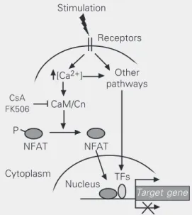
Figure 1. Schematic view of NFAT transcription factor signal-ing pathway in gene expression regulation. NFAT proteins are activated upon different stimuli that lead to increased intracellu-lar calcium levels. When de-phosphorylated by calcineurin, NFAT rapidly translocates to the nucleus where it regulates the expression of several target genes. NFATs may function as transcriptional activators or re-pressors and cooperate with several other transcription fac-tors. Ca2+ = calcium; CaM =
cal-modulin; Cn = calcineurin; CsA = cyclosporin A; NFAT = nuclear factor of activated T cells; P = phosphorylation; TFs = trans-cription factors.
P
CaM/Cn
CsA FK506
Other pathways Receptors Stimulation
[Ca2+]
Cytoplasm
NFAT NFAT
TFs Nucleus
Recently, it has been demonstrated that the promoter of CDK4 presents a consensus-bind-ing site for NFAT proteins and is negatively regulated by NFAT1 without AP-1 coopera-tion (4). This finding demonstrates that NFAT1 directly controls the expression of CDK4 at the transcription level. Furthermore, NFAT1 has been implicated in the control of some cyclin genes, acting as a negative regulator of cyclins A2, E and B1 during lymphocyte acti-vation (3). These results support the idea that the hyperproliferative phenotype observed in NFAT1-/- lymphocytes is related to a deregu-lated cell cycle progression, suggesting that NFAT1 transcription factor plays a central role in controlling cell cycle during T cell activation (3). Several other data also support this evidence. It has been shown that cyclo-sporin A and FK506 inhibit the expression of cyclins A and E in 3T3 fibroblasts (31). An-other evidence for the involvement of NFAT on cell growth has been shown in keratinocyte cells, where cyclosporin A suppressed the expression of p21WAF1/Cip1 and p27KIP1, two
CDK inhibitors (32). Moreover, transactiva-tion of p21 promoter is upregulated by calci-neurin and is dependent of Sp1/Sp3 in syner-gism with NFAT1 and NFAT2 (32). In addi-tion, it has also been demonstrated that over-expression of NFAT2 in preadipocyte 3T3-L1 cells promoted cell cycle progression even under low serum concentrations and induced altered expression of cell cycle-related genes, such as cyclin D1, cyclin D2, pRB, and c-Myc (6). Finally, preliminary data from our labora-tory suggest that NFAT1 directly regulates the expression of cyclin A2 gene (Carvalho LDS and Viola JPB, unpublished observations). This evidence supports the hypothesis that NFAT transcription factors control the expres-sion of cell cycle-related genes and are central regulators of cell cycle progression.
Cell cycle and CD4+ T cell differentiation
Upon primary stimulation, naive CD4+ T
lymphocytes (T helper, Th) differentiate into two classes of effector cells with distinct functional abilities (33). During Th differen-tiation, these cells commit to a specific and mutually exclusive pattern of cytokine se-cretion (33). Th1 cells are characterized by the production of IFN-γ, contributing to cel-lular immune responses against intracelcel-lular pathogens, while Th2 cells produce 4, IL-5 and IL-13, participating on the response against extracellular pathogens (33). Several factors can influence the differentiation path-way of Th cells, specially the conditions prevailing within the microenvironment where T cells encounter antigen (34). How-ever, the molecular basis for the cell-specific and mutually exclusive expression of Th1 or Th2 cytokines is not yet completely defined. Genetic experiments have implicated sev-eral transcription factors in Th cell differen-tiation, including NFAT, STAT4, STAT6, T-Bet and GATA-3. The proteins T-Bet and GATA-3 have been implicated as critical lineage-specific transcription factors which are sufficient to program Th1 and Th2 cy-tokine expression profiles, respectively (35,36). Conversely, gene disruption of STAT4 and STAT6 indicated that these trans-cription factors also play major roles in Th differentiation (37,38). However, in this sec-tion we will focus on reviewing data regard-ing the role of NFAT family proteins in Th differentiation.
NFAT proteins are present in both Th1 and Th2 cells, and can activate cytokine promoters in both cell types (1,15). Also, NFAT transcriptional activity appeared to be similarly regulated during Th1/Th2 de-velopment, although it was enhanced in dif-ferentiated Th2 cells compared with differ-entiated Th1 cells (39). These similarities suggested that the activity of NFAT trans-cription factors were likely to overlap sub-stantially. However, data generated by in vivo gene disruption of the different NFAT
developed normally, and did not exhibit any obvious behavioral deficiencies (40,41). However, NFAT1-/- mice consistently showed a marked increase in their immune response. NFAT1-/- mice showed an en-hanced Th2 development in both in vivo and in vitro models of Th differentiation, as
evi-denced by increased levels of IL-4 produc-tion (40,42,43). Furthermore, cells from lymph nodes and spleen hyperproliferated in response to different antigen stimulations (41). Likewise, CD4+ T cells hyperproliferated
in vitro in response to anti-CD3 (40). On the
other hand, targeted disruption of NFAT2 resulted in an embryonic lethal phenotype (9,15). Complementing RAG-1- or RAG-2-deficient blastocysts with homozygous NFAT2-/- mutant ES cell lines demonstrated that T and B cells from chimeric mice showed reduced proliferation of peripheral lympho-cytes when compared to wild-type mice (44,45). In an in vitro model of Th
differen-tiation, the chimeric mice displayed a de-creased production of IL-4 but normal pro-duction of IFN-γ, demonstrating an impaired Th2 response in NFAT2-/- T cells (44,45). Taken together, the results observed in NFAT-deficient mice demonstrated that these transcription factors play an important role in Th differentiation, and suggest that NFAT1 and NFAT2 have opposite roles in regulat-ing gene expression in Th cell activation and differentiation. However, the molecular ba-sis of these effects remains to be identified. Cell cycle entry is one of the first events that occur during lymphocyte activation. Upon T cell receptor (TCR) activation, naive T lymphocytes, which are in a quiescent stage, proliferate, differentiate and acquire their effector functions. It has been shown that T cell differentiation is dependent on the cell cycle entry (46,47). Although prolifera-tion and differentiaprolifera-tion of CD4+ T cells
oc-cur concomitantly during cellular activation, it is still not clear whether they are effec-tively interconnected. Reiner and colleagues (46) have argued that cell cycle is an
impor-tant regulator of Th differentiation. IL-4 and IFN-γ production by naive CD4+ T cells could only be detected after one and three rounds of cellular division, respectively, be-ing blocked by cell cycle-arrestbe-ing drugs (46). Furthermore, it has been suggested that DNA synthesis is essential for the acquisi-tion of IL-4 producacquisi-tion competence in acti-vated CD4+ T cells, since it is blocked by
specific drugs that prevent cell cycle pro-gression to S phase, but not to G2/M phase (48). In contrast, it has been proposed that cell cycling only increases the frequency of cytokine-secreting cells, showing a correla-tion rather than dependence between cell cycle and differentiation (47,49). In fact, non-cycling cells are capable of IFN-γ secre-tion, suggesting that acquisition of compe-tence to produce IFN-γ can be achieved with-out entering into S phase (49). Other studies have argued that cell cycle progression is fundamental to the commitment of a Th cell with its cytokine-secreting profile, including the heritable activation/silencing of lineage-defining players (50). Actually, the reversi-bility of Th1 and Th2 populations is lost along the increasing number of cellular divi-sions and after long-term stimulation (47). Moreover, it has been shown that T cell differentiation is associated with a dynamic process of histone acetylation and chromatin remodeling at regulatory regions of IL-4 and IFN-γ genes, suggesting that cell cycle could provide a window of opportunity to rear-range genes from an inactive to an active state (47,51).
cycle control, such as cyclin genes, and in Th1/Th2 differentiation, such as IFN-γ and IL-4 cytokines (3). It has been demonstrated that NFAT1 is able to bind to IFN-γ pro-moter regions and to the IL-4 enhancer in T cells stimulated under Th1 or Th2 condi-tions, respectively (51). Since TCR stimula-tion activates an immediate and global chro-matin derepression program in naive T cells, it has been suggested that TCR-inducible transcription factors, such as the preexisting cytoplasmic NFAT1 protein, may bind to regulatory regions of these cytokine genes, initiating localized chromatin remodeling (51). Finally, we indicate here that NFAT proteins regulate central events during lym-phocyte activation, such as cell proliferation and cytokine expression, and may represent an informative model to elucidate the real correlation between cell cycle and Th differ-entiation.
NFAT and malignant cell transformation
As we discussed until here, NFAT pro-teins are involved in a variety of cellular mechanisms, and thus represent broader con-trollers of cell growth and development of normal cells. Several studies have already demonstrated definitive evidence for the in-volvement of NFAT proteins in the regula-tion of cell cycle progression, cell differen-tiation and cell death in different tissues (3,6,52,53). These findings point to NFAT activation as an essential pathway in normal cell physiology, and therefore suggest that deregulation of NFAT signaling may occur during cellular aberrations and tumorigenic processes.
NFAT proteins have been shown to regu-late cell cycle progression by modulating cyclin/CDK gene expression profile (3,4). In mouse models, deficiency of NFAT1 trans-cription factor caused hyperproliferative cel-lular responses, altered cell cycle control, and increased stage-specific cyclin
expres-sion in lymphocytes (3,40,41). NFAT1 pro-tein also repressed the CDK4 gene in T cells, dictating cell cycle re-entry from quiescence (4). Out of the immune system, NFAT1 defi-ciency also led to uncontrolled cell growth and differentiation of the connective tissue and skeletal muscle cells (52,54). These re-sults clearly implicate NFAT1 as a negative controller of early-inducible genes that regu-late cell cycle progression and differentia-tion, and thus suggest a putative tumor sup-pressor role for this protein in different or-gan systems.
process, such as caspases, kinases and trans-cription factors. In fact, NFAT proteins have shown to bind to promoter regions and up-regulate the expression of some effectors of apoptosis, such as TNF-α and FasL (1). It seems that NFAT1 presents a pro-apoptotic role in T lymphocytes since cells lacking this protein showed a decreased expression of FasL and TNF-α and were resistant to apop-tosis (53). Moreover, overexpression of some NFAT members in primary T cells led to increased rates of apoptosis (53). These re-sults thus suggest that NFAT deregulation may rescue normal cells from apoptosis and enable tumor cell survival. Conversely, some reports have also indicated anti-apoptotic roles for NFAT members. Overexpression of the NFAT2 protein in adipocytes pro-tected cells from undergoing apoptosis in response to growth factor withdrawal (6). Mice lacking NFAT4 presented impaired expression of the anti-apoptotic protein Bcl-2 and increased thymocyte apoptosis during T cell development in the thymus (56). Also, NFAT1 showed to rescue lymphocytes from activated-induced cell death upon antigen stimulation in vitro (3). In spite of the
con-troversial data discussed, NFAT proteins are known regulators of apoptotic mechanisms and may take part in tumor development by sustaining cell survival.
Besides indirect evidence, there is plenty of data demonstrating the effective partici-pation of NFAT proteins in tumor-related processes. Several in vitro and also in vivo
reports support NFAT transcription factors as essential targets for better understanding tumor progression pathways. In an elegant work, Neal and Clipstone (6) have investi-gated the role of NFAT2 in tumorigenesis. A constitutively active form of the NFAT2 pro-tein expressed in the murine 3T3-L1 preadi-pocyte cell line was able to induce cell trans-formation and inhibit cell differentiation. Upregulated NFAT2 activity induced the acquisition of well-defined hallmarks of tu-morigenesis, including: 1) loss of
contact-mediated growth inhibition, 2) reduced se-rum growth requirements, 3) protection from apoptosis, 4) formation of colonies in semi-solid media, 5) acquisition of growth au-tonomy, and 6) formation of tumors in athymic nude mice (6). Moreover, authors speculate that sustained NFAT2 activity in vivo could be achieved by the continuous
presence of a soluble promitogenic/prosur-vival factor, which establishes an autocrine regulatory growth loop and leads to cell autonomy. These results provide strong evi-dence for the oncogenic potential of NFAT2 transcription factor and elicit its participa-tion during tumor progression in vivo. Still,
the expression of functional NFAT2 has been also identified in proliferating glioma cells, a nervous system-derived cell line. In line with this, cyclosporin A inhibited the prolifera-tion of these cells and induced apoptotic cell death in a dose-dependent manner (57). Thus, the correlation between the presence of NFAT proteins and the proliferative status of glioma cells might converge to the participation of NFAT transcription factors in the transformed glioma cell phenotype by regulating an auto-crine growth factor stimulation.
human breast and colon cancer. Integrins are extracellular matrix ligand receptors widely described as controllers of tumor invasion and migration. These results have highlighted NFAT proteins as relevant players in pro-moting tumor metastasis.
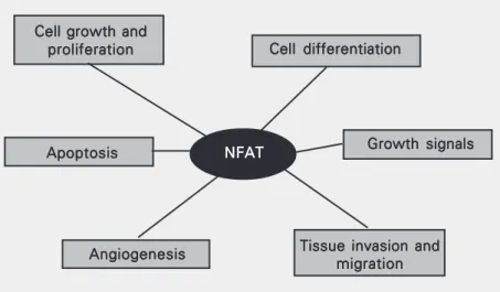
It is evident the involvement of NFAT transcription factors during tumorigenic pro-cesses (Figure 2). In accordance, antagonists of NFAT proteins inhibit tumor formation, supporting an oncogenic potential for these family of transcription factors (59). When binding to promoter regions and regulating gene expression, NFATs have shown to co-operate with several protooncogene prod-ucts, such as c-Fos, c-Jun (AP-1), and Egr
proteins (1,60). These interactions may re-sult in transcriptional activators or repres-sors in different moments (Figure 1). How-ever, the subset of genes regulated by NFAT proteins and its importance during the differ-ent stages of tumor progression remain to be elucidated. Strong effort must focus on clari-fying this still puzzling participation of NFATs during tumorigenesis.
Final remarks
NFAT transcription factors act as ubiqui-tous regulators of gene expression during cellular activation. NFAT proteins have di-verse effects not only on cytokine expres-sion, but also on cell cycle entry and apopto-sis. Cumulative evidence has recently dem-onstrated that NFAT transcription factors exert important roles in the physiology of different cell types, and not only in cells related to the immune system. As such, NFAT has the potential to participate in malignant cell transformation related to cancer. As we discussed here, NFAT proteins regulate genes involved in cell cycle control, cell death and angiogenesis. Given the importance of these phenomena in the development of cell ma-lignancies, it is of considerable interest to understand the mechanisms by which NFAT affects cell growth, differentiation and func-tion. Finally, understanding more about the cell cycle regulation by NFAT proteins is central to precisely indicate the involvement of these transcription factors in malignant cell transformation and oncogenic processes.
Figure 2. Tumor-related processes regulated by NFAT transcription factors. NFAT proteins have shown to directly regulate the expression of genes related to cellular proliferation (e.g., CDK4) and differentiation (e.g., cytokines and growth factors), apoptosis (e.g., FasL), angiogenesis (e.g., cyclooxygenase-2) and tissue invasion mechanisms among others. NFAT = nuclear factor of activated T cells.
References
1. Rao A, Luo C & Hogan PG (1997). Transcription factors of the NFAT family: regulation and function. Annual Review of Immunology, 15: 707-747.
2. Hernández GL, Volpert OV, Íñiguez MA, Lorenzo E, Martínez-Martínez S, Grau R, Fresno M & Redondo JM (2001). Selective inhibition of vascular endothelial growth factor-mediated angiogen-esis by cyclosporin A: roles of the nuclear factor of activated T cells and cyclooxygenase 2. Journal of Experimental Medicine, 193: 607-620.
3. Caetano MS, Vieira-de-Abreu A, Teixeira LK, Werneck MBF, Barcinski MA & Viola JPB (2002). NFATC2 transcription factor regu-lates cell cycle progression during lymphocyte activation: evidence of its involvement in the control of cyclin gene expression. FASEB Journal, 16: 1940-1942.
4. Baksh S, Widlund HR, Frazer-Abel AA, Du J, Fosmire S, Fisher DE, DeCaprio JA, Modiano JF & Burakoff SJ (2002). NFATc2-mediated repression of cyclin-dependent kinase 4 expression. Molecular Cell, 10: 1071-1081.
Cell growth and Cell growth and Cell growth and Cell growth and Cell growth and proliferation proliferation proliferation proliferation
proliferation Cell differentiationCell differentiationCell differentiationCell differentiationCell differentiation
Growth signals Growth signals Growth signals Growth signals Growth signals
Tissue invasion and Tissue invasion and Tissue invasion and Tissue invasion and Tissue invasion and
migration migration migration migration migration Angiogenesis
Angiogenesis Angiogenesis Angiogenesis Angiogenesis
NFAT NFATNFAT NFATNFAT Apoptosis
5. Graef IA, Wang F, Charron F, Chen L, Neilson J, Tessier-Lavigne M & Crabtree GR (2003). Neurotrophins and netrins require calcineu-rin/NFAT signaling to stimulate outgrowth of embryonic axons. Cell, 113: 657-670.
6. Neal JW & Clipstone NA (2003). A constitutively active NFATc1 mutant induces a transformed phenotype in 3T3-L1 fibroblasts.
Journal of Biological Chemistry, 278: 17246-17254.
7. Zaichuk TA, Shroff EH, Emmanuel R, Filleur S, Nelius T & Volpert O (2004). Nuclear factor of activated T cells balances angiogenesis activation and inhibition. Journal of Experimental Medicine, 199:
1513-1522.
8. Shaw JP, Utz PJ, Durand DB, Toole JJ, Emmel EA & Crabtree GR (1988). Identification of a putative regulator of early T cell activation genes. Science, 241: 202-205.
9. Macian F, Lopez-Rodriguez C & Rao A (2001). Partners in transcrip-tion: NFAT and AP-1. Oncogene, 20: 2476-2489.
10. McCaffrey PG, Luo C, Kerppola TK et al. (1993). Isolation of the cyclosporin-sensitive T cell transcription factor NFATp. Science, 262: 750-754.
11. Luo C, Burgeon E, Carew JA, Badalian TM, McCaffrey PG, Lane WS, Hogan PG & Rao A (1996). Recombinant NFAT1 (NFATp) is regulated by calcineurin in T cells and mediates the transcription of several cytokine genes. Molecular and Cellular Biology, 16: 3955-3966.
12. Northrop JP, Ho SN, Chen L, Thomas DJ, Timmerman LA, Nolan GP, Admon A & Crabtree GR (1994). NF-AT components define a family of transcription factors targeted in T-cell activation. Nature, 369: 497-502.
13. Ho SN, Thomas DJ, Timmerman LA, Li X, Francke U & Crabtree GR (1995). NFATc3, a lymphoid-specific NFATc family member that is calcium-regulated and exhibits distinct DNA binding specificity. Jour-nal of Biological Chemistry, 270: 19898-19907.
14. Masuda E, Naito Y, Tokumitsu H, Campbell D, Saito F, Hannum C, Arai K-I & Arai N (1995). NFATx, a novel member of the NFAT family that is expressed predominantly in the thymus. Molecular and Cellular Biology, 15: 2697-2706.
15. Crabtree GR & Olson EN (2002). NFAT signaling: choreographing the social lives of cells. Cell, 109 (Suppl): S-67-S-79.
16. Horsley V & Pavlath GK (2002). NFAT: ubiquitous regulator of cell differentiation and adaptation. Journal of Cell Biology, 156: 771-774. 17. Jain J, Burgeon E, Badalian TM, Hogan PG & Rao A (1995). A similar DNA-binding motif in NFAT family proteins and the Rel homology region. Journal of Biological Chemistry, 270: 4138-4145.
18. Shaw KTY, Ho AM, Raghavan A, Kim J, Jain J, Park J, Sharma S, Rao A & Hogan PG (1995). Immunosuppressive drugs prevent a rapid dephosphorylation of the transcription factor NFAT1 in stimu-lated immune cells. Proceedings of the National Academy of Sci-ences, USA, 92: 11205-11209.
19. Loh C, Shaw KTY, Carew J, Viola JPB, Luo C, Perrino BA & Rao A (1996). Calcineurin binds the transcription factor NFAT1 and revers-ibly regulates its activity. Journal of Biological Chemistry, 271: 10884-10891.
20. Baksh S, DeCaprio JA & Burakoff SJ (2000). Calcineurin regulation of the mammalian G0/G1 checkpoint element, cyclin dependent kinase. Oncogene, 19: 2820-2827.
21. Iniguez MA, Martinez-Martinez S, Punzon C, Redondo JM & Fresno M (2000). An essential role of the nuclear factor of activated T cells in the regulation of the expression of the cyclooxygenase-2 gene in human T lymphocytes. Journal of Biological Chemistry, 275: 23627-23635.
22. Koepp DM, Harper JW & Elledge SJ (1999). How the cyclin became
a cyclin: regulated proteolysis in the cell cycle. Cell, 97: 431-434. 23. Sherr CJ & Roberts JM (1999). CDK inhibitors: positive and negative
regulators of G1-phase progression. Genes and Development, 13: 1501-1512.
24. Ren S & Rollins BJ (2004). Cyclin C/Cdk3 promotes Rb-dependent G0 exit. Cell, 117: 239-251.
25. Ohtani K, DeGregori J & Nevins JR (1995). Regulation of the cyclin E gene by transcription factor E2F1. Proceedings of the National Academy of Sciences, USA, 92: 12146-12150.
26. Murray AW (2004). Recycling the cell cycle: cyclins revisited. Cell, 116: 221-234.
27. Anraku Y, Ohya Y & Iida H (1991). Cell cycle control by calcium and calmodulin in Saccharomyces cerevisiae. Biochimica et Biophysica Acta, 1093: 169-177.
28. Tomono M, Toyoshima K, Ito M & Amano H (1996). Calcineurin is essential for DNA synthesis in Swiss 3T3 fibroblasts. Biochemical Journal, 317: 675-680.
29. Lipskaia L & Lompré AM (2004). Alteration in temporal kinetics of Ca2+ signaling and control of growth and proliferation. Biology of the Cell, 96: 55-68.
30. Terada N, Lucas JJ & Gelfand EW (1991). Differential regulation of the tumor suppressor molecules, retinoblastoma susceptibility gene product (Rb) and p53, during cell cycle progression of normal hu-man T cells. Journal of Immunology, 147: 698-704.
31. Tomono M, Toyoshima K, Ito M, Amano H & Kiss Z (1998). Inhibitors of calcineurin block expression of cyclins A and E induced by fibroblast growth factor in Swiss 3T3 fibroblasts. Archives of Bio-chemistry and Biophysics, 353: 374-378.
32. Santini MP, Talora C, Seki T, Bolgan L & Dotto GP (2001). Cross talk among calcineurin Sp1/Sp3 and NFAT in control of p21WAF1/CIP1
expression in keratinocyte differentiation. Proceedings of the Na-tional Academy of Sciences, USA, 98: 9575-9580.
33. Abbas AK, Murphy KM & Sher A (1996). Functional diversity of helper T lymphocytes. Nature, 383: 787-793.
34. Constant SL & Bottomly K (1997). Induction of Th1 and Th2 CD4+ T
cells response: the alternative approaches. Annual Review of Im-munology, 15: 297-322.
35. Zheng W & Flavell RA (1997). The transcription factor GATA-3 is necessary and sufficient for Th2 cytokine gene expression in CD4 T cells. Cell, 89: 587-596.
36. Szabo SJ, Kim ST, Costa GL, Zhang X, Fathman CG & Glimcher LH (2000). A novel transcription factor T-bet directs Th1 lineage com-mitment. Cell, 100: 655-669.
37. Kaplan MH, Schindler U, Smiley ST & Grusby MJ (1996). Stat6 is required for mediating responses to IL-4 and for development of Th2 cells. Immunity, 4: 313-319.
38. Kaplan MH, Sun YL, Hoey T & Grusby MJ (1996). Impaired IL-12 responses and enhanced development of Th2 cells in Stat4-defi-cient mice. Nature, 382: 174-177.
39. Rincon M & Flavell RA (1997). T-cell subsets: transcriptional control in the Th1/Th2 decision. Current Biology, 7: R729-R732.
40. Hodge MR, Ranger AM, Charles de la Brousse F, Hoey T, Grusby MJ & Glimcher LH (1996). Hyperproliferation and dysregulation of IL-4 expression in NF-ATp-deficient mice. Immunity, 4: 397-405. 41. Xanthoudakis S, Viola JP, Shaw KT, Luo C, Wallace JD, Bozza PT,
43. Viola JPB, Kiani A, Bozza PT & Rao A (1998). Regulation of allergic inflammation and eosinophil recruitment in mice lacking the trans-cription factor NFAT1: Role of interleukin-4 (IL-4) and IL-5. Blood, 91: 2223-2230.
44. Ranger AM, Hodge MR, Gravallese EM, Oukka M, Davidson L, Alt FW, Charles de la Brousse F, Hoey T, Grusby M & Glimcher LH (1998). Delayed lymphoid repopulation with defects in IL-4-driven responses produced by inactivation of NF-ATc. Immunity, 8: 125-134.
45. Yoshida H, Nishina H, Takimoto H et al. (1998). The transcription factor NF-ATc1 regulates lymphocyte proliferation and Th2 cytokine production. Immunity, 8: 115-124.
46. Bird JJ, Brown DR, Mullen AC, Moskowitz NH, Mahowald MA, Sider JR, Gajewski TF, Wang C-R & Reiner SL (1998). Helper T cell differentiation is controlled by the cell cycle. Immunity, 9: 229-237. 47. Grogan JL, Mohrs M, Harmon B, Lacy DA, Sedat JW & Locksley RM (2001). Early transcription and silencing of cytokine genes un-derlie polarization of T helper cell subsets. Immunity, 14: 205-215. 48. Richter A, Löhning M & Radbruch A (1999). Instruction for cytokine
expression in T helper lymphocytes in relation to proliferation and cell cycle progression. Journal of Experimental Medicine, 190: 1439-1450.
49. Ben-Sasson SZ, Gerstel R, Hu-Li J & Paul WE (2001). Cell division is not a “clock” measuring acquisition of competence to produce
IFN-γ or IL-4. Journal of Immunology, 166: 112-120.
50. Mullen AC, Hutchins AS, Villarino AV, Lee HW, High FA, Cereb N, Yang SY, Hua X & Reiner SL (2001). Cell cycle controlling the silencing and function of mammalian activators. Current Biology, 11: 1695-1699.
51. Avni O, Lee D, Macian F, Szabo S, Glimcher L & Rao A (2002). Th cell differentiation is accompanied by dynamic changes in histone
acetylation of cytokine genes. Nature Immunology, 3: 643-651. 52. Ranger AM, Gerstenfeld LC, Wang J, Kon T, Bae H, Gravallese EM,
Glimcher MJ & Glimcher LH (2000). The nuclear factor of activated T cells (NFAT) transcription factor NFATp (NFATc2) is a repressor of chondrogenesis. Journal of Experimental Medicine 191: 9-22. 53. Chuvpilo S, Jankevics E, Tyrsin D et al. (2002). Autoregulation of
NFATc1/A expression facilitates effector T cells to escape from rapid apoptosis. Immunity, 16: 881-895.
54. Horsley V & Pavlath GK (2003). Prostaglandin F2α stimulates growth
of skeletal muscle cells via an NFATC2-dependent pathway. Journal of Cell Biology, 161: 111-118.
55. Musaro A, McCullagh KJ, Naya FJ, Olson EN & Rosenthal N (1999). IGF-1 induces skeletal myocyte hypertrophy through calcineurin in association with GATA-2 and NF-ATc1. Nature, 400: 581-585. 56. Oukka M, Ho IC, De la Brousse FC, Hoey T, Grusby MJ & Glimcher
LH (1998). The transcription factor NFAT4 is involved in the genera-tion and survival of T cells. Immunity, 9: 295-304.
57. Mosieniak G, Figiel I & Kaminska B (1997). Cyclosporin A, an immu-nosuppressive drug, induces programmed cell death in rat C6 glioma cells by a mechanism that involves the AP-1 transcription factor.
Journal of Neurochemistry, 68: 1142-1149.
58. Jauliac S, López-Rodriguez C, Shaw LM, Brown LF, Rao A & Toker A (2002). The role of NFAT transcription factor in integrin-mediated carcinoma invasion. Nature Cell Biology, 4: 540-544.
59. Jiang H, Yamamoto S, Nishikawa K & Kato R (1993). Anti-tumor-promoting action of FK506 a potent immunosuppressive agent.
Carcinogenesis, 14: 67-71.
60. Rengarajan J, Mittelstadt PR, Mages HW, Gerth AJ, Kroczek RA, Ashwell JD & Glimcher LH (2000). Sequential involvement of NFAT and Egr transcription factors in FasL regulation. Immunity, 12: