MicroRNAs in veterinary cardiology
MicroRNAs em cardiologia veterinária Marcela Wolf1* Eloísa Muehlbauer1 Marlos Gonçalves Sousa1
ISSNe 1678-4596
INTRODUCTION
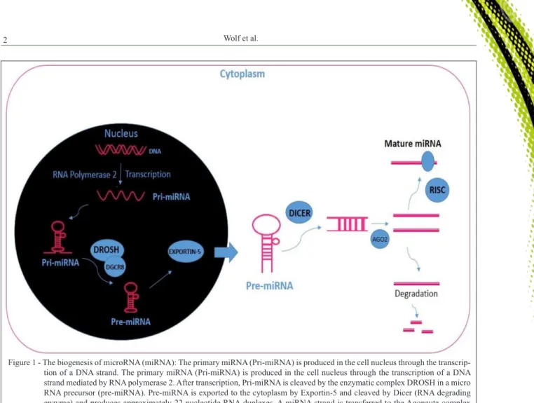
MicroRNAs are small, non-coding, endogenous RNAs that modulate a large and complex set of gene expression. MicroRNAs are potentially involved in a wide range of biological and pathological events. They are composed of approximately 19 to 25 nucleotides (ZHANG et al., 2015), transcribed from intranuclear DNA and transported to the cytoplasm, where a pre-microRNA is cleaved into mature microRNAs (Figure 1) (CREEMERS et al., 2012). Increasingly, new evidence points to the role of microRNAs in the pathogenesis of several diseases. In recent years, there has been an increased interest in genetic and genomic markers of heart diseases
and the evaluation of microRNAs expression may provide information for their use as biomarkers, since such molecules act in many ways on cardiomyocytes, with roles from basic maintenance to the promotion of remodeling in congestive heart failure (IKEDA e PU, 2010; ORENES-PIÑERO et al., 2015).
Given that the pathogenesis of some diseases involves under or overexpression of microRNAs (Table 1), the evaluation of microRNA expression may be used in therapeutic planning. Thus, therapy aims to antagonize microRNAs that are overexpressed and mimic the action of those that are
underexpressed. However, the low specificity of the
gene in question may provide problems for therapeutic targetting (XIAO et al., 2007; ROMAINE et al., 2015). 1Departamento de Medicina Veterinária, Universidade Federal do Paraná (UFPR), 80035-050, Curitiba, PR, Brasil. E-mail: [email protected]. *Corresponding author.
ABSTRACT: The use of biomarkers is an important recent development in veterinary medicine. Biomarkers allow non-invasive quantification
of substances with diagnostic and prognostic potential in several diseases. The microRNAs are small, non-coding RNAs that regulate gene expression and are expressed in different forms in many diseases. Reduced or over-expression of microRNAs showed to be part of the pathogenesis of some heart diseases in humans and animals. Diagnostic and therapeutic value of measuring microRNAs in veterinary cardiology is increased because abnormal expression can be managed by the use of antagonists (in the case of overexpression) and mimicking (in the case of underexpression). Thus, this literature review aimed to compile scientific evidence of dysregulation of microRNAs expression in different cardiac diseases being one of the promises in the therapeutic field and diagnosis of veterinary cardiology. MicroRNAs not only have potential as a biomarker but may also help in elucidation of aspects of the pathogenesis of a variety of diseases.
Key words: molecular biology, genetic, biomarker, heart failure.
RESUMO: Os biomarcadores vêm crescendo na medicina veterinária, pois permitem, de forma não invasiva, a quantificação de substâncias
com potencial para avaliação prognóstica e diagnóstica em diversas doenças. Os microRNAs são pequenos RNAs não codificantes que regulam a expressão de genes e são expressos de diferentes formas em diversas doenças. A sua super ou subexpressão já foi evidenciada como parte da patogênese de cardiopatias em seres humanos e animais. O controle dessa anormalidade de expressão pode ser obtido pela utilização de antagonistas (em casos de superexpressão) e mimetizadores (em casos de subexpressão). Dessa forma, esta revisão de literatura tem como objetivo compilar as evidências científicas da desregulação da expressão de microRNAs nas diferentes doenças cardíacas, sendo essa uma das promessas no campo terapêutico e diagnóstico da cardiologia veterinária, permitindo não só a sua utilização como biomarcador, mas também com aspecto elucidativo da patogênese de diversas doenças.
Palavras-chave: biologia molecular, genética, biomarcador, insuficiência cardíaca.
In veterinary cardiology, the study of new biomarkers is important because in general, the diseases are progressive and many have a genetic basis, such as feline hypertrophic cardiomyopathy (WEBER et al., 2015). The study of the microRNAs can facilitate the understanding of the etiopathogenesis, diagnosis and collaborate with the treatment of these diseases (XIAO et al., 2007; BOSWOOD, 2009). This article reviews the literature to demonstrate the dysregulation of the expression of microRNAs in various cardiac diseases in veterinary medicine and its potential as a biomarker and therapeutic approach.
DEVELOPMENT
Most studies that have evaluated microRNA
expression have performed serum or tissue quantification
(FUJIWARA-IGARASHI et al., 2015; WEBER et al., 2015; DIRKSEN et al., 2016). Interestingly, some studies have tried to measure microRNA expression in other locations including cardiac cells
(ORENES-PIÑERO et al., 2015; POWERS et al., 2015). CHEN et al. (2014), for example, reported that some microRNAs were expressed differently in the left atrium and ventricle
in dogs with CHF, and identified eight microRNAs that were selectively expressed in fibroblasts and
three in cardiomyocytes. Understanding where these microRNAs are located may facilitate understanding of their role in the pathogenesis of various heart conditions.
The possibilities offered by microRNAs make it one of the most exciting areas of modern veterinary medicine. Changes in expression are correlated with pathogenesis of several diseases, such
as viral infections, inflammatory, neoplastic, metabolic
and autoimmune diseases and they may have utility as biomarkers (FUJIWARA-IGARASHI et al., 2015; WEBER et al., 2015). Several cardiac biomarkers have already been shown to be useful in the evaluation of a variety of diseases (BOSWOOD, 2009) and, similarly, the evaluation of microRNA expression in heart diseases may well provide a noninvasive technique for gathering information to aid diagnosis and prognosis which could
lead to the development of new therapeutic protocols. In cardiology, microRNA expression has an important role in the development of heart failure and cardiac remodeling (CONDORELLI et al., 2014). Congestive heart failure (CHF) triggers a series of changes in the expression of various microRNAs, such as 1, 21, 23, 133, 199, 208 and miR-320, but the complexity of the expression system makes understanding the action of individual microRNAs a great challenge(TOPKARA & MANN, 2011).
MicroRNAs have also been used as biomarkers in other areas of veterinary medicine. A
recent study investigated the use of microRNAs, in particular miR-122, as a biomarker to assess hepatocyte damage in Labrador Retriever dogs (DIRKSEN et al., 2016). Evaluation of 5 of 277 microRNAs demonstrated different expression in dogs with lymphoma, with let-7b, miR-223, miR-25 and miR-92a being under expressed while miR-423A was overexpressed compared with the control group (FUJIWARA-IGARASHI et al., 2015).
The study conducted by THUM et al. (2008), demonstrated that the use of miR-21 antagonists in rats with concentric cardiac hypertrophy induced by pressure
overload, significantly reduced remodeling, improving
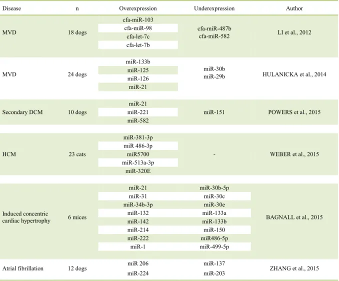
Table 1 - Summary of the literature review evidencing dysregulation of the expression of microRNAs in various diseases.
Disease n Overexpression Underexpression Author
MVD 18 dogs
cfa-miR-103
cfa-miR-487b
cfa-miR-582 LI et al., 2012
cfa-miR-98 cfa-let-7c cfa-let-7b
MVD 24 dogs
miR-133b
miR-30b
miR-29b HULANICKA et al., 2014
miR-125 miR-126
miR-21
Secondary DCM 10 dogs
miR-21
miR-151 POWERS et al., 2015
miR-221 miR-582
HCM 23 cats
miR-381-3p
- WEBER et al., 2015
miR 486-3p miR5700 miR-513a-3p
miR-320E
Induced concentric
cardiac hypertrophy 6 mices
miR-21 miR-30b-5p
BAGNALL et al., 2015
miR-31 miR-30c
miR-34b-3p miR-30e
miR-132 miR-133a
miR-142 miR-133b
miR-214 miR-150
miR-222 miR486-5p
miR-1 miR-499-5p
Atrial fibrillation 12 dogs miR 206
miR-224
miR-137
miR-203 ZHANG et al., 2015
systolic function. Furthermore, histopathologic evaluation of the myocardium demonstrated reduction
of fibrosis in treated animals. MicroRNA mimics can
be used to treat diseases where there is subexpression microRNAs. MicroRNA mimics are synthesized from nucleic acids, and have the same effect as the
microRNAs being mimicked (XIAO et al., 2007).
Despite a promise in veterinary cardiology exists, there are some obstacles to the use of microRNAs in the clinical routine, such as their instability in the sample and a degradation of the molecules, which may affect the results of the expression analyzes. For this, there are several extraction protocols aiming to minimize these effects (EIKMANS et al., 2013).
Degenerative mitral valve disease
Mitral valve disease (MVD) is a degenerative heart disease with a high incidence in small dogs (ATKINS et al., 2009). Although the echocardiographic
and histopathologic findings and some biomarkers
(e.g. natriuretic peptides) have already been well documented, genetic and molecular changes need to be better investigated (REYNOLDS et al., 2012).
Altered expressions of 11 microRNAs have been shown in MVD, and since these changes vary according to disease progression they also pro-vide information on gene regulation in this disease. Symptomatic dogs (American College of Veterinary Internal Medicine (ACVIM) C and D) appear to ex-hibit different gene expression from asymptomatic dogs (ACVIM B1 and B2). In symptomatic animals there is underexpression of microRNAs miR-487b and miR-582 and overexpression of cfa-miR-103, cfa-miR-98, cfa-let-7c and cfa-let-7b (LI et al., 2012). This changes in gene expression may ex-plain the genetic component of disease progression.
Another research, in Dachshunds showed that the expression of microRNAs is different between dogs with MVD in different phases. The miR-30b was under expressed in dogs with class B and C disease when compared to those with class A, while the microRNAs miR-133b, miR-125, miR-126, miR-21, miR-29b and miR-30b were less expressed in dogs with class C disease. These results emphasized the potential of miR-133b as a marker for stage C congestive heart failure (HULANICKA et al., 2014). In another study, that used an experimental model of mitral regurgitation in pigs, levels of miR-21 microRNAs and miR-133a expression were associated with the severity of mitral regurgitation (CIRERA et al., 2013).
There is evidence that the signaling pathway of transforming growth factor β (TGF-β) has an important role in the pathogenesis of MVD (DISATIAN
et al., 2009). MicroRNA 302 inhibits the TGF-βII receptors consequently decreasing the production
of fibronectin and spondine which cause fibrosis.
Reduced expression of miR-302 in disease and its role
in the production of these fibrotic substances supports
the theory of its close involvement in the pathogenesis of MVD (LI et al., 2015). Other microRNAs, such as miR-30 and miR-133 also regulated the action of
TGF, a key molecule in the fibrosis process, and may
represent an attractive target for the treatment of heart disease (HULANICKA et al., 2014).
The study of diagnostic and therapeutic approaches in MVD is extremely important because it is the most common cardiac disease in dogs, and has a progressive nature. The microRNAS are intended to be used as biomarkers, contributing to the diagnostic procedure and a better understanding of the pathophysiology of the disease, and may in the future help in the therapeutic approach with antagonists and agonists substances.
Cardiomyopathies in dogs
Expressions of different microRNAs have also been evaluated in hypertrophic cardiomyopathy and primary and secondary dilated cardiomyopathy (DCM) (STEUDEMANN et al., 2013; JEANSON-LEH et al., 2014; WEBER et al., 2015).
DCM is the primary cardiopathy of large dogs and has high rates of morbidity and mortality (TIDHOLM et al., 2001). Studies in man have shown that several microRNAs are involved in the pathogenesis of DCM and are used as biomarkers for the disease in human patients, such as miR-423-5p microRNA for diagnosis of heart failure secondary to DCM (IKEDA & PU, 2010; FAN et al., 2013). A study with Doberman Pinscher dogs evaluated the expression of 401 microRNAs in serum and reported no differences in expression in any animal with DCM (STEUDEMANN et al., 2013). This may be explained in part by the small number of animals in the study or by the type of sample analyzed. In contrast, several studies in people have used cardiac tissue samples instead of blood samples, since microRNAs are expressed differently in various tissues and cells (SATOH et al., 2010; POWERS et al., 2015).
MicroRNA 208 is encoded by the heavy chain myosin gene and is closely involved in myosin
regulation and myocardial fibrosis, as well as being
compared to the samples from the control group. Levels of α-heavy chain myosin messenger RNA were
lower in DCM patients, whereas the β-heavy chains
levels were higher and were associated with greater volumes of collagen in the myocardium (SATOH et al., 2010). In other research, ZHOU et al. (2010)
observed a significant increase in the risk of dilated
cardiomyopathy in people with overexpression of some pre-microRNAs, as a consequence of common polymorphism of a single nucleotide, including
miR-196a2 and HSA-miR-499, which influenced several
genes with different functions.
An experimental model in dogs with induced dilated cardiomyopathy demonstrated that the expression of 20 microRNAs differed from that in control animals. There were differences in right ventricular (RV) expression of miR-211, miR-221, miR-582 and miR-151 compared to the left ventricle, a fact that may play a fundamental
role in RV insufficiency in CHF (POWERS et al.,
2015). Another study observed the expression of microRNAs in dogs with cardiomyopathy secondary to Duchenne muscular dystrophy. In this disease the 1, 133, 206, 378, 208, miR-208b, miR-499, miR-539 and miR-95 microRNAs are deregulated (JEANSON-LEH et al., 2014).
Feline cardiomyopathy
Hypertrophic cardiomyopathy (HCM) is the most common cardiopathy in cats, although its pathogenesis is not fully understood, it is known to have a strong genetic background. According to WEBER et al. (2015), 49 microRNAs are related to cardiac hypertrophy in feline species. For example, greater expression of miR-381-3p, miR 486-3p, miR5700, miR-513a-3p and miR-320E has already been recognized in cats with stable congestive heart failure secondary to HCM compared to the control group. In contrast, in mice with concentric left ventricle hypertrophy there is a decrease in the expression of miR-1 and miR-133 in the early phase of the disease, while in the terminal phase 16 microRNAs are deregulated. This dysregulation is associated with
cardiac hypertrophy, calcium signaling, fibrosis and
TGF-β signaling pathway (BAGNALL et al., 2015).
Arrhythmias
Heart failure results in remodeling of the atrium and left ventricle, creating substrates for the appearance
of arrhythmias. Atrial fibrillation (AF) is a common
arrhythmia in dogs and is histologically associated with
atrial fibrosis (LI et al., 2012). In arrhythmias the expression
of microRNAs can also be altered. Changes in miR-1 for
example, may lead to the development of arrhythmias, because miR-1 is closely related to genes regulating the ion channels and alterations in its expression may alter the ionic balance. Other microRNAs, such as miR-21, are overexpressed in cardiac remodeling, and miR-208
and miR-29, which are expressed primarily in fibroblasts,
also have arrhythmogenic potential (ORENES-PIÑERO et al., 2015).
According to ZHANG et al. (2015), micro-cfa-miR 206 is overexpressed 9.5 times more in dogs with AF when compared to dogs without arrhythmia. Expression of 16 other microRNAs is also dysregulated in dogs with AF. Another study that included 42 dogs showed that miR-133 and miR-30, which are
antifibrotic markers, were underexpressed in dogs with atrial fibrillation (LI et al., 2012). In human patients,
RADTKE et al. (2014) showed that microRNA 208 is
associated with fibrosis of the conduction tissue and
allows the clinical categorization of individuals with
atrial fibrillation, due to the observation of similar levels
of microRNA 208 in individuals with paroxysmal AF
and longstanding persistent AF, whereas significantly
higher levels were found in patients with persistent AF. In 260 dogs with pacemaker-induced ventricular tachycardia, the evaluation of microRNAs in the atrial and ventricular extracellular matrix revealed alterations in the expression of 99 microRNAs in the left atrium and 36 in the left ventricle. There was lower expression of miR-1 in the atrial myocardium in animals with tachycardia when compared to the control group, although no alteration in its expression
was identified in the left ventricle, nor in expression of
miR-26, miR-29b and miR-30a (CHEN et al., 2014). The different expression of the microRNAs in arrhythmias can facilitate the understanding of the origin, duration and severity of these rhythm disturbances, besides having potential to clarify unknown pathways of arrhythmogenesis.
CONCLUSION
The understanding of the mechanisms involved in the expression of microRNAs and their role in the pathogenesis of the various cardiopathies
is a promising new field in veterinary medicine. As
involved extraction and quantification. It is likely that
further studies will extend the application of these molecules in veterinary cardiology.
ACKNOWLEDGEMENTS
The authors are thankful for the financial support
from Coordenação de Aperfeiçoamento de Pessoal de Nível Superior (CAPES).
REFERENCES
ATKINS, C. et al. Guideline for the diagnosis and treatment of canine chronic valvular heart disease. Journal of Veterinary Internal
Medicine, v.23, n.6, p.1142-1150, 2009. Available from: <http://
onlinelibrary.wiley.com/doi/10.1111/j.1939-1676.2009.0392.x/full>. Accessed: Sept. 14, 2016. doi: 10.1111/j.1939-1676.2009.0392.x.
BAGNALL, R.D. et al. Global microRNAs profiling of the
mouse ventricles during development of severe hypertrophic cardiomyopathy and heart failure. PloS one, v.7, n.9, p.1-8, 2012. Available from: <http://dx.doi.org/10.1371/journal.pone.0044744>. Accessed: Sept. 14, 2016. doi: 10.1371/journal.pone.0044744.
BOSWOOD, A. Biomarkers in cardiovascular disease: beyond natriuretic peptides. Journal of Veterinary Cardiology, v.11, p.s23-s32, 2009. Available from: <http://dx.doi.org/10.1016/j.jvc.2009.01.003>. Accessed: Sept. 14, 2016. doi: 10.1016/j.jvc.2009.01.003.
CHEN, Y. et al. Detailed characterization of microRNA changes in a canine heart failure model: relationship to arrhytmogenic structural remodeling. Journal of Molecular and Cellular
Cardiology, v.77, p.113-124, 2014. Available from: <http://dx.doi.
org/10.1016/j.yjmcc.2014.10.001>. Accessed: Sept. 14, 2016. doi: 10.1016/j.yjmcc.2014.10.001.
CIRERA, S. et al. Plasma proANP and SDMA and microRNAs are associated with chronic mitral regurgitation in a pig model.
Endocrine connetions, v.2, p.161-171, 2013. Available from:
<http://www.endocrineconnections.com/content/2/3/161.short>. Accessed: Sept. 14, 2016. doi: 10.1530/EC-13-0051.
CONDORELLI, G. et al. MicroRNAs in cardiovascular diseases: current knowledge and the road ahead. Journal of the American
College of Cardiology, v.63, p.2177-2187, 2014. Available from:
<http://content.onlinejacc.org/article.aspx?articleid=1838321>. Accessed: Sept. 14, 2016. doi: 10.1016/j.jacc.2014.01.050.
CREEMERS, E.E. et al. Circulating MicroRNAs: novel biomarkers and extracellular communications in cardiovascular disease?
Circulation, v.110, p.483-495, 2012. Available from: <http://dx.doi.
org/10.1161/CIRCRESAHA.111.247452>. Accessed: Sept. 14, 2016. doi: 10.1161/CIRCRESAHA.111.247452.
DIRKSEN, K. et al. Hepatocyte-derived microRNAs as sensitive serum biomarkers of hepatocellular injury in Labrador retrievers.
Veterinary Journal, v.211, p.75-81, 2016. Available from: <http://
dx.doi.org/10.1016/j.tvjl.2016.01.010>. Accessed: Sept. 14, 2016. doi: 10.1016/j.tvjl.2016.01.010.
DISATIAN, S.; ORTON, E.C. Autocrine serotonin and transforming growth factor beta 1 signaling mediates spontaneous myxomatous mitral valve disease. Journal of Heart Valve Disease, v.18, p.44-51, 2009. Available from: <http://journals.plos.org/plosone/
article?id=10.1371/journal.pone.0042527>. Accessed: Sept. 14, 2016. doi: 10.1371/journal.pone.0042527.
EIKMANS, M. et al. Blood cell mRNAs and microRNAs: optimized protocols for extraction and preservation. Blood Journal, 1.ed. 2013. Available from: <http://www.bloodjournal.org/content/bloodjournal/ early/2013/01/17/blood-2012-06-438887.full.pdf>. Accessed: Jan. 22, 2017. doi: 10.1182/blood-2012-06-438887.
FAN, K. et al. Circulating microRNAs levels in Chinese heart failure patients caused by dilated cardiomyopathy. Indian Heart Journal, v.65, p.12-16, 2013. Available from: <http://dx.doi.org/10.1016/j. ihj.2012.12.022>. Accessed: Oct. 18, 2016. doi: 10.1016/j.ihj.2012.12.022.
FUJIWARA-IGARASHI, A. et al. Expression profile of circulating
serum microRNAs in dogs. Veterinary Journal, v.205, p.317-321, 2015. Available from: <http://dx.doi.org/10.1016/j.tvjl.2015.04.029>. Accessed: Sept. 14, 2016. doi: 10.1016/j.tvjl.2015.04.029.
HULANICKA, M. et al. Plasma miRNAs as potential biomarkers of chronic degenerative valvular disease in Dachshunds. BMC
Veterinary Research, v.10, p.1, 2014. Available from: <http://
bmcvetres.biomedcentral.com/articles/10.1186/s12917-014-0205-8>. Accessed: Sept. 14, 2016. doi: 10.1186/s12917-014-0205-8.
IKEDA, S.; PU, W.T. Expression and function of MicroRNAs in heart disease. Current Drug Targets, v.11, n.8, p.913-925, 2010. Available from: <http://www.ncbi.nlm.nih.gov/pubmed/20415651>. Accessed: Sept. 14, 2016. doi: 1389-4501/10 $55.00+.00.
JEANSON-LEH, L. et al. Serum profiling identifies novel muscle
miRNA and cardiomyopathy-related miRNA biomarkers in golden retriever muscular dystrophy dogs and duchenne muscular dystrophy patients. American Journal of Pathology, v.184, n.11, p.2885-2898, 2014. Available from: <http://dx.doi.org/10.1016/j.ajpath.2014.07.021>. Accessed: Sept. 14, 2016. doi: 10.1016/j.ajpath.2014.07.021.
LI, H. et al. Expression of miR-133 and miR-30 in chronic atrial fibrillation
in canines. Molecular Medicine Reports, v.5, p.1457-1460, 2012. Available from: <https://www.spandidos-publications.com/10.3892/ mmr.2012.831>. Accessed: Sept. 14, 2016. doi: 10.3892/mmr.2012.831.
ORENES-PIÑERO, E. et al. Novel biomarkers in cardiology:
microRNAs in atrial fibrillation. Archivos de Cardiología de
México, v.85, n.3, p.225-229, 2015. Available from: <http://www.
ncbi.nlm.nih.gov/pubmed/25957926>. Accessed: Sept. 14, 2016. doi: 10.1016/j.acmx.2015.01.005.
POWERS, J. et al. Differential interventricular expression of microRNAs-21, -93, -151, -221 and -582, in dilated cardiomyopathy with biventricular dysfunction. Journal of the American College
of Cardiology, v.65, n.106, p.A904 2015. Available from: <http://
content.onlinejacc.org/article.aspx?articleid=2198513>. Accessed: Sept. 14, 2016. doi: 10.1016/S0735-1097(15)60904-4.
RADTKE, A. et al. MicroRNA 208 in atrial fibrillation. Clinical
& Experimental Cardiology, v.5, n.6, p.1-4, 2014. Available
from:
<http://www.omicsonline.org/open-access/microrna-in-atrial-fibrillation-2155-9880-5-325.php?aid=28182>. Accessed: Sept. 14, 2016. doi: 10.4172/2155-9880.1000325.
REYNOLDS, C.A. et al. Prediction of first onset of congestive heart
ROMAINE, S.P.R. et al. MicroRNAs in cardiovascular disease: an introduction for clinicians. Heart, v.101, p.921-928, 2015. Available from: <http://heart.bmj.com/content/101/12/921.short>. Accessed: Sept. 14, 2016. doi: 10.1136/heartjnl-2013-305402.
SATOH, M. et al. Expression of microRNAs 208 is associated with adverse clinical outcomes in human dilated cardiomyopathy. Journal
of Cardiac Failure, v.16, n.5, p.404-410, 2010. Available from:
<http://dx.doi.org/10.1016/j.cardfail.2010.01.002>http://dx.doi. org/10.1016/j.cardfail.2010.01.002. Accessed: Sept. 14, 2016. doi: 10.1016/j.cardfail.2010.01.002.
STEUDEMAN, C. et al. Detection and comparison of microRNAs expression in the serum of Doberman Pinchser with dilated cardiomiopathy and healthy controls. BMC Veterinary Research, v.9, n.12, p.1-14, 2013. Available from: <http://bmcvetres.biomedcentral. com/articles/10.1186/1746-6148-9-12>. Accessed: Sept. 14, 2016. doi: 10.1186/1746-6148-9-12.
THUM, T. et al. MicroRNA-21 contributes to myocardial disease
by stimulating MAP kinase signaling in fibroblast. Nature, v.456, p.980-984, 2008. Available from: <http://www.nature.com/nature/ journal/v456/n7224/abs/nature07511.html>. Accessed: Sept. 14, 2016. doi: 10.1038/nature07511.
TIDHOLM, A. et al. Canine idiopathic dilated cardiomyopathy. Part I: aetiology, clinical characteristics, epidemiology and pathology.
Veterinary Journal, v.162, n.2, p.92-107, 2001. Available from: <http://
www.sciencedirect.com/science/article/pii/S1090023301905714>. Accessed: Sept. 14, 2016. doi: 10.1053/tvjl.2001.0571.
TOPKARA, V.K.; MANN, D.L. Role of microRNAs in cardiac remodeling and heart failure. Cardiovascular Drugs Therapy, v.25, p.171-182, 2011. Available from: <http://link.springer.com/ article/10.1007/s10557-011-6289-5>. Accessed: Sept. 14, 2016. doi: 10.1007/s10557-011-6289-5.
WEBER, K. et al. Serum microRNA profiles in cats with
hypertrophic cardiomyopathy. Molecular and Cellular
Biochemistry, v.402, p.171-180, 2015. Available from: <http://
link.springer.com/article/10.1007/s11010-014-2324-8>. Accessed: Sept. 14, 2016. doi: 10.1007/s11010-014-2324-8.
XIAO, J. et al. Novel approaches for gene-specific interference via
manipulating actions of microRNAs: examination on the pacemaker channel genes HCN2 and HCN4. Journal of Cellular Physiology, v.212, n.2, p.285-292, 2007. Available from: <https://www.ncbi.nlm. nih.gov/pubmed/17516552http://onlinelibrary.wiley.com/doi/10.1002/ jcp.21062/full>. Accessed: Sept. 14, 2016. doi: 10.1002/jcp.21062.
ZHANG, Y. et al. MicroRNA profiling of atrial fibrillation in canines:
MiR-206 modulates intrinsic cardiac autonomic nerve remodeling by regulating SOD1. PloS one,v.10, n.3, p.1-16, 2015. Available from: <http://dx.doi.org/10.1371/journal.pone.0122674>. Accessed: Sept. 14, 2016. doi: 10.1371/journal.pone.0122674.
ZHOU, B. et al. Common genetic polymorphisms in pre-microRNAs were associated with increased risk of dilated cardiomyopathy.
Clinica Chimica Acta, v.411, n.17, p.1287-1290, 2010. Available