dimensional-ultrasound in primiparous women
according to delivery mode: initial experience
from a single reference service in Brazil
Avaliação do assoalho pélvico por meio da ultrassonografia tridimensional de
mulheres primíparas de acordo com o tipo de parto: experiência inicial de um
centro de referência do Brasil
rogério CaixEta MoraEs dE FrEitas2
ZsuZsanna ilona Katalin dE járMy di BElla2
sandra Maria alExandrE3
Mary uChiyaMa naKaMura3
luCiano MarCondEs MaChado nardoZZa1
antonio FErnandEs Moron4
Abstract
PURPOSE: To evaluate changes to the pelvic loor of primiparous women with different delivery modes, using three-dimensional ultrasound. METHODS: A prospective cross-sectional study on 35 primiparae divided into groups according to the delivery mode: elective cesarean delivery (n=10), vaginal delivery (n=16), and forceps delivery (n=9). Three-dimensional ultrasound on the pelvic loor was performed on the second postpartum day with the patient in a resting position. A convex volumetric transducer (RAB4-8L) was used, in contact with the large labia, with the patient in the gynecological position. Biometric measurements of the urogenital hiatus were taken in the axial plane on images in the rendering mode, in order to assess the area, anteroposterior and transverse diameters, average thickness, and avulsion of the levator ani muscle. Differences between groups were evaluated by determining the mean differences and their respective 95% conidence intervals. The proportions of levator ani muscle avulsion were compared between elective cesarean section and vaginal birth using Fisher’s exact test.RESULTS: The mean areas of the urogenital hiatus in the cases of vaginal and forceps deliveries were 17.0 and 20.1 cm2, respectively, versus 12.4 cm2 in the Control Group
(elective cesarean). Avulsion of the levator ani muscle was observed in women who underwent vaginal delivery (3/25), however there was no statistically signiicant difference between cesarean section and vaginal delivery groups (p=0.5).
CONCLUSION: Transperineal three-dimensional ultrasound was useful for assessing the pelvic loor of primiparous women, by allowing pelvic morphological changes to be differentiated according to the delivery mode.
Resumo
OBJETIVO: Avaliar as mudanças no assoalho pélvico de mulheres primíparas em diversos tipos de partos por meio da ultrassonograia tridimensional.MÉTODOS: Estudo de corte transversal prospectivo com 35 primigestas, divididas em grupos com relação ao tipo de parto: cesariana eletiva (n=10), parto vaginal (n=16) e fórceps (n=9). A ultrassonograia tridimensional do assoalho pélvico foi realizada no segundo dia pós-parto com a paciente em repouso. Utilizou-se transdutor convexo volumétrico (RAB4-8L) em contato com os grandes lábios vaginais, estando a paciente em posição ginecológica. Medidas biométricas do hiato urogenital foram tomadas no plano axial da imagem renderizada para avaliar a área, os diâmetros anteroposterior e transverso, a espessura média e a avulsão do músculo elevador do ânus. Diferenças entre os grupos foram avaliadas pela determinação da média das diferenças com seus respectivos intervalos de coniança de 95%. As proporções de avulsão do músculo elevador do ânus foram comparadas entre a cesárea eletiva e o parto vaginal pelo teste exato de Fisher. RESULTADOS: As áreas médias do hiato urogenital dos partos vaginais e fórceps foram 17,0 e 20,1 cm2, respectivamente, contra 12,4 cm2 do Grupo Controle (cesárea eletiva).
Avulsão do músculo elevador do ânus foi observado em mulheres submetidas ao parto vaginal (3/25); no entanto, não houve diferença signiicativa entre os grupos cesárea e parto vaginal (p=0,5). CONCLUSÃO: A ultrassonograia tridimensional por via perineal foi útil na avaliação do assoalho pélvico de mulheres primíparas, diferenciando alterações pélvicas de acordo com o tipo de parto.
Study carried out at the Department of Obstetrics, Universidade Federal de São Paulo – UNIFESP – São Paulo (SP), Brazil.
1Fetal Medicine Discipline, Department of Obstetrics, Universidade Federal de São Paulo – UNIFESP – São Paulo (SP), Brazil.
2Department of Gynecology, Universidade Federal de São Paulo – UNIFESP – São Paulo (SP), Brazil.
3Physiological and Experimental Obstetrics Discipline, Department of Obstetrics, Universidade Federal de São Paulo – UNIFESP – São
Paulo (SP), Brazil.
4Department of Obstetrics, Universidade Federal de São Paulo – UNIFESP – São Paulo (SP), Brazil.
Conlict of interest: none
Keywords
Pelvic loor/ultrasonography Parity Natural childbirth Cesarean section Imaging, three-dimensional
Palavras-chave
Diafragma da pelve/ultrassonograia Paridade Parto normal Cesárea Imagem tridimensional
Correspondence
Edward Araujo Júnior Rua Napoleão de Barros, 875 – Vila Clementino Zip code: 04024-002 São Paulo (SP), Brazil
Received
01/07/2013
Accepted with modiications
Introduction
Over recent years, because of greater stimulation aimed at increasing the vaginal delivery rates in many countries, discussion about its potential negative effects on the pelvic loor is becoming more widely disseminated. On the other hand, performing cesarean section without any formal indication may contribute towards increased maternal and neonatal morbidity and mortality, even though this is associated with lower need for corrective surgery for prolapse or incontinence, and it protects against prolapse symptoms1.
With regard to some pelvic loor alterations, it is unclear whether pregnancy or delivery is the real predis-posing factor1. Nevertheless, epidemiological evidence for an association between vaginal delivery, prolapse, and urinary incontinence exists currently. It remains unclear whether pelvic loor lesions due to vaginal delivery are caused by strain or avulsion, and whether the changes observed are primary (directly resulting from delivery) or are medium and long-term consequences of damage to the levator ani muscle2. Several mechanisms may coexist in the same woman. The risk factors are operative vaginal delivery, prolonged second stage, and possibly high-birth weight. However, the extent of the trauma clearly varies from one woman to another2.
Three-dimensional (3D) ultrasonography provides images similar to those obtained using magnetic reso-nance imaging (MRI). It has the capacity for image postprocessing and improved standardization of the evaluation and measurement planes3, along with proven reproducibility of its measurements4. Several studies us-ing 3D ultrasonography in order to evaluate predictions related to pelvic loor lesions during or after delivery have recently been published5-12. However, only two of them evaluated the inluence of the delivery mode on predic-tions of pelvic loor lacerapredic-tions during the immediate postpartum period8,11.
Due to the importance of evaluating the integrity of the pelvic loor during the postpartum period, as a means of predicting the future risk of disorders such as genital prolapses, we have presented here our initial experience at our service, concerning postpartum evaluation of the pelvic loor by means of transperineal 3D ultrasonogra-phy, with comparisons between different delivery modes.
Methods
This was a cross-sectional study conducted on 37 primiparous women who gave birth at the São Paulo Hospital, Federal University of São Paulo (UNIFESP), between October 2010 and January 2011. The study was approved by the Research Ethics Committee of UNIFESP,
and the patients who volunteered to participate signed an informed consent form. The participants were divided into three groups, according to delivery type: elective cesarean, vaginal delivery (with or without episiotomy), or forceps delivery. To meet the inclusion criteria, the patients had to be 18 years of age or older, primiparous, and with a single pregnancy and live birth. The exclusion criteria were the following: newborns with structural abnormalities or chromosome disorders; prematurity (under 37 weeks); nonselective cesarean section during labor; time period of more than 48 hours between the ultrasound examination and the birth; pain symptoms that imposed limits on the ultrasound scan; and low-quality ultrasound images that prevented adequate evaluation of the parameters.
The maternal parameters t analyzed were: mother’s
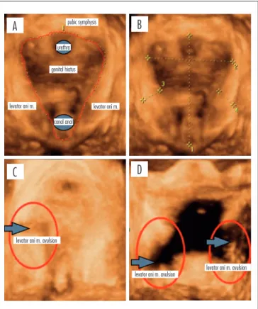
age, body mass index (BMI), delivery mode, and ges-tational age at delivery. The fetal parameters evaluated included gender, birth weight, and head circumference. The biometric variables of the pelvic loor that were taken into consideration were area and anteroposterior and transverse diameters of the urogenital hiatus; aver-age thickness of the levator ani muscle, and echographic signs of levator avulsion. The hiatal area was measured on the plane of minimum hiatal dimensions, which was referenced as midsagittal, comprising the area between the posterior region of the pubic symphysis and the anterior and posterior borders of the muscles and of the levator ani, including only the anorectal muscle. This transverse section in the axial plane enables measurements of the hiatal dimension, such as area (Figure 1A) and transverse and anteroposterior diameters (Figure 1B), as described by Dietz et al.13. The mean thickness of the levator ani muscle was deined in the axial plane as the mean of the levator ani thicknesses measured bilaterally (Figure 1B). The echo-graphic sign of levator avulsion was stipulated as a loss of continuity between the muscle and the pelvic sidewall, as obtained in the axial plane (Figures 1C and D) and shows a schematic image of the anatomical structures of the female pelvic loor.
levator ani m. avulsion levator ani m. avulsion levator ani m. avulsion
genital hiatus urethra
canal anal
levator ani m. levator ani m.
pubic symphysis
from right to left, a view of the pubic symphysis, blad-der neck, urethra, vaginal length, and distal portion of the rectum with the anorectal junction and the proximal part of the anal canal.
The opening angle was standardized to 70º in the sagit-tal plane and 75º in the axial one. After automatic scanning (four seconds), the image was displayed on the screen in the multiplanar (axial, sagittal, and coronal planes) and rendering modes. The sagittal plane was selected as the reference as to obtain measurements of the chosen parameters. The green line (region of interest, ROI) was placed in the upper portion of the sagittal plane, in order that all the pelvic loor structures became visible in the rendering image. Three volumes were acquired for each patient and stored in the memory of the machine. Subsequently, the volume with the highest deinition image quality was selected for off-line analysis, which was then transferred to a personal computer, and the parameters were analyzed by the same examiner (RCMF) using version 9.0 of the 4D Viewsoftware (General Electric Medical Systems, Zipf, Austria). At the time of parameters’ analysis, the examiner did not have any access to postnatal data.
Figure 1. Axial plane of the female pelvic floor on the second postpartum day in rendering mode. (A) measurement of the hiatal area; (B) anteroposterior diameter (measurement 1), transverse di-ameter (measurement 2), mean thickness of the bilateral levator ani muscles (measurements 3 and 4); (C) avulsion of the unilateral levator ani muscle (red circle and blue arrow); (D) avulsion of the bilateral leva-tor ani muscles (red circles and blue arrows).
The sample size calculation was based on data published by Falkert et al.6. Considering that the estimated hiatal area is 16.2±3.2 cm2 for women undergoing cesarean section, and 22.2±4.7 cm2 for those undergoing vaginal birth, evaluations on a total of nine subjects per group would be required in order to have a statistical power of 90% and to identify this difference.
Data were written down using a speciic protocol, trans-ferred to an Excel 2003 spreadsheet (Microsoft, Redmond, WA, USA), and analyzed using version 13.0 for Windows of the Statistical Package for the Social Sciences – SPSS (SPSS Inc., Chicago, IL, USA). The quantitative variables were subjected to the Kolmogorov-Smirnov’s test to check for normal distribution. Differences between groups were evaluated by determining the mean differences and their respective 95% conidence intervals (95%CI). The proportions of levator ani muscle avulsion were compared between elective cesarean section and vaginal birth by means of Fisher’s exact test. The signiicance level was set at 5% (p<0.05).
Results
Thirty-seven primiparous patients with a single preg-nancy underwent pelvic loor evaluation by means of 3D ultrasound on the second postpartum day. Two of them were excluded from the study: one due to signiicant pain in the episiotomy scar at the time of volume measurement, and the other because of preterm labor (<37 weeks). The remaining 35 patients were allocated to three groups according to the delivery mode, as follows: elective cesarean delivery (n=10), vaginal delivery (n=16) and forceps delivery (n=9). Episiotomy was performed in only three women. The quantitative variables tested for normal distribution using Kolmogorov-Smirnov’s test did not depart from normality (p>0.05). Considering the total sample, the maternal age was 24.5±6.3 years-old (mean±standard deviation – SD), BMI was 27.3±4.5 kg/m², gestational age at delivery was 38.9±1.3 weeks, head circumference was 34.6±1.0 cm, and birth weight was 3.251±418 g.
When comparing the ultrasound measurements on the pelvic loor among the groups (Table 1), it was observed that women who underwent elective cesarean section had a smaller hiatal area and anteroposterior di-ameter than those that underwent either non-forceps or forceps-assisted vaginal delivery.
Avulsion of the levator ani muscle was observed only in women who underwent vaginal delivery: 3/25 women (one case in the non-forceps group and two in the forceps group). However, no signiicant differences between elective cesarean section and vaginal delivery were observed (p=0.54). A
C D
Discussion
The preliminary data from this study indicate that there are signiicant differences in the changes to the pelvic loor corresponding to the delivery mode. The results show that women who had a vaginal or a forceps delivery presented greater biometric parameters than those who had an elective cesarean section. Similar data have been reported in several other published papers, in which it is stated that the hiatal area is directly inluenced by age, parity, and pelvic organ prolapse14. DeLancey et al.15 reported that alterations to the muscles are most likely caused by vaginal delivery and are more evident in both forceps- and vacuum-assisted deliveries16. Their data are compatible with our indings, as demonstrated by a larger hiatal area in the forceps delivery group and a higher maternal mean age.
In turn, macrotrauma may be responsible for more pronounced changes to the pelvic loor structure. It can be observed that levator ani muscle avulsion alters the anatomical V shape of the posterior pelvic loor structure to an H one, thus further increasing the dimensions of the pelvic hiatal area17. Data from our study showed that such increase in pelvic hiatal area occurred primarily in the forceps delivery group, in which the muscle avulsion rate was higher. Our results indicated that there was an increase of up to 37% for vaginal delivery and 62% for
forceps one, compared with the Control Group at rest. From correlating our results with those reported in the literature, it was observed that the increase in urogenital hiatus area in the literature ranged from 13 to 37% for vaginal delivery and from 28 to 39% for forceps, with the patients at rest. However, it was unclear whether the Control Groups comprised elective cesarean section cases. We believe that the signiicant difference between our results and those in the literature for the forceps group is partly due to the small number of cases in this group and to the avulsion and maternal age rates, which pushed our results up higher. Developing this study will help us to come up with more concrete answers in the future and thus better compare our data from a miscegenated population with those published in the worldwide literature.
There was a 12% avulsion rate among patients postvaginal and forceps deliveries taken together, cor-responding to 22.2% for the forceps delivery group alone and 6.2% for the non-forceps vaginal deliveries. These data are consistent with those in the literature, which show an avulsion rate of 10 to 30% for vaginal deliveries18. It should be emphasized that the incidence of avulsion has been reported to be up to 63.3% for rotational forceps deliveries6,and 47% for surgical deliveries among patients over 30 years of age19. Other studies have indicated that forceps use correlates with a three to fourfold greater risk of macrotrauma of the pelvic structures (levator ani muscle avulsion)5,18,20.
Table 1. Pelvic loor measurements from three-dimensional ultrasonography
MD: mean difference; 95%CI: 95% conidence interval; *statistically signiicant difference in the comparison; – no statistically signiicant difference in the comparison.
Elective C-section Vaginal
n=10 Non-forceps (n=16) Forceps (n=9)
Mean SD Mean SD Mean SD
Hiatal area (cm²) 12.4 1.6 17.0 2.1 20.1 3.2
Hiatal anteroposterior diameter (cm) 5.0 0.4 6.2 0.4 6.5 0.9
Hiatal transverse diameter (cm) 4.0 0.3 4.2 0.5 4.6 0.5
Levator ani thickness (mm) 7.5 0.8 8.4 10 8.3 0.8
Pairwise comparison
MD 95%CI Signiicance
Hiatal area (cm²)
Elective C-section versus vaginal (non-forceps) -4.6 -7.0– -2.2 *
Elective C-section versus vaginal (forceps) -7.7 -10.4– -5.0 *
Vaginal (non-forceps) versus vaginal (forceps) -3.1 -5.6– -0.6 *
Hiatal anteroposterior diameter (cm)
Elective C-section versus vaginal (non-forceps) -1.1 -1.7– -0.5 *
Elective C-section versus vaginal (forceps) -1.5 -2.2– -0.8 *
Vaginal (non-forceps) versus vaginal (forceps) -0.4 -1.0–0.2 –
Hiatal transverse diameter (cm)
Elective C-section versus vaginal (non-forceps) -0.2 -0.7–0.3 –
Elective C-section versus vaginal (forceps) -0.6 -1.2– -0.1 *
Vaginal (non-forceps) versus vaginal (forceps) -0.4 -0.9–0.1 –
Levator ani thickness (mm)
Elective C-section versus vaginal (non-forceps) -0.9 -1.8–0.0 –
Elective C-section versus vaginal (forceps) -0.8 -1.8–0.2 –
1. MacLennan AH, Taylor AW, Wilson DH, Wilson D. The prevalence of pelvic loor disorders and their relationship to gender, age, parity and mode of delivery. BJOG. 2000;107(12):1460-70. 2. Dietz HP. Pelvic loor trauma following vaginal delivery. Curr Opin
Obstet Gynecol. 2006;18(5):528-37.
3. Valsky DV, Yagel S. Three-dimensional transperineal ultrasonography of the pelvic loor: improving visualization for new clinical applications and better functional assessment. J Ultrasound Med. 2007;26(10):1373-87.
4. Majida M, Braekken IH, Umek W, Bø K, Saltyte Benth J, Ellstrøm Engh M. Interobserver repeatability of three- and four-dimensional transperineal ultrasound assessment of pelvic loor muscle anatomy and function. Ultrasound Obstet Gynecol. 2009;33(5):567-73.
5. Krofta L, Otcenásek M, Kasiková E, Feyereisl J. Pubococcygeus-puborectalis trauma after forceps delivery: evaluation of the levator ani muscle with 3D/4D ultrasound. Int Urogynecol J Pelvic Floor Dysfunct. 2009;20(10):1175-81.
Furthermore, other risk factors such as the occiput posterior position during the second stage of labor, prolonged expulsion period and episiotomy may contribute towards avulsion. However, the role of such factors is still rather unclear and not as well-established as that of forceps delivery17. Shek and Dietz17 attempted to determine antenatal predictive factors that could signal an increased risk of pelvic floor avulsion, but they did not find any factors. Curiously, these authors found that patients with a BMI less than 30 kg/m2 had a greater chance of avulsion, and they hypothesized that nutritional status was related to this protective biomechanical effect of the pelvic muscles. However, their finding is still considered inconclusive17,21. Despite the small number of cases included in this study, 66% of the patients with levator ani muscle avulsion had a BMI less than 30 kg/m2, which coincide with the already presented data. Our results are the opposite of what was found when studying urinary incontinence mechanisms related to maternal overweight, i.e., that there is a direct relationship between weight and the chance that the patient will be incontinent. The latter finding seems obvious, since there is a different pathophysiological mechanism for this situation22.
The present study shows that there was no differ-ence among groups concerning the mean thickness of the levator ani muscle. These results may be explained by the fact that there had not been enough time to recover from the immediate and temporary delivery-related trauma. Some authors had stated that the postpartum period is not the most appropriate time for evaluating avulsions or average muscle thickness23. Based on what was reported, we would suggest that forceps and vaginal deliveries are important risk fac-tors for morphological alterations to the pelvic floor. However, many other factors such as prolonged ex-pulsion period, avulsion of the perineal muscles, fetal weight, maternal BMI, hormone variations, variations
in interpersonal relations, fetal position, surgical team behavior, and so on, should not be overlooked.
Only two articles in the literature carried out assessments on the pelvic floor by means of 3D ul-trasound during the postpartum period, according to different delivery modes8,11. The first of these was conducted by Cassadó Garriga et al.8 and assessed 164 women: 20 nulliparae, 20 primigravidae, and 124 postpartum women (62 at one month and 62 at nine-month postpartum). They observed that levator ani avulsion was diagnosed in 59.5% of the forceps deliveries. Nevertheless, there were no significant differences in postnatal hiatal dimensions between normal vaginal deliveries at nine postpartum months, while the levator hiatal area was significantly greater after forceps delivery. In another recent study, Albrich et al.11 evaluated 157 women’s after vaginal deliver-ies (70), forceps (11) and cesarean sections (76). They observed that 27 (38.5%) and 5 (45.4%) presented laceration of the levator ani muscle, respectively. In comparison with those studies, the incidence of lac-eration of the levator ani muscle after forceps delivery among our patients was smaller, probably due to the small sample.
In summary, we have presented the initial expe-rience of our group from using 3D ultrasonography for pelvic floor evaluations among primiparae during the immediate postpartum period, according to the delivery mode. These initial results prove that there is a need for routine evaluation of the pelvic floor by means of 3D ultrasonography, among puerperae who underwent either spontaneous or operative vaginal delivery. Because of the low sensitivity of the clinical assessment, 3D ultrasonography may contribute to-wards identifying lacerations of the levator ani muscle that might otherwise have gone unnoticed, and thus may contribute towards diminishing the future risk of genital dystopia. Studies with larger samples are needed in order to prove this assertion.
6. Falkert A, Endress E, Weigl M, Seelbach-Göbel B. Three-dimensional ultrasound of the pelvic loor 2 days after irst delivery: inluence of constitutional and obstetric factors. Ultrasound Obstet Gynecol. 2010;35(5):583-8.
7. Shek KL, Dietz HP. Can levator avulsion be predicted antenatally? Am J Obstet Gynecol. 2010;206(6):586.e1-6.
8. Cassadó Garriga J, Pessarrodona Isern A, Espuña Pons M, Durán Retamal M, Felgueroso Fabregas A, Rodriguez-Carballeira M. Tridimensional sonographic anatomical changes on pelvic loor muscle according to the type of delivery. Int Urogynecol J. 2011;22(8):1011-8.
9. Blasi I, Fuchs I, D’Amico R, Vinci V, La Sala GB, Mazza V, et al. Intrapartum translabial three-dimensional ultrasound visualization of levator trauma. Ultrasound Obstet Gynecol. 2011;37(1):88-92.
10. Chan SS, Cheung RY, Yiu AK, Lee LL, Pang AW, Choy KW, et al. Prevalence of levator ani muscle injury in Chinese women after irst delivery. Ultrasound Obstet Gynecol. 2012;39(6):704-9.
11. Albrich SB, Laterza RM, Skala C, Salvatore S, Koelbl H, Naumann G. Impact of mode of delivery on levator morphology: a prospective observational study with three-dimensional ultrasound early in the postpartum period. BJOG. 2012;119(1):51-60.
12. Falkert A, Willmann A, Endreß E, Meint P, Seelbach-Göbel B. Three-dimensional ultrasound of the pelvic loor 18-24 months after the irst delivery: is there a correlation to delivery mode and persisting pelvic loor disorders? Ultrasound Obstet Gynecol. 2012 Jun 29. [ahead of print]
13. Dietz HP, Shek C, Clarke B. Biometry of the pubovisceral muscle and levator hiatus by three-dimensional pelvic loor ultrasound. Ultrasound Obstet Gynecol. 2005;25(6):580-5.
14. Steensma AB, Dietz HP. 3D pelvic loor ultrasound in the assessment of the levator ani muscle complex. Ultrasound Obstet Gynecol. 2004;24(3):258.
15. DeLancey JO, Kearney R, Chou Q, Speights S, Binno S. The appearance of levator ani muscle abnormalities in magnetic resonance images after vaginal delivery. Obstet Gynecol. 2003;101(1):46-53.
16. Dietz HP. Pelvic loor trauma following vaginal delivery. Curr Opin Obstet Gynecol. 2006;18(5):528-37.
17. Shek K, Dietz HP. Intrapartum risk factors for levator trauma. BJOG. 2010;117(12):1485-92.
18. South MM, Stinnett SS, Sanders DB, Weidner AC. Levator ani denervation and reinnervation 6 months after childbirth. Am J Obstet Gynecol. 2009;200(5):519.e1-7.
19. Dietz HP, Haylen BT, Broome J. Ultrasound in the quantiication of female pelvic organ prolapse. Ultrasound Obstet Gynecol. 2001;18(5):511-4.
20. Dietz HP, Lanzarone V. Levator trauma after vaginal delivery. Obstet Gynecol. 2005;106(4):707-12.
21. Baumann P, Hammoud AO, McNeeley SG, DeRose E, Kudish B, Hendrix S. Factors associated with anal sphincter laceration in 40,923 primiparous women. Int Urogynecol J Pelvic Floor Dysfunct. 2007;18(9):985-90.
22. Glazener CM, Herbison GP, MacArthur C, Lancashire R, McGee MA, Grant AM, et al. New postnatal urinary incontinence: obstetric and other risk factors in primiparae. BJOG. 2006;113(2):208-17.