1 School of Nursing, Nutrition and Physical Therapy, Pontifícia Universidade Católica do Rio Grande do Sul (PUCRS), Porto Alegre, RS, Brazil 2 School of Medicine, PUCRS, Porto Alegre, RS, Brazil
3 Institute of Geriatrics and Gerontology, PUCRS, Porto Alegre, RS, Brazil Received: 07/20/2012 Revised: 01/07/2013 Accepted: 03/15/2013
a r t i c l e
The effect of outpatient physical therapy intervention on
pelvic floor muscles in women with urinary incontinence
Mara R. Knorst1, Thais L. Resende1, Thaís G. Santos2, José R. Goldim3ABSTRACT | Objective: To assess the effect of a weekly, short-term physical therapy intervention on the pelvic loor
muscles and urinary incontinence (UI) among patients of the public health system. Method: Quasi-experimental before-and-after study. Clinical history and function evaluation were performed using perineal bidigital maneuvers and
perineometry. The intervention consisted of transvaginal electrical stimulation and pelvic loor kinesiotherapy. Data were analyzed using the paired t test or Wilcoxon signed-rank test, Pearson product-moment correlation coeficient or Spearman’s rank correlation coeficient. A value of P<0.05 was considered signiicant. Results: Eight-two women
55.1±10.9 years-old were evaluated. Mixed urinary incontinence (MUI), stress urinary incontinence (SUI) and urge urinary incontinence (UUI) were observed in 52.4%, 36.6% and 11%, respectively. The length of UI was 6.0 years (3.0-10). Approximately 13.64 physical therapy sessions were held on average. There was no difference in perineometry measurements following the intervention (40.6±24.1 versus 41.7±25.4, P=0.098). Muscle function signiicantly increased (P<0.01) in the bidigital maneuver. The patients reported being continent or satisied with the treatment in 88.9% of
cases. Conclusions: The results demonstrated an increase in muscle function and the attainment of urinary continence or treatment satisfaction in most cases.
Keywords: physical therapy; women’s health; muscular strength; electrical stimulation therapy; exercise therapy.
Article registered in the Brazilian Clinical Trials Registry (Registro Brasileiro de Ensaios Clínicos, REBEC), number RBR-3P5S66.
HOW TO CITE THIS ARTICLE
Knorst MR, Resende TL, Santos TG, Goldim JR. The effect of outpatient physical therapy intervention on pelvic loor muscles in women with urinary incontinence. Braz J Phys Ther. 2013 Sept-Oct; 17(5):442-449. http://dx.doi.org/10.1590/ S1413-35552012005000117
Introduction
Urinary incontinence (UI) affects over 200
million people worldwide and is considered a public health problem1. It is classiied as stress UI (SUI) if
there is involuntary loss of urine when coughing or sneezing; urge UI (UUI) if there is an abrupt and sudden urge to urinate that cannot be postponed;
or mixed UI (MUI) if it is associated with both
situations mentioned above, which may also result from emotional aspects2.
The factors involved in the pathophysiology of
UI include the following: extra-abdominal pressure
on the bladder neck, the presence of a short urethra, decreased estrogen and injury of the sphincter
mechanism, pudendal nerve, fascia and pelvic loor muscles (PFM)1.
UI is often associated with aging3. However,
urinary problems are not natural or exclusive consequences of aging3,4 as mild to moderate UI is
more common in younger women, whereas older women have moderate to severe UI5. UUI occurs in
extreme age groups, and SUI is observed in women
in their 50 s6. Notwithstanding, some changes related
to aging occur in the lower urinary tract, even in the absence of disease, including decreased detrusor muscle contraction strength, bladder capacity and ability to postpone urination. Involuntary contractions of the vesical muscle and increased post-void residual volume and tissue aging also occur6, which may lead
to muscle hypotrophy, decreasing the ability of the
pelvic loor muscles (PFM) to contribute effectively
to urinary continence7.
The clinical history and physical examination are part of the routine assessment of incontinent women to gather the vesicle history; reproduce and characterize incontinence8; assess the motility,
and pelvic support using the Valsalva maneuver with an empty bladder; and to exclude other pelvic diseases and neurological disorders10. Palpation
of the vaginal canal enables the assessment of the level of contraction of the perineal muscles using different scales8,11,12 and teaches patients the correct PFM contraction13.
The conservative treatment is performed using
techniques aimed at strengthening the PFM, including
strengthening exercises, electrical stimulation, biofeedback and the use of vaginal cones14.
PFM training is indicated to strengthen the
support of pelvic organs and to improve the closing mechanism of the urethral sphincter. The pelvic muscle work in SUI aims to improve the support muscle function during stress and teaches women to contract the muscle before and during stress, when the intra-abdominal pressure increases, including coughing. In UUI, the training aims to inhibit the
detrusor contraction relex15.
The conservative procedure should be the irst
choice treatment for the different causes of UI, starting with kinesiotherapy, and if it does not lead to the expected results, transvaginal electrotherapy should be considered8. Surgical treatment is an
invasive resource, which may be accompanied by complications, has no guaranteed success and may be followed by relapse7.
Kegel16 emphasized the importance of supervision
and encouragement in the treatment of patients with UI and recommended weekly check-ups. He reported that restoration of tone and muscle function could
occur 20 to 60 days after the start of treatment1. In Brazil, a country where the public health system
faces chronic funding problems17, UI affects 30 to 43% of women7,15, generating the need to seek
low-cost, low-risk forms of evaluation and treatment with
proven eficacy and to make such measures accessible
to public health system users. Therefore, the present study was developed with the objectives of assessing the effect of short-term, weekly physical therapy
on PFM and UI and of comparing two methods of functional assessment of the PFM in public health
system patients.
Method
This is a quasi-experimental before-and-after-type
study conducted at São Lucas Hospital (Hospital São Lucas, HSL), Pontiical Catholic University of Rio Grande do Sul (Pontifícia Universidade
Católica do Rio Grande do Sul, PUCRS), Porto Alegre, Rio Grande do Sul (RS), Brazil. The study was conducted between 2006 and 2011 and was approved by the PUCRS Research Ethics Committee under record 06/03194. The patients signed informed
consent forms.
Eighty-two women with the medical (clinical)
diagnosis of UI (SUI, MUI and UUI) were included
in the study, consecutively referred from the
HSL-PUCRS urogynecology outpatient clinic for physical therapy care. According to the care protocol
of the urogynecology outpatient clinic, women with UI are only forwarded to physical therapy, and thus were only available for the present study, after three months without of unsuccessful home exercises to
strengthen the pelvic muscles (ifteen repetitions, three times/day), guided by the medical staff.
The patients who performed any additional type of physical therapy, who started any type of structured or planned physical activity other than the exercises planned in the protocol, or those who submitted to surgery for the correction of UI during the study period were excluded. Patients with diseases including severe heart or lung disease, neurological diseases or malignancies were also excluded.
The study was divided into three stages: initial evaluation, intervention and inal evaluation. All
evaluations and interventions were performed at the
HSL-PUCRS Physical Therapy Department. Each
patient’s procedures during the initial evaluation
and inal evaluation were performed by the same
examiner, who did not have access to the data from
the irst evaluations during the inal evaluation.
The initial evaluation, which consisted of a clinical history and pre-treatment measurements
(perineometry and functional evaluation of the PFM), were performed during the irst meeting before the
patient was submitted to the intervention. Up to
ifteen sessions (one per week) were performed in
the intervention stage using transvaginal electrical
stimulation and perineal exercises. Once the patient
reported satisfaction with the results of the treatment and explicitly declared that she would not return, the sessions were discontinued, and she was reevaluated. Therefore, not all patients completed a maximum
of 15 sessions. This procedure was adopted in light of the indings of a pilot study conducted by the
authors, wherein women would no longer return
once they considered themselves satisied with the gains resulting from treatment. In the inal evaluation,
treatment was examined based on four categories:
no loss of urine (continence), satisfaction with the treatment (satisfaction), improvement (improvement), or no improvement as a result of the intervention (no improvement). This outcome (the level of patient satisfaction with the treatment) was chosen because the cure or improvement of symptoms reported by women with UI was identified as a primary outcome to be used in studies evaluating the effect of therapeutic interventions in that population18.
For the clinical history, data were gathered on age, education level, marital status, type and length of UI, situations of urine loss, amount of urine loss, number of pregnancies, type of delivery, episiotomy, menopausal status, obesity, prolapse, constipation, and performance of physical activity.
A d i g i t a l p e r i n e o m e t e r w a s u s e d (Kroman – T.I.U. – KG 40; São Paulo/SP/Brazil)
which had a sensor for measuring pressures ranging
from 0 to 1.75 cmH2O. The test was conducted three
times, and the highest value was used as the reference.
The functional evaluation of the pelvic loor was
performed by bidigital maneuver with the patient
in the supine position and lower limbs lexed. The patient was asked to contract and maintain the PFM contraction around the examiner’s inger. The ability
of that muscle to contract was graded based on the
Ortiz’ scale12, which grades the vaginal occlusion
pressure and levator ani muscle contraction with
values from zero to ive, where zero represents the absence of muscle contraction and ive represents a strong contraction sustained for more than ive
seconds.
The intervention involved Kinesiotherapy and transvaginal electrical stimulation. Kinesiotherapy
consisted of PFM activation exercises using a ball
and elastic band. The exercises performed were hip abduction and adduction (supine position and sitting)
and pelvic bridge (supine). All exercises involved
isotonic and isometric contractions (maintained for six seconds), with a series of ten repetitions for each type of exercise used.
The electrotherapy was performed for ten
minutes using a device (Dualpex 961 URO model; manufacturer QUARK – Piracicaba, SP, Brazil)
connected to an electrode inserted into the vagina, whose intensity was set according to the patient’s
tolerance, reaching a maximum current of 60 mA.
The current parameters varied according to the type
of UI: Heterodyne 2 K/10 Hz was used for UUI; and Kots 2 K/50 Hz, for SUI. The previous parameters were used interchangeably for MUI; that is, they
received current with a frequency of 10 Hz (UUI) in one week and of 50 Hz (SUI) in the other.
Data analysis was performed using the statistical software SPSS 11.0. Data distribution was assessed
using the Kolmogorov-Smirnov test. The before-and-after comparison was performed using the paired t test for variables with normal distribution and Wilcoxon signed-rank test for asymmetric variables. The association between variables was examined using
the Spearman’s rank correlation coeficient. A value of P<0.05 was considered signiicant.
Results
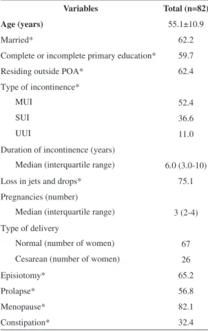
The demographic and clinical characteristics of the
82 patients are shown in Table 1. Half of the patients
had between two to four pregnancies, and there were two nulliparous women in the total sample.
The patients performed on average 13.64 physical therapy sessions (range: 5-15).
Table 1. Sample characteristics.
Variables Total (n=82)
Age (years) 55.1±10.9
Married* 62.2
Complete or incomplete primary education* 59.7
Residing outside POA* 62.4
Type of incontinence*
MUI 52.4
SUI 36.6
UUI 11.0
Duration of incontinence (years)
Median (interquartile range) 6.0 (3.0-10)
Loss in jets and drops* 75.1
Pregnancies (number)
Median (interquartile range) 3 (2-4)
Type of delivery
Normal (number of women) 67
Cesarean (number of women) 26
Episiotomy* 65.2
Prolapse* 56.8
Menopause* 82.1
Constipation* 32.4
Regarding the results of the PFM function evaluation (Table 2), no statistically significant
difference was found in the two measurements performed using the perineometer, while the post physical therapy intervention measurements by the
bidigital maneuver yielded signiicantly higher values than the initial ones (P<0.01).
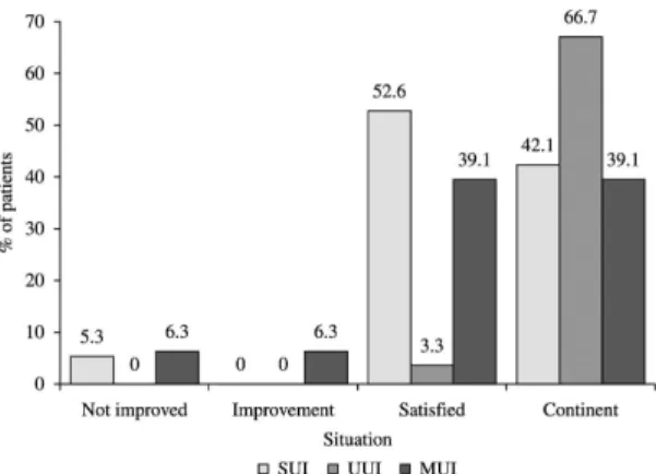
After the intervention, 88.9% of the patients
reported being continent or satisfied with the
treatment (Figure 1). In comparing the distribution
between the type of incontinence and the patients’
satisfaction, no statistically signiicant association was found (p>0.05), indicating an independent
relationship between both variables. However, the
frequency distribution demonstrates that 60.0% (n=18) of 30 patients with SUI reported being satisfied, whereas 30.0% (n=9) reported being continent. Approximately 48.6% (n=17) of the patients with MUI reported being satisied, and 37.1% (n=13) reported being continent. Continent patients predominated (71.4%; n=5) in the group with UUI.
The values observed in the perineometry were associated with the bidigital test measurements
(Table 3), wherein the correlations (before=0.57; after=0.59) were positive and statistically signiicant (P<0.001).
Discussion
The results observed demonstrate that the most prevalent form of UI was mixed urinary incontinence
(52.4%), followed by SUI (36.6%). These results
corroborate data published by Figueiredo et al.7, who also evaluated women from the Brazilian
public health system in a similar age group as the participants of this study. In contrast, Isherwood and
Rane19 reported a higher prevalence of SUI. This
discrepancy between results is most likely explained by those researchers’ recruitment of patients who exhibited urine loss due to stress and a lower age group than the present study patients.
We have also observed in our study that 50% of
the subjects exhibited a time of incontinence ranging
from three to ten years, corroborating the inding by Lewis20, who reported that 75% of the 827 women with
UI in their study took approximately three years to seek medical attention and physical therapy. Women with mild and moderate degrees of incontinence do not seek help, nevertheless, the search for treatment increases in the post-menopausal, when the degree of urinary loss increases8. However, the long period of
time between the onset of urinary loss and the search for help not only results in worsening of the loss itself
but also of the patients’ quality of life. According to
different authors, many women are unaware that UI
Table 2. The values of perineometry and bidigital tests obtained before and after the physical therapy intervention.
Measurements Assessment period P
Before After
Perineometry
0.098* Mean ± standard
deviation
40.6±24.1 41.7±25.4
Minimum - Maximum 4-100 9-100
Bidigital test
<0.001§ Median (interquartile) 4 (3-5) 4 (4-5)
Minimum - Maximum 0-5 1-5
*Student t-test for paired samples; §: Wilcoxon test.
Table 3. Relationship between results of the bidigital test and of
perineometry obtained in the assessments performed before and after the physical therapy intervention.
Bidigital test
Perineometer
Before After
Coefficient
(r)* p
Coefficient
(r)* p
Before 0.569 <0.001 0.468 <0.001
After 0.364 0.001 0.591 <0.001
*Spearman’s correlation coeficient.
Figure 1. Distribution of the type of urinary incontinence with
respect to patients’ treatment satisfaction. SUI: stress urinary incontinence, UUI: urge urinary incontinence and MUI: mixed
is treatable and consider it a natural result of the aging process4,20. Such indings may explain the delayed
search for a treatment for UI observed in this study
and in that by Lewis20.
In the present study, approximately 32% of the
subjects reported constipation. Constipation may stimulate bladder receptors, which may reduce its contractility and cause incomplete bladder emptying, a common complaint among women with UI8.
Therefore, the importance of treating constipation alongside the treatment for UI is clear because the risk for all types of UI increases with constipation21.
In our study, 75.1% of the subjects reported urine losses in drops and jets. A similar result was found
by Figueiredo et al.7, wherein 46% of the women reported losses in jets, and 28% reported losses in drops. Given that the urinary loss volume in SUI is
usually small (droplets), this fact could explain the results of our study since most of the patients had a
diagnosis of SUI and MUI. The literature emphasizes
that the greater the volume of urine loss, the greater the negative effects on patient’ lives22.
According to Neumann and Morrison23,
improvement in muscle function resulting from the conservative treatment of UI leads to a better pelvic viscera positioning, which may lead to a reduction in the mass present inside the vaginal canal, thereby causing a reduction in the size of the prolapse, which, in turn, may lead to lower readings recorded by the perineometer in the post-treatment period. This
inding may explain the fact that, despite a signiicant
difference observed in the values of the bidigital maneuver and the patients’ reports of having achieved
continence, there was no statistically signiicant
difference between the perineometer measurements obtained before and after therapeutic intervention, as
56.8% of subjects in the present study had prolapse.
Hundley et al.11 compared the use of perineometer with the bidigital maneuver in the evaluation of PFM
function and reported similar levels of reproducibility
and a strong linear correlation (0.66>r<0.71) between
the two measurements used. In our study, although the reproducibility was not examined because of its
design, the correlation coeficients obtained were similar (0.57>r<0.59). Hundley et al.11 also reported
a greater interobserver variability in the data obtained
using the bidigital test. Due to this inding, in the
present study all volunteers were examined by the
same researcher at the initial and inal phases of
the treatment. This methodological precaution is
particularly important as it has been shown that the
inal ratings from assessments performed by different evaluators presented signiicant differences8.
Although the Royal College of Obstetricians and Gynecologists (RCOG) reports that the use of PFM digital assessment still lacks scientiic evidence,
their expert advice is that the determination of
the contractile capacity of the PFM may direct
treatment decisions and that this assessment can be used to determine the effect of physical therapy intervention8. Furthermore, perineometry, regarded
as the most objective measure, exhibits similar levels of reliability and accuracy if the measurements are conducted by the same observer on different occasions and if all observers are trained to conduct the measurements10, albeit at a much higher cost. The cost of the device (R$ 880.00) and its maintenance, as
well as the longer time and greater labor involved in the process, must be considered, with the exception of the expenses common to both methods (gloves, lubricant and condom). Therefore, the results of the present study and those from previous studies reported in literature11,19 conirm the clinical use
of the bidigital maneuver because it is a making it the instrument of choice for use in public health for
determining the contractile capacity of the PFM,
which guides the physical therapy treatment and the effect of this intervention.
There are several indications in the international literature regarding the frequency and length of treatment sessions for UI. In our study, we performed a single, weekly session of treatment because most subjects lived in the State hinterland and were
dependent on free public transportation and/or could
not afford to bear the costs of travel to our unit more
times per week. Bo et al.24 indicate daily sessions of electrical stimulation for 30 minutes and 12 series of contractions for the pelvic loor. The RCOG8
suggested three months of exercises repeated three to four times daily and, if necessary, application of electrical stimulation two to three times per week,
lasting 15 to 30 minutes. However, the results of the
present study demonstrate that satisfactory gains may be reached through sessions of much shorter duration
and lower frequency: this study’s subjects attended an average of 13.6 weekly sessions, with ten minutes of electrostimulation and approximately 15 minutes
of kinesiotherapy, with no indication for additional
duration of the treatment program of the present study, the success rate was similar to that obtained
in other studies: 88.9% of subjects reported being continent or satisied with the treatment undertaken,
requiring no other type of additional treatment. These results corroborate those found by Herrmann et al.25, who reported cure or improvement in 81.7% of the women with UI following treatment. Other authors reported a cure rate of 84% in women with different types of UI after training the pelvic loor18,21.
Therefore, the success rate in the present study falls within the success rate ranges described in the international literature, despite the lower frequency and shorter duration of treatment sessions.
In our study, we found that the success rate varied among the different types of UI, as has been described in previous studies25,26. Approximately 90% of the 30 patients with SUI reported being satisied
or continent. This result is similar to that found by
Barroso et al.27, wherein 88% of the women with SUI
reported cure or satisfaction with the result reached.
Approximately 80% of the patients with MUI treated with electrical stimulation were satisied in a study conducted by Amaro et al.28. Superior results were found in the present study, wherein 86.1% of patients reported being satisied or continent.
Seven of the nine patients with UUI in the present study completed the study reaching continence, whereas another study on women with the same type of UI reported lower success rates, with a reduction
of episodes of urinary urgency in 52.4% of patients who performed electrical stimulation and in 57.1% of
patients who performed perineal exercises26.
These high success rates obtained with the conservative treatment of UI explain the position of
the RCOG8 in suggesting that the primary surgical
treatment for UI should only be considered when conservative treatment fails or if the patient declines to perform it. Thus, given the high success rates obtained with the conservative treatment, it becomes
the irst indication in the treatment of UI. The pelvic loor exercises are a low-cost, low-risk treatment and,
when necessary, may include transvaginal electrical
stimulation, as seen in this study, with proven eficacy
even when conducted in short weekly sessions. Significant technological resources will not be required to implement this treatment program in public health units; instead, it will require the presence of a physical therapist trained to conduct the
evaluation and correct treatment for such dysfunction. The implementation of this program would meet the
National Policy on Primary Care (Política Nacional de Atenção Básica) and the principles and guidelines
of the National Policy for Comprehensive Healthcare
for Women (Política Nacional de Atenção Integral à Saúde da Mulher)17. Furthermore, this treatment
program would avoid the patients’ travel from the interior of the state to the capital and its inherent costs,
as occurred with 62.4% of the present study subjects,
who had to be directed to a reference unit specialized in that treatment to successfully conduct a simple treatment that could be held in their home town.
Considering the possible abnormal and harmful functioning that this condition brings to health and quality of life of those who are affected by it20, the
results observed in this study clearly reinforce the need to conduct preventive studies on UI, as well as the implementation of a strengthening pelvic
loor exercise program for pregnant women and all
other women. Thus, based on the results found in this study, the physical therapy treatments proposed could easily be implemented within the primary healthcare system, given its low complexity, the low cost of the bidigital test and treatment, and the high success rate reached. The treatment could be started
with PFM exercises, which could be developed in
groups, subsequently adding transvaginal electrical stimulation if necessary. Therefore, the cost and length of sessions could be even further reduced.
In conclusion, a treatment program with short-duration, weekly sessions resulted in the increase in
pelvic loor muscle function and urinary continence
or satisfaction with treatment for most women studied. The bidigital maneuver exhibited better results than the perineometer in detecting increased muscle function and continence resulting from the outpatient physical therapy.
References
1. Zanetti MR, Castro RA, Rotta AL, Santos PD, Sartori M, Girão MJ. Impact of supervised physiotherapeutic pelvic loor exercises for treating female stress urinary incontinence. Med J. 2007;125(5):265-9.
2. Abrams P, Cardoso L, Fall M, Grifiths D, Rosier P,
Ulmsten U. The standardisation of terminology of lower
urinary tract function: report from the standardisation
sub-committee of International Continence Society.
3. Dos Reis RB, Cologna AJ, Martins ACP, Paschoalin EL, Tucci S Jr, Suaid HJ. Incontinência urinária no idoso. Acta Cir Bras. 2003;18(supl5):47-51.
4. Ko Y, Lin SJ, Salmon JW, Bron MS. The impact of urinary incontinence on quality of life of the elderly. Am J Manag Care. 2005;11(4 Suppl):S103-11. PMid:16161383. 5. Hannestad YS, Rortveit G, Sandvik H, Hunskaar S. A
community-based epidemiological survey of female
urinary incontinence: the Norwegian EPINCONT study.
Epidemiology of Incontinence in the County of
Nord-Trondelag. J Clin Epidemiol. 2000;53(11):1150-7. http:// dx.doi.org/10.1016/S0895-4356(00)00232-8
6. Bent AE, Ostergard DR, Cundiff GW, Swift SE. Ostergard: Uroginecologia e disfunções do assoalho pélvico. 5a ed. Rio de Janeiro: Guanabara Koogan; 2006.
7. Figueiredo EM, Lara JO, Cruz MC, Quintão DMG, Monteiro MVC. Peril sociodemográico e clínico de usuárias de Serviço de Fisioterapia Uroginecológica da rede pública. Rev Bras Fisioter. 2008;12(2):136-42. http:// dx.doi.org/10.1590/S1413-35552008000200010 8. Adams E, Bardsley A, Crumlin L, Currie I, Evans L,
Haslam J. Urinary incontinence the management of urinary incontinence in women. RCOG Press at the Royal College of Obstetricians and Gynaecologists; 2006 [cited 2008 Oct 28]. Available from: http://www.nice.org. uk/nicemedia/pdf/CG40fullguideline.pdf.
9. Cardozo L, Staskin D. Textbook of female urology and urogynecology. 2nd ed. London: Informa Healtcare; 2006. 10. Feldner JR, Sartori MGF, Lima GR, Baracat EC, Girão
MJBC. Diagnóstico clínico e subsidiário da incontinência urinária. Rev Bras Ginecol Obstet. 2006;28(1):54-62. http://dx.doi.org/10.1590/S0100-72032006000100010 11. Hundley AF, Wu JM, Visco AG. A comparison of
perineometer to brink score for assessment of pelvic loor muscle strength. Am J Obstet Gynecol. 2005;192(5):1583-91. PMid:15902162. http://dx.doi.org/10.1016/j. ajog.2004.11.015
12. Ortiz O. Valoración dinâmica de la disfunción perineal da classiicación. Boletim de La Sociedad Latino Americana de Uroginecologia y Cirurgia vaginal. 1994;1(2):7-9. 13. Uyar Y, Baytur YB, Inceboz U. Perineometer and digital
examination for assessment of pelvic loor strength. Int J Gynaecol Obstet. 2007;98(1):64-5. PMid:17466302. http://dx.doi.org/10.1016/j.ijgo.2007.03.015
14. Schoueri J, Ramos LR, Papaléo Netto M. Crescimento populacional: Aspectos Demográficos e Sociais. In: Carvalho ET Fº, Papaléo M Nº, Editores. Geriatria - Fundamentos, Clínica e Terapêutica. São Paulo: Atheneu; 2000. p. 9-29.
15. Guarisi T, Pinto AM Nº, Osis MJ, Pedro AO, Paiva LH, Faundes A. Incontinência urinária entre mulheres climatéricas brasileiras: inquérito domiciliar. Rev Saude Publica. 2001;35(5):428-35. PMid:11723513. http:// dx.doi.org/10.1590/S0034-89102001000500004
16. Kegel A. Proressive resistance exercise in the functional restration of muscles. Am J Obstet Gynecol. 1948;56:238-49. PMid:18877152.
17. Brasil. Ministério da Saúde. secretaria de Gestão Estratégica e Participativa. Considerações Finais. In: Ministério da Saúde, Secretaria de Gestão Estratégica e Participativa. A construção do SUS: histórias da reforma Sanitária e do processo Participativo. Brasília: Ministério da Saúde; 2006. p. 261-278.
18. Dumoulin C, Hay-Smith J. Pelvic loor muscle training
versus no treatment, or inactive control treatments, for urinary incontinence in women. Cochrane
Database Syst Rev. 2006;(1):CD005654. http://dx.doi. org/10.1002/14651858.CD005654
19. Isherwood PJ, Rane A. Comparative assessment of pelvic loor strength using a perineometer and digital examination. BJOG. 2000;107(8):1007-11. http://dx.doi. org/10.1111/j.1471-0528.2000.tb10404.x
20. Lewis D. Incontinence survey report. In: Getliffe K, Dolman M, editors. Promoting Continence: A clinical and Research resource. London: Bailliere Tindall; 1997. 21. Sobhgol SS, Charandabee SMA. Related factors of
urge, stress, mixed urinary incontinence and overactive
bladder in reproductive age women in Tabriz, Iran: a cross-sectional study. Int Urogynecol J. 2008;19:367-373. PMid:17704857. http://dx.doi.org/10.1007/ s00192-007-0437-2
22. Lopes MHBM, Higa R. Restrições causadas pela incontinência urinária da mulher. Rev Esc Enferm USP. 2006;40(1):34-41. http://dx.doi.org/10.1590/ S0080-62342006000100005
23. Neumann P, Morrison S. Physiotherapy for urinary incontinence. Aust Fam Physician. 2008;37(3):118-21. PMid:18345359.
24. Bo K, Talseth T, Holme I. Single blind, randomised
controlled trial of pelvic floor exercises, electrical stimulation, vaginal cones, and no treatment in management of genuine stress incontinence in women.
BMJ. 1999;318(7182):487-93. PMid:10024253 PMCid:PMC27740. http://dx.doi.org/10.1136/ bmj.318.7182.487
25. Herrmann V, Potrick BA, Palma PCR, Zanettini CL, Marques A, Rodrigues NJ. Eletroestimulação transvaginal do assoalho pélvico no tratamento da incontinência urinária de esforço: avaliação clínica e ultra-sonográica. Rev Assoc Med Bras. 2003;49(4):401-5. PMid:14963592. http://dx.doi.org/10.1590/S0104-42302003000400031 26. Arruda RM, Sousa GO, Castro RA, Sartori MGF, Baracat
EC, Girão MJBC. Hiperatividade do detrusor: comparação entre oxibutinina, eletroestimulação funcional do assoalho pélvico e exercícios perineais. Estudo randomizado. Rev Bras Ginecol Obstet. 2007;29(9):252-8. http://dx.doi. org/10.1590/S0100-72032007000900003
Int. 2004;93:319-323. PMid:14764129. http://dx.doi. org/10.1111/j.1464-410X.2004.04608.x
28. Amaro JL, Gameiro MO, Padovani CR. Effect of
intravaginal electrical stimulation on pelvic floor
muscle strength. Int Urogynecol J Pelvic Floor Dysfunct. 2005;16(5):355-8. PMid:15647885. http:// dx.doi.org/10.1007/s00192-004-1259-0
Correspondence
Mara Regina Knorst
Faculdade de Enfermagem, Nutrição e Fisioterapia Av. Ipiranga, 6681, Prédio 12