Compartments of Human Monocytes
Karim J. Brandt, Céline Fickentscher, Egbert. K. O. Kruithof, Philippe de Moerloose
*Division of Angiology and Hemostasis, University Hospital of Geneva and Faculty of Medicine, Geneva, Switzerland
Abstract
Localization of Toll-like receptors (TLR) in subcellular organelles is a major strategy to regulate innate immune responses. While TLR4, a cell-surface receptor, signals from both the plasma membrane and endosomal compartments, less is known about the functional role of endosomal trafficking upon TLRβ signaling. Here we show that the bacterial TLRβ ligands PamγCSK4 and LTA activate NF-κB-dependent signaling from endosomal compartments in human monocytes and in a NF-κB sensitive reporter cell line, despite the expression of TLRβ at the cell surface. Further analyses indicate that TLRβ-induced NF-κB activation is controlled by a clathrin/dynamin-dependent endocytosis mechanism, in which CD14 serves as an important upstream regulator. These findings establish that internalization of cell-surface TLRβ into endosomal compartments is required for NF-κB activation. These observations further demonstrate the need of endocytosis in the activation and regulation of TLRβ-dependent signaling pathways.
Citation: Brandt KJ, Fickentscher C, Kruithof EKO, de Moerloose P (β01γ) TLRβ Ligands Induce NF-κB Activation from Endosomal Compartments of Human Monocytes. PLoS ONE 8(1β): e8074γ. doi:10.1γ71/journal.pone.008074γ
Editor: Jörg Hermann Fritz, McGill University, Canada
Received July 18, β01γ; Accepted October 6, β01γ; Published December 1β, β01γ
Copyright: © β01γ Brandt et al. This is an open-access article distributed under the terms of the Creative Commons Attribution License, which permits unrestricted use, distribution, and reproduction in any medium, provided the original author and source are credited.
Funding: This work was supported by two grants: the Swiss National Fonds (n°γ100γ0–1β76γ9) and the ISTHβ007 Presidential Fund. The funders had no role in study design, data collection and analysis, decision to publish, or preparation of the manuscript.
Competing interests: The authors have declared that no competing interests exist. * E-mail: [email protected]
Introduction
The innate immune system provides a first line of defense against various pathogens without the requirement of prior exposure to foreign antigens. Among the members of the pattern-recognition receptors, Toll-like receptors (TLR) play a central role in innate immunity. A fundamental principle that governs all aspects of TLR signal transduction is that the mechanisms that ensure the fidelity of signaling are determined by their cellular localization and selective regulators of TLR signal transduction [1]. Endocytosis of plasma membrane– localized TLRs was initially thought to attenuate ligand-induced responses, but it is now widely accepted that receptor internalization permits both the propagation of the signaling cascade from endosomal compartments and the generation of distinct signaling events [β-4]. Although less is known about the regulators that control TLR endocytosis after microbial detection, new accumulating evidence indicates that accessory proteins, such as CD14, are not only critical in ligand recognition but may also fulfill additional functions [5]. TLRβ recognizes various ligands, and makes use of different mechanisms to provide specificity to each of them. In this context, recognition by receptor heterodimerization and/or endocytosis gives rise to broader ligand specificity [γ,6,7]. In particular, by association with TLR1 and/ or TLR6, TLRβ is able
to recognize bacterial triacylated or diacylated lipopeptides [8]. Indeed, the role of TLR1 and TLR6, as well as that of CD14, in cell activation in response to lipoteichoic acid (LTA) and the synthetic bacterial lipopeptide Pamγ-Cys-Ser-Lys4 (PamγCSK4) is well established [7,9]. Like TLR4, TLRβ has
been demonstrated to associate with CD14 to increase cellular responses to ligands [7,10,11]. CD14 regulates TLR4 endocytosis and hence the subsequent endosomal-dependent signaling pathway [5]. In addition to the TLR4 signaling that takes place at the plasma membrane through MyD88/TIRAP adaptor proteins leading to early NF-κB activation, a second signaling event was demonstrated to be initiated from the endosomal compartments through TRAM/TRIF adaptor proteins mediating late NF-κB signaling and phosphorylation of the transcription factor Interferon Regulatory Factor-γ (IRFγ), which in turn regulates type I interferon (IFN) [1β]. While an intracellular localization of TLRβ has been observed [γ,1γ,14], its relevance remains to be clearly established in TLRβ signaling. Indeed, Nilsen et al. have shown that a dominant negative form of Dynamin had no effect on TLRβ signaling whereas several other studies have shown that production of TNF and IL-6 is altered by inhibition of TLRβ internalization [11,15].
demonstrate that NF-κB activation in primary monocytes in response to LTA and PamγCSK4 requires internalization of
TLRβ through clathrin-coated pits, and that this mechanism is regulated by CD14.
Materials and Methods
Ethics statement
Buffy coats of blood of healthy donors were provided by the Geneva Hospital Blood Transfusion Center. In accordance with the ethical committee of the Geneva Hospital and with the Declaration of Helsinki, the blood bank obtained informed consent from the donors, who are thus informed that part of their blood will be used for research purposes.
Reagents
Ultra-pure LPS K1β, LPS conjugated-biotin; lipoteichoic acid (LTA) from Staphylococcus aureus, PamγCSK4
conjugated-biotin, blocking peptide against MyD88 and TRIF were from InvivoGen (San Diego, CA). PamγCSK4 was from Alexis
Corporation (San Diego, CA). IFN was from Laboraoire Roussel et Cie, SNC, Paris. Chloroquine diphosphate, Chlorpromazine hydrochloride, Dynasore and Ammonium chloride are from Sigma (Sigma, St. Louis, MO). Monoclonal anti-human CD14 blocking antibody, AF488-conjugated mouse anti-human TLRβ and TLR4, anti-human CDγβ antibodies were from Biolegend. Secondary antibodies were from Jackson ImmunoResearch Laboratories, Inc (West Grove, PA).
Cell culture
Monocytes were isolated from blood buffy coats of healthy volunteers as previously described [16]. Monocyte purity routinely consisted of >90% CD14+ cells, <1% CDγ+ cells, and
<1% CD19+ cells as assessed by flow cytometry. Cells were
cultured in RPMI containing 10% Fetal Bovine Serum (FBS; Gibco BRL-Life Technologies).
Human embryonic kidney β9γ (HEKβ9γ) cells stably transfected with human TLR4, MDβ and CD14 (HEK-Blue4™) or with human TLRβ and CD14 (HEK-Blueβ™) were obtained from InvivoGen (San Diego, CA) and grown in Dulbecco’s Modified Eagle’s Medium containing 10% FBS. The HEK-Blue cells express the secreted embryonic alkaline phosphatase reporter genes (SEAP) under the control of promoter containing five NF-κB binding sites, which enables to quantify cell activation by measuring SEAP activity in media containing specific enzyme substrate. HEK-β9γ cells expressing only TLRβ were obtained as previously described [17].
TNF production
Blocking antibodies and peptides were used at 10μg/ml and 50μM or pharmacological inhibitors were added prior to incubation for β4h with 1μg/ml LTA, 100ng/ml PamγCSK4 or
IFN (500U/ml). Culture supernatants were tested for the production of TNF by a commercially available enzyme immunoassay (R&D Systems, Minneapolis, MN, USA).
mRNA Silencing
HEK-Blueβ™ and HEK-Blue4™ were transfected for 7βh with 100nM of Stealth siRNA against heavy chain clathrin 17 (CHC17) designed by the supplier (Life Technologies, Grand Island, NY) or with 100nM of negative control duplex. Transfections were done using TransIT-TKO, according to the supplier's protocol (Mirus, Madison, WI). CHC17 silencing was ascertained by Western blot. Production of SEAP enzyme by silenced HEK-Blueβ™ was measured with HEK-Blue™ Detection Medium (InvivoGen, San Diego, CA). Cell’s activation was assessed by measuring the absorbance at 650 nm.
Beads assay
β00ng/ml of a solution of LTA conjugated-biotin, PamγCSK4
-biotin or LPS--biotin were bound to Streptavidin-Agarose Resin (Thermo Fisher). After ligand binding, the supernatants were conserved and the beads washed γ times. HEK-Blueβ™ cells or HEK-Blue4™ cells were treated with soluble LTA-biotin (β00ng), PamγCSK4-biotin or LPS-biotin (β0ng), with
beads-bound ligands at corresponding quantities or with the supernatants remaining after incubation of biotin-conjugated ligands with Streptavidin-Agarose. The capacity of ligand treated HEK-Blueβ™ or HEK-Blue4™ cells to produce SEAP enzyme was measured with HEK-Blue™ Detection Medium. Cells activation was assessed by measuring the absorbance at 650 nm. The ability of the conjugated-beads to interact with respective receptors is evaluated by flow cytometry. Briefly, HEK-Blueβ™ cells or HEK-Blue4™ cells were treated with beads-bound ligands for 1h. Beads were isolated and washed γ times with PBS then HEK-Blueβ™ cells or HEK-Blue4™ cells associated to the beads through interaction between ligand and receptors are quantified using antibodies against TLRβ and LTR4 by flow cytometry.
Real-time quantitative PCR
mRNA was prepared by TriReagent® (Molecular Research
Center) according to the provided protocol. qPCR was done on a StepOne™ instrument (Life Technologies, Grand Island, NY). TNF and IFN- TaqMan probes and master mix were from Life Technologies, Grand Island, NY. Targets mRNA expression were normalized against the expression of 18S ribosomal mRNA analyzed simultaneously. Data were analyzed using the comparative ΔCT method.
Western blot
Flow cytometry
For staining of extracellular proteins, cells were stained with mouse anti-biotin/ goat anti-mouse-phycoerythrin (Jackson, West Grove, PA). For intracellular staining, cells were activated as described above, trypsinized, fixed and permeabilized. Staining was assessed with ACCURI C6 flow cytometer (BD Biosciences, San Jose, CA).
Statistical analysis
Where indicated, significance of differences between groups was assessed using Student’s paired t test. *:p ≤ 0.05; **:p ≤ 0.005; ***:p ≤ 0.0005. All data are represented as mean +/- SD of at least γ independent experiments.
Results
Clathrin-dependent endocytosis controls TNF expression induced by LTA and Pam3CSK4
Recent data indicate a key role for the internalization of TLR4 and TLRβ molecules expressed at the plasma membrane by a process dependent on clathrin and the GTPase dynamin in the triggering of specific signals from early endosomes [β,18]. The previously described localization of TLRβ ligands within endosomal compartments [18,19] suggests a possible role of endocytic pathways in TLRβ signaling. To assess the role of TLRβ internalization in the induction of TNF in monocytes, cells were treated with different inhibitors: Chlorpromazine (CPZ), an inhibitor of clathrin-dependent endocytosis; Chloroquine (CHQ), an inhibitor of endosomal maturation; and Dynasore (Dyn), a specific inhibitor of dynamin which is crucial for pinching off of clathrin-coated pits and other vesicular trafficking processes. In addition, we used a non-specific inhibitor of vesicular acidification: NH4Cl.
This weak base inhibits endosomes maturation by increasing the vesicular pH [β0,β1]. As shown in Figure 1 and Figure S1A, CPZ, CHQ and Dyn significantly decreased the expression of TNF in LTA- and PamγCSK4-activated monocytes (Figures
1A-B and S1A). The effects of CPZ, CHQ, Dyn and NH4Cl were
not due to direct cytotoxic effects of the drugs on monocytes (Figure S1B), nor to a non-specific inhibition of cellular responses, because these drugs had no effect on the TNF expression in IFN -activated monocytes (Figure 1C) meaning that translation and release of TNF are not affected by endocytosis inhibitors. Our results suggest that internalization of TLRβ is required to increase TNF expression in LTA- and PamγCSK4-activated monocytes. The mean ± SD of all
experiments are presented in Table 1. The variations were due to difference in response of the different monocytes preparations used.
Clathrin mediated endocytosis controls NF-κB activation by LTA and Pam3CSK4
Taking in consideration the primary role of NF-κB in regulating monocytic TNF expression in response to LPS [ββ,βγ] and the data in Figure 1 suggesting a key function of endocytosis in favoring TNF production by monocytes, we hypothesized that TLRβ internalization is required for NF-κB
activation. To this end, we took advantage of HEK-Blueβ™ cells which express TLRβ, CD14 and a secreted embryonic alkaline phosphatase (SEAP) reporter gene under the control of a promoter containing five NF-κB binding sites. As shown in Figures βA-C and S1C, the treatment of HEK-Blueβ™ cells with CPZ, CHQ, Dyn and NH4Cl decreased the activation of
NF-κB in response to TLRβ ligands and subsequent secretion of SEAP.
As an alternative approach to the use of pharmacological inhibitors, we analyzed the functional consequences of gene silencing of the clathrin heavy chain (HC-Clathrin) on TLRβ ligand-induced NF-κB activation. As shown in Figure γA, left panel, the silencing of HC-Clathrin expression in HEK-Blueβ™ cells decreased NF-κB activity induced by LTA and PamγCSK4
confirming the results obtained with CPZ (Figures 1A and βA). Furthermore, the silencing of Clathrin expression in HEK-Blue4™ cells does not affect significantly NF-κB activity induced by LPS (Figure γA). The partial inhibition by clathrin siRNA of NF-κB activity is consistent with the partial silencing of the clathrin heavy chain, as assessed by immunoblot quantification (Figure γA, right panel). Our results indicate that the clathrin-dependent internalization of TLR molecules expressed at the cell surface (i.e. TLR1/β/6) lead to NF-κB activation by their respective ligands.
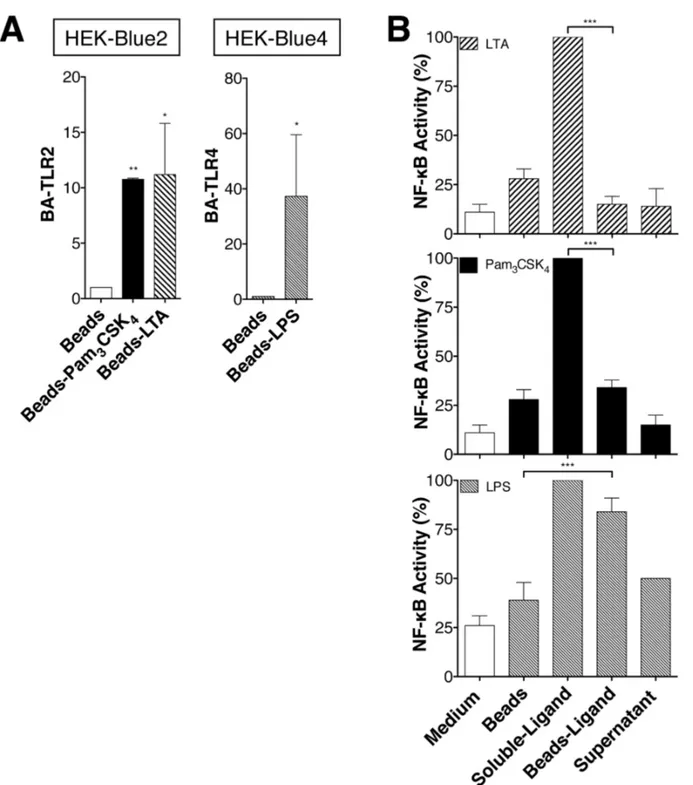
LTA and Pam3CSK4 induce NF-κB activation from endosomal compartments
To determine whether the internalization is important for NF-κB activation, PamγCSK4-conjugated biotin and
LTA-conjugated biotin were fixed to Streptavidin-coated beads (bead size 50-80 μm). The size of the beads prevents their endocytosis but does not interfere with binding of LTA and PamγCSK4 or LPS to TLRβ and TLR4 [15,19]. Indeed,
HEK-Blueβ™ cells are associated to LTA- and PamγCSK4-coated
beads as similar manner than HEK-Blue4™ cells to LPS-coated beads (Figure 4A). When bound to the beads, LTA and PamγCSK4 were unable to induce NF-κB activation in
Blueβ™ cells (Figure 4B). In contrast, NF-κB activation in HEK-Blue4™ cells induced by LPS (a TLR4 ligand) which is known to activate NF-κB from the plasma membrane and endosomal compartment, was hardly affected when fixed to the beads (Figure 4B, third panel).
Data indicate that vaccinia virus and bacterial ligands are not only able to produce type I interferon through TLRβ and IRF family members but are also capable of activating NF-κB [γ,β4]. In contrast to studies suggesting that TLRβ can activate IFN- from endosomal compartments and pro-inflammatory cytokines from the plasma membrane [β4], our data indicate no IRF-γ phosphorylation or IFN- production in LTA- or PamγCSK4-activated monocytes. However, as previously
described, LPS induced IRF-γ phosphorylation, IFN-production and NF-κB activation (Figure 5A-B). In accordance with previous results [β5], MyD88 and TRIF are required for TNF production by LPS-treated monocytes from plasma membrane or endosome, respectively, but MyD88 only is required for TNF production induced by LTA and PamγCSK4 in
of TLRβ within an endosomal compartment is required to trigger NF-κB activation in response to PamγCSK4 or LTA. This
additional mechanism is not functionally coupled to the
triggering of an IRFγ/ IFN response. In conclusion, while ligands bind to TLRβ at the cell surface, they initiate NF-κB
Figure 1. Effect of endocytosis inhibitors on TLR2 mediated induction of TNF in primary human monocytes. Monocytes
were treated with pharmacological endocytosis inhibitors during 45min prior to treatment for 4h or β4h with LTA (1μg/ml) and PamγCSK4 (100ng/ml).
(A) Dose response of the effect of chlorpromazine (CPZ), chloroquine (CHQ) and Dynasore (Dyn) on TNF secretion in TLRβ ligand-activated monocytes. TNF response to LTA and PamγCSK4 after β4h is strongly reduced by all three endosomal pathway inhibitors.
(B) Effect of CPZ, CHQ and Dyn on TNF mRNA expression in LTA- and PamγCSK4-activated monocytes at 4h. TNF mRNA
response to LTA and PamγCSK4 is reduced by all three endosomal pathway inhibitors.
(C) Effect of CPZ, CHQ and Dyn on TNF mRNA and TNF production after 4h and β4h, respectively, in IFN -activated monocytes. Data were normalized to the TNF production observed in the absence of inhibitors.
For all panels data are represented as mean +/- SD of at least γ independent experiments. *:p ≤ 0.05; ** p ≤ 0.005; *** p ≤ 0.0005.
activation from endosomal vesicles by a MyD88-dependent mechanism.
CD14 controls internalization and NF-κB activation in LTA- and Pam3CSK4-activated cells
Previous studies indicate that LTA [14] and PamγCSK4 [7,10]
interact with CD14 and that the internalization of TLR4 is controlled by CD14 [5]. Using the loss of cell surface expression and intracellular accumulation of LTA and PamγCSK4 as a read-out for efficient endocytosis, we show that
CD14 controls LTA and PamγCSK4 internalization. While the
amount of LTA and PamγCSK4 bound to on the cell surface of
CD14 negative cells is the same that of CD14 positive cells
Table 1. 1TNF production by isolated human monocytes.
Stimulus TNF (pg/ml)
Medium 0.6 ± 1.5
LTA (1 µg/ml) 4956 ± β709
PamγCSK4 (100 ng/ml) 157γ ± 808
LPS (100 ng/ml) 8071 ± γ664
Due to the great variation of TNF and TF mediator production among the different monocyte preparations (blood donors) used in this study, data are expressed as mean ± SD of all experiments. We therefore present all results as percentages of TNF production observed in the absence of inhibitors.
doi: 10.1γ71/journal.pone.008074γ.t001
(Figures SγA and SγB), internalization of TLRβ ligands depends of the expression of CD14 on the surface of HEK-Blueβ™ cells (Figure 6A-D). In agreement with Figures 1, γ and 4 indicating that endocytosis is important for TNF expression induced by LTA and PamγCSK4, CD14 negative
(CD14-) cells produce a lower level of IL-8 compared to CD14
positive (CD14+) cells in response to LTA and Pam γCSK4
(Figure 6E). Moreover, specific monoclonal blocking Abs against CD14 reduces the TNF responses in LTA- and PamγCSK4-activated monocytes (Figure 6F). In further
experiments, IκB was degraded in CD14+ cells, but not in
CD14- cells upon stimulation with LTA and Pam
γCSK4 (Figure
6G). Interestingly, responses were markedly reduced, but not completely absent, in CD14 knockout cells or in monocytes treated with blocking antibodies (Figure 6E and 6F). This effect could not be explain by the participation of the scavenger receptors CDγ6, that was show to participate to TLRβ signaling, because CDγ6 is not expressed by CD14+ or CD14
-cells (Figure SγC).
Altogether, these results demonstrate that CD14 controls TLRβ internalization and facilitates NF-κB activation and resulting TNF expression in response to LTA and PamγCSK4.
Discussion
TLRs are classified according to the ligands they recognize and their cellular location. Due to the cell surface localization of TLR4 and TLRβ, it was originally assumed that cell signaling
Figure 2. Effect of endocytosis inhibitors on TLR2 mediated induction of NF-κB in HEK-Blue2™ cells. (A-C) Dose response of the effect of CPZ, CHQ and Dyn on NF-κB activity in LTA- and PamγCSK4-activated HEK-Blueβ™ cells, which express soluble alkaline phosphatase (SEAP) under control of a promoter containing five NF-κB binding sites. LTA- and PamγCSK4-induced NF-κB activity was reduced by all endocytic pathway inhibitors. NF-κB activity was monitored in cells supernatant by SEAP enzyme activity measured with HEK-Blue™ Detection Medium, which contains a specific SEAP substrate. Data are represented as mean +/- SD of at least γ independent experiments. *:p ≤ 0.05; **:p ≤ 0.005; ***:p ≤ 0.0005
from these receptors took place from the cell surface of many cell types. However, recent studies have challenged this assumption by demonstrating that LPS binding by cell-surface expressed TLR4 was able to induce a specific signaling pathway from an endolysosomal compartment [β]. Here, we demonstrate that TLRβ, together with its co-receptors, TLR1 and TLR6, requires internalization to trigger NF-κB activation in response to LTA and PamγCSK4, providing a novel
understanding on how TLRs coordinate ligand recognition and subsequent triggering of a specific signalization. In two separate experimental systems, i.e. primary human monocytes and HEK-Blueβ™ cells, our data further indicate that clathrin-dependent endocytosis mediates NF-κB activation, and that this mechanism is controlled by CD14.
The effect of internalization on TLR4 signaling has been well characterized. While TLR4 induces early NF-κB activation from the cell surface through TIRAP/MyD88-dependent pathways, a TRAM/TRIF-dependent pathway triggers late NF-κB activation and IFN- expression from endosomal compartments [β]. Although proinflammatory cytokine production depends on both signaling from the cell surface and endosomal compartments, several lines of evidence suggest that the
endosomal-dependent signaling pathway is more important. In particular, recent advances have demonstrated that peritoneal macrophages derived from TRAM-/- or TRIF-/- mice produce
neither TNF nor IL-6 in response to high dose of LPS [β5], and that, pharmacological inhibition of TRAM and endocytosis impaired NF-κB activation and IL-6 production in LPS-treated cells [β,β6]. With regard to TLRβ, while numerous studies have identified the presence of TLRβ in endosomal compartments [γ,1γ,14], only few studies have investigated the functional implication of endocytosis in TLRβ signaling [11,β4]. Of specific importance, TLRβ-TLR1 and TLRβ-TLR6, which signal through TIRAP and MyD88, are expressed on the cell surface and are recruited to the phagosome to induce IFN- expression in monocytes upon viral infection [γ] However, it remains unknown whether the cellular localization of TLRβ is important for NF-κB activation induced by LTA and PamγCSK4 in
monocytes. While TLRβ internalization, via a CDγ6-dependent mechanism, was reported to be required for TNF expression, solid data demonstrating an effect of this mechanism on NF-κB activation are still missing [β7,β8]. Supportive insights were however provided by the following data: (i) FSL-1, a TLRβ and TLR6 ligand analogue to LTA, is taken up by murine
Figure 3. Effects of clathrin knockdown on NF-κB responses to LTA and Pam3CSK4. (A) The right panel shows quantification
by western blot of the heavy chain of clathrin in Blueβ™ and representative experience obtains with Blueβ™ and HEK-Blue4™cells treated with stealth siRNA for the heavy chain of clathrin or Stealth RNAi™ negative control duplex. The left panel shows NF-κB activity of HEK-Blueβ™ and HEK-Blue4™cells siRNA-treated for 7βh and then activated with PamγCSK4, LTA or LPS (100ng/ml, 1μg/ml, 100ng/ml, respectively) for β4h. Cells were tested for NF-κB activity by measuring SEAP activity in cell supernatants. NF-κB activity is presented as percentage of production in mock-siRNA transfected HEK-Blueβ™or HEK-Blue4™ cells. Data are represented as mean +/- SD of at least γ independent experiments.
Figure 4. LTA and Pam3CSK4 internalization allows NF-κB activation. (A) HEK-Blueβ™ or HEK-Blue4™ cells were treated
with LTA-biotin, PamγCSK4-biotin, LPS-biotin beads-bound ligands as well as Streptavidin-beads. After beads isolation, the amount
of beads-associated HEK-Blueβ™ (BA-TLRβ) and HEK-Blue4™ (BA-TLR4) cells was measured by flow cytometry using anti-TLRβ and anti-TLR4 antibodies. Results are expressed as the ratio of the geometric mean of TLRβ or TLR4 fluorescence (GMEAN) ± SD of three experiments. Final GMEAN values are the result of GMEAN subtraction from isotype control.
(B) HEK-Blueβ™ or HEK-Blue4™ cells for LPS, were treated with soluble LTA-biotin (1μg/ml), PamγCSK4-biotin (100ng/ml),
LPS-biotin (100ng/ml) or LTA-LPS-biotin, PamγCSK4-biotin, LPS-biotin beads-bound ligands (containing the same concentrations of TLR
ligands as the soluble forms) and ligands depleted supernatants for β4h. NF-κB activity is monitored by SEAP enzyme activity measured with HEK-Blue™ Detection Medium, which contains a specific SEAP substrate. Data are represented as mean +/- SD of at least γ independent experiments.
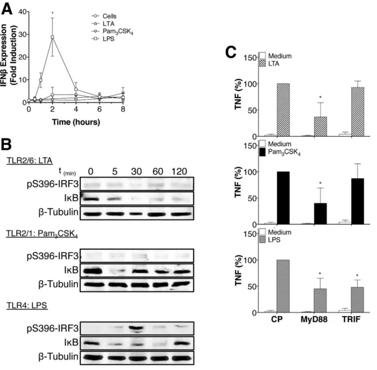
Figure 5. LTA and Pam3CSK4 do not induce IRF-3 phosphorylation or IFN-β production. (A) Human monocytes were treated
with LTA, PamγCSK4, or LPS (1μg/ml, 100ng/ml and 100ng/ml respectively) for the indicated time periods. IFN- expression was
assayed by quantitative PCR. Data are represented as mean +/- SD of results obtained with monocytes from at least γ different blood donors.
(B) Human monocytes were treated with LTA, PamγCSK4 or LPS (1μg/ml, 100ng/ml and 100ng/ml respectively) for indicated time
and the degradation of IκB (NF-κB) and phosphorylation of IRFγ analyzed by Western blot. Data are representative of γ independent experiments.
(C) Human monocytes were treated with 50μM of blocking peptides for MyD88 and TRIF or control peptide (CP) during 60min prior to be activated for β4h with LTA (1μg/ml), PamγCSK4 (100ng/ml) or LPS (100ng/ml). MyD88 blocking peptide reduces TNF
response to LTA and PamγCSK4 after β4h. Data are represented as mean +/- SD of results obtained with monocytes from at least γ
different blood donors.
macrophage cell lines [18], (ii) the signaling triggered by PamγCSK4 (TLRβ and TLR1 ligand) was detected from
endosomal compartments although the pathways involved were not defined [15], (iii) Staphylococcus aureus, from which LTA is purified, induced pro-inflammatory cytokines only after internalization [β7], and (iv) it was reported that a TLRβ-P6γ1H mutant (TLRβ-P6γ1H is a single nucleotide polymorphism) affects the rate of internalization of the wild type TLRβ and NF-κB activation in a dominant negative fashion [β9].
Localization of TLRβ into endosomal vesicles raises the question of the nature/identity of the pathway used for its internalization. Thus, our results suggest that the clathrin/ dynamin-dependent endocytic pathway (Figures β and γA) is involved in TLRβ internalization and NF-κB activation. While TLRβ is reported to be associated to lipid rafts [14,γ0], their disruption does not significantly affect LTA or FSL-1 internalization [15,18]. However, one could also assume that both specific pathways might be involved to fulfill different functions, i.e. the lipid raft pathway for passive recycling of receptors and the clathrin/dynamin pathway for active endocytosis-dependent signaling.
While our data indicate (Figure 4A) that both free LPS and LPS-coated beads are capable of activating NF-κB, only free LTA and PamγCSK4, but not LTA- and PamγCSK4-coated
beads could activate NF-κB. These results are consistent with data indicating the inability of PamγCSK4 to induce IL-6
secretion from U9γ7 macrophages when immobilized on a cell culture plate [15] and the observation that internalization is critical for TLRβ-mediated recognition of LTA [19]. Similar to the requirement of NF-κB in driving TNF and IL-6 expression in LPS-activated monocytes [ββ,βγ], our results and results from other groups show that endocytosis inhibition decreases TNF and IL-6 expression induced by TLRβ ligands in primary human monocytes (Figure 1A-1B), RAWβ64.7 cells [β4] and U9γ7 cells, respectively [15]. Taken together these results demonstrate that endosomal localization of TLRβ is important for NF-κB activation.
CD14 has been described as an important TLR4 co-receptor in LPS recognition and was shown to control TLR4 internalization and type I IFN expression [5,γ1]. While CD14 was also found to serve as a co-receptor for the recognition of TLRβ ligands [7,10,11], its role in NF-κB activation triggered by TLRβ ligands remained to be established. Here, we further demonstrate that LTA and PamγCSK4 internalization is
CD14-dependent. This suggests a comparable role for CD14 in cell activation by both TLR4 and TLRβ ligands. Interestingly, other evidence also indicates that CD14 is required for DNA uptake and delivery to TLR7 and TLR9 [γβ] and that CD14 knockdown decreased FSL-1 internalization in macrophages [18]. In support of a functional implication of endocytosis in NF-κB activation downstream TLRβ signaling (Figures β and γA), our results demonstrate that CD14-dependent internalization of
Figure 6. CD14 controls LTA and Pam3CSK4 internalization and NF-κB activation. HEK-Blueβ™ cells are TLRβ+ and CD14+
(CD14) while HEK-TLRβ cells are TLRβ+ and CD14- cells (no CD14).
(A and C) HEK-Blueβ™ cells ((red dot) and HEK-TLRβ cells (■ were treated with LTA-biotin 1μg/ml or PamγCSK4-biotin 100ng/ml
and TLRβ ligands endocytosis was measured by flow cytometry at the times indicated. Displayed are the mean +/- SD of the percentage of corrected fluorescence index (MFI) of specific extracellular TLRβ ligands staining at each time point.
(B and D) HEK-Blueβ™ cells ((red dot) and HEK-TLRβ cells (■ were treated with LTA-biotin 1μg/ml or PamγCSK4-biotin 100ng/ml
and intracellular accumulation of TLRβ ligands was measured by flow cytometry at the times indicated. Displayed are the mean +/-SD of the percentage of corrected fluorescence index (MFI) of specific intracellular TLRβ ligands staining at each time point. (E) HEK-Blueβ™ cells and HEK-TLRβ cells were activated by indicated TLRβ ligands (LTA 1μg/ml or PamγCSK4 100ng/ml) and IL-8
production was assayed by ELISA. Data are represented as mean +/- SD of at least 5 independent experiments.
(F) Monocytes were treated with blocking antibody against CD14 (10μg/ml) during 45min prior to be activated with PamγCSK4
100ng/ml or LTA 1μg/ml. After β4h, TNF secretion was assayed by ELISA. CD14 blocking antibody decreases significantly TNF production. Data are represented as mean +/- SD of at least 4 independent experiments.
(G) HEK-Blueβ™ cells (CD14+ cells) and HEK-TLRβ cells (no CD14 cells) were activated with LTA 1μg/ml or Pam
γCSK4 100ng/ml.
The presence of CD14 and activation of IκB (NF-κB) were analyzed by Western blot. CD14 controls NF-κB activation in TLRβ ligands-activated cells. Data are representative of γ independent experiments.
LTA and PamγCSK4 regulates NF-κB activation (Figures 6A-D
and 6G). Our data, however, indicate that CD14 may only be partially required for TLRβ-mediated TNF and IL-8 expression (Figure 6E and 6F) or enhance the efficacy of ligand TLRβ interaction as stated below and that other mechanisms are likely to be required to generate a maximal response. Possibly, CDγ6 could participate to such additional mechanisms. However, whereas CDγ6 was shown to increases the response to LTA in HEKβ9γ transfected cells, it was not essential [6]. Furthermore, CDγ6 is not required for cell activation by PamγCSK4 [6,γγ]. Thus, CDγ6 seems to be implicated in some
aspects of TLRβ activity dependent on the ligand and the cell type. Thus, HEKβ9γ Blue cells (CD14+) and HEKβ9γ TLRβ
cells (CD14-) do not express CDγ6, but respond to LTA and
PamγCSK4 (Figures 6 and Sγ). As cellular responses of HEK
cells are dependent on ligand internalization, we conclude is not required for endocytosis of LTA and PamγCSK4.
It is also possible that CD14 is required for early uptake of ligands at low concentration by clathrin-dependent endocytosis, while other regulators may contribute to late uptake of ligands at higher concentrations. In support of this hypothesis, it was shown that TLRβ signaling may vary depending on ligand concentration [γ4] and that CD14 is required for recognition of low concentrations of LPS [γ5]. Furthermore, Zanoni et al. have shown that while CD14 is important for TLR4-MyD88-dependent signal transduction only at low concentrations of LPS, the function of CD14 is independent of the signaling triggered by TLR4 obtained at high LPS concentrations [5]. Likewise, while MyD88 was shown to be critical for TNF production in macrophages in response to S.Aureus, LTA and PamγCSK4, it did not play a significant role in pathogen
internalization, indicating that TLRβ-mediated endocytosis and selective signaling are functionally separated (Figure 5C) [β7].
In conclusion, our study reveals that NF-κB activation by LTA and PamγCSK4 is regulated via a clathrin- and
CD14-dependent endocytosis of TLRβ into primary human monocytes. Thus, although present at the plasma membrane, TLRβ ligands induce NF-κB activation from endosomal compartment by a MyD88-dependent mechanism.
Supporting Information
Figure S1. Effects of weak base NH4Cl and Endosomal inhibitors on TNF secretion and viability of human monocytes. Related to Figures 1 and β. (A-B) Dose response of the effect of weak base NH4Cl on TNF secretion in TLRβ
ligand-activated monocytes. The TNF response to LTA and
PamγCSK4 after β4h was strongly reduced by NH4Cl. Data are
represented as mean +/- SD of γ independent experiments. (C) Viability of human monocytes at 4h and β4h and Viability of HEK-Blueβ™ cells at β4h treated with highest endocytosis inhibitor concentration used in the experiments, as assessed by Trypan blue exclusion. Data are represented as mean +/-SD of β independent experiments.
(TIF)
Figure S2. Blocking peptide effects on Poly I:C-induced TNF production. Related to Figure 5. Human monocytes were treated with blocking peptides for MyD88 and TRIF or control peptide (CP) during 60min prior to be activated for β4h with Poly I:C (10μg/ml). TRIF blocking peptide reduces TNF response to Poly I:C after β4h. Data are represented as mean +/- SD of results obtained with monocytes from at least γ different blood donors.
(TIF)
Figure S3. Expression and quantification of TLR2 ligands and TLR2 coreceptors on cell surface. Related to Figure 6. (A) Representative experiment of the presence of LTA-biotin and PamγCSK4-biotin on HEK-Blueβ™ (CD14+) cells and
HEK-TLRβ cells (CD14-) cells surface.
(B) HEK-Blueβ™ cells and HEK-TLRβ cells were treated with LTA-biotin 1μg/ml or PamγCSK4-biotin 100ng/ml on ice and
their presence on cells surface was quantified by flow cytometry. Displayed are the mean +/- SD of MFIs of specific cell surface TLRβ ligands staining of at least γ independent experiments.
(C) Cells surface expression of CDγ6 and CD14 in human monocytes, HEK-Blueβ™ (CD14+) cells and HEK-TLRβ
(CD14-) cells. Data are representative of γ independent
experiments. (TIF)
Acknowledgements
We thank Dr. Sylvie Dunoyer-Geindre, Dr. Danielle Burger, Dr. Richard Fish and Dr. Nicolas Molnarfi for helpful discussions and advices.
Author Contributions
Conceived and designed the experiments: KJB. Performed the experiments: KJB CF. Analyzed the data: KJB EKOK PdM. Wrote the manuscript: KJB EKOK PdM.
References
1. Barton GM, Kagan JC (β009) A cell biological view of Toll-like receptor function: regulation through compartmentalization. Nat Rev Immunol 9: 5γ5-54β. doi:10.10γ8/nriβ587. PubMed: 19556980.
β. Kagan JC, Su T, Horng T, Chow A, Akira S et al. (β008) TRAM couples endocytosis of Toll-like receptor 4 to the induction of interferon-beta. Nat Immunol 9: γ61-γ68. doi:10.10γ8/ni1569. PubMed: 18β9707γ. γ. Barbalat R, Lau L, Locksley RM, Barton GM (β009) Toll-like receptor β
on inflammatory monocytes induces type I interferon in response to viral but not bacterial ligands. Nat Immunol 10: 1β00-1β07. doi: 10.10γ8/ni.179β. PubMed: 19801985.
4. Sadowski L, Pilecka I, Miaczynska M (β009) Signaling from endosomes: Location makes a difference. Exp Cell Res γ15: 1601-1609. doi:10.1016/j.yexcr.β008.09.0β1. PubMed: 189γ0045. 5. Zanoni I, Ostuni R, Marek LR, Barresi S, Barbalat R et al. (β011) CD14
controls the LPS-induced endocytosis of Toll-like receptor 4. Cell 147: 868-880. doi:10.1016/j.cell.β011.09.051. PubMed: ββ07888γ. 6. Triantafilou M, Gamper FG, Haston RM, Mouratis MA, Morath S et al.
7. Manukyan M, Triantafilou K, Triantafilou M, Mackie A, Nilsen N et al. (β005) Binding of lipopeptide to CD14 induces physical proximity of CD14, TLRβ and TLR1. Eur J Immunol γ5: 911-9β1. doi:10.100β/eji. β004β5γγ6. PubMed: 15714590.
8. Jin MS, Lee JO (β008) Structures of the toll-like receptor family and its ligand complexes. Immunity β9: 18β-191. doi:10.1016/j.immuni. β008.07.007. PubMed: 1870108β.
9. Kang JY, Nan X, Jin MS, Youn SJ, Ryu YH et al. (β009) Recognition of lipopeptide patterns by Toll-like receptor β-Toll-like receptor 6 heterodimer. Immunity γ1: 87γ-884. doi:10.1016/j.immuni.β009.09.018. PubMed: 199γ1471.
10. Nakata T, Yasuda M, Fujita M, Kataoka H, Kiura K et al. (β006) CD14 directly binds to triacylated lipopeptides and facilitates recognition of the lipopeptides by the receptor complex of Toll-like receptors β and 1 without binding to the complex. Cell Microbiol 8: 1899-1909. doi: 10.1111/j.146β-58ββ.β006.00756.x. PubMed: 16848791.
11. Nilsen NJ, Deininger S, Nonstad U, Skjeldal F, Husebye H et al. (β008) Cellular trafficking of lipoteichoic acid and Toll-like receptor β in relation to signaling: role of CD14 and CDγ6. J Leukoc Biol 84: β80-β91. doi: 10.1189/jlb.0907656. PubMed: 18458151.
1β. Kawai T, Akira S (β011) Toll-like Receptors and Their Crosstalk with Other Innate Receptors in Infection and Immunity. Immunity γ4: 6γ7-650. doi:10.1016/j.immuni.β011.05.006. PubMed: β16164γ4. 1γ. Underhill DM, Ozinsky A, Hajjar AM, Stevens A, Wilson CB et al.
(1999) The Toll-like receptor β is recruited to macrophage phagosomes and discriminates between pathogens. Nature 401: 811-815. doi: 10.10γ8/44605. PubMed: 10548109.
14. Triantafilou M, Manukyan M, Mackie A, Morath S, Hartung T et al. (β004) Lipoteichoic Acid and Toll-like Receptor β Internalization and Targeting to the Golgi Are Lipid Raft-dependent. J Biol Chem β79: 4088β-40889. doi:10.1074/jbc.M400466β00. PubMed: 15β47β7γ. 15. Marre ML, Petnicki-Ocwieja T, DeFrancesco AS, Darcy CT, Hu LT
(β010) Human Integrin aγB1 Regulates TLRβ Recognition of Lipopeptides from Endosomal Compartments. PLOS ONE 5: e1β871. doi:10.1γ71/journal.pone.001β871. PubMed: β0877569.
16. Brandt KJ, Carpintero R, Gruaz L, Molnarfi N, Burger D (β010) A novel MEKβ/PIγKdelta pathway controls the expression of IL-1 receptor antagonist in IFN-beta-activated human monocytes. J Leukoc Biol 88: 1191-1β00. doi:10.1189/jlb.0510γ1β. PubMed: β08γ7746.
17. Satta N, Kruithof EK, Fickentscher C, Dunoyer-Geindre S, Boehlen F et al. (β011) Toll-like receptor β mediates the activation of human monocytes and endothelial cells by antiphospholipid antibodies. Blood 117: 55βγ-55γ1. doi:10.118β/blood-β010-11-γ16158. PubMed: β1γγ0474.
18. Shamsul HM, Hasebe A, Iyori M, Ohtani M, Kiura K et al. (β010) The Toll-like receptor β (TLRβ) ligand FSL-1 is internalized via the clathrin-dependent endocytic pathway triggered by CD14 and CDγ6 but not by TLRβ. Immunology 1γ0: β6β-β7β. doi:10.1111/j. 1γ65-β567.β009.0γβγβ.x. PubMed: β011γγ68.
19. Bunk S, Sigel S, Metzdorf D, Sharif O, Triantafilou K et al. (β010) Internalization and Coreceptor Expression Are Critical for TLRβ-Mediated Recognition of Lipoteichoic Acid in Human Peripheral Blood. J Immunol 185: γ708-γ717. doi:10.4049/jimmunol.0901660. PubMed: β071γ89γ.
β0. Clague MJ, Urbé S, Aniento F, Gruenberg J (1994) Vacuolar ATPase activity is required for endosomal carrier vesicle formation. J Biol Chem β69: β1-β4. PubMed: 8β76796.
β1. Huynh KK, Grinstein S (β007) Regulation of Vacuolar pH and Its Modulation by Some Microbial Species. Microbiol Mol Biol Rev 71: 45β-46β. doi:10.11β8/MMBR.0000γ-07. PubMed: 17804666.
ββ. Udalova IA, Knight JC, Vidal V, Nedospasov SA, Kwiatkowski D (1998) Complex NF-κB Interactions at the Distal Tumor Necrosis Factor Promoter Region in Human Monocytes. J Biol Chem β7γ: β1178-β1186. doi:10.1074/jbc.β7γ.γγ.β1178. PubMed: 9694874. βγ. Collart MA, Baeuerle P, Vassalli P (1990) Regulation of tumor necrosis
factor alpha transcription in macrophages: involvement of four kappa B-like motifs and of constitutive and inducible forms of NF-kappa B. Mol Cell Biol 10: 1498-1506. PubMed: β181β76.
β4. Dietrich N, Lienenklaus S, Weiss S, Gekara NO (β010) Murine Toll-Like Receptor β Activation Induces Type I Interferon Responses from Endolysosomal Compartments. PLOS ONE 5: e10β50. doi:10.1γ71/ journal.pone.0010β50. PubMed: β04ββ0β8.
β5. Yamamoto M, Sato S, Hemmi H, Uematsu S, Hoshino K et al. (β00γ) TRAM is specifically involved in the Toll-like receptor 4-mediated MyD88-independent signaling pathway. Nat Immunol 4: 1144-1150. doi:10.10γ8/ni986. PubMed: 14556004.
β6. Fitzgerald KA, Rowe DC, Barnes BJ, Caffrey DR, Visintin A et al. (β00γ) LPS-TLR4 Signaling to IRF-γ/7 and NF-κB Involves the Toll Adapters TRAM and TRIF. J Exp Med 198: 104γ-1055. doi:10.1084/ jem.β00γ10βγ. PubMed: 14517β78.
β7. Ip WKE, Sokolovska A, Charriere GM, Boyer L, Dejardin S et al. (β010) Phagocytosis and Phagosome Acidification Are Required for Pathogen Processing and MyD88-Dependent Responses to Staphylococcus aureus. J Immunol 184: 7071-7081. doi:10.4049/jimmunol.1000110. PubMed: β048γ75β.
β8. Stuart LM, Deng J, Silver JM, Takahashi K, Tseng AA et al. (β005) Response to Staphylococcus aureus requires CDγ6-mediated phagocytosis triggered by the COOH-terminal cytoplasmic domain. J Cell Biol 170: 477-485. doi:10.108γ/jcb.β0050111γ. PubMed: 16061696.
β9. Etokebe GE, Skjeldal F, Nilsen N, Rodionov D, Knezevic J et al. (β010) Toll-Like Receptor β (P6γ1H) Mutant Impairs Membrane Internalization and is a Dominant Negative Allele. Scand J Immunol 71: γ69-γ81. doi: 10.1111/j.1γ65-γ08γ.β010.0βγ79.x. PubMed: β0500688.
γ0. Triantafilou M, Gamper FGJ, Lepper PM, Mouratis MA, Schumann C et al. (β007) Lipopolysaccharides from atherosclerosis-associated bacteria antagonize TLR4, induce formation of TLRβ/1/CDγ6 complexes in lipid rafts and trigger TLRβ-induced inflammatory responses in human vascular endothelial cells. Cell Microbiol 9: β0γ0-β0γ9. doi:10.1111/j.146β-58ββ.β007.009γ5.x. PubMed: 17419716.
γ1. Jiang Z, Georgel P, Du X, Shamel L, Sovath S et al. (β005) CD14 is required for MyD88-independent LPS signaling. Nat Immunol 6: 565-570. doi:10.10γ8/ni1β07. PubMed: 15895089.
γβ. Baumann CL, Aspalter IM, Sharif O, Pichlmair A, Blüml S et al. (β010) CD14 is a coreceptor of Toll-like receptors 7 and 9. J Exp Med β07: β689-β701. doi:10.1084/jem.β0101111. PubMed: β1078886.
γγ. Hoebe K, Georgel P, Rutschmann S, Du X, Mudd S et al. (β005) CDγ6 is a sensor of diacylglycerides. Nature 4γγ: 5βγ-5β7. doi:10.10γ8/ nature0γβ5γ. PubMed: 1569004β.
γ4. Kenny EF, Talbot S, Gong M, Golenbock DT, Bryant CE et al. (β009) MyD88 Adaptor-Like Is Not Essential for TLRβ Signaling and Inhibits Signaling by TLRγ. J Immunol 18γ: γ64β-γ651. doi:10.4049/jimmunol. 0901140. PubMed: 197175β4.