What Role Do Annelid Neoblasts Play? A Comparison of
the Regeneration Patterns in a Neoblast-Bearing and a
Neoblast-Lacking Enchytraeid Oligochaete
Maroko Myohara*
Insect Growth Regulation Research Unit, National Institute of Agrobiological Sciences, Tsukuba, Ibaraki, Japan
Abstract
The term ‘neoblast’ was originally coined for a particular type of cell that had been observed during annelid regeneration, but is now used to describe the pluripotent/totipotent stem cells that are indispensable for planarian regeneration. Despite having the same name, however, planarian and annelid neoblasts are morphologically and functionally distinct, and many annelid species that lack neoblasts can nonetheless substantially regenerate. To further elucidate the functions of the annelid neoblasts, a comparison was made between the regeneration patterns of two enchytraeid oligochaetes,Enchytraeus japonensisandEnchytraeus buchholzi, which possess and lack neoblasts, respectively. InE. japonensis, which can reproduce asexually by fragmentation and subsequent regeneration, neoblasts are present in all segments except for the eight anterior-most segments including the seven head-specific segments, and all body fragments containing neoblasts can regenerate a complete head and a complete tail, irrespective of the region of the body from which they were originally derived. InE. japonensis, therefore, no antero-posterior gradient of regeneration ability exists in the trunk region. However, when amputation was carried out within the head region, where neoblasts are absent, the number of regenerated segments was found to be dependent on the level of amputation along the body axis. InE. buchholzi, which reproduces only sexually and lacks neoblasts in all segments, complete heads were never regenerated and incomplete (hypomeric) heads could be regenerated only from the anterior region of the body. Such an antero-posterior gradient of regeneration ability was observed for both the anterior and posterior regeneration in the whole body of E. buchholzi. These results indicate that the presence of neoblasts correlates with the absence of an antero-posterior gradient of regeneration ability along the body axis, and suggest that the annelid neoblasts are more essential for efficient asexual reproduction than for the regeneration of missing body parts.
Citation:Myohara M (2012) What Role Do Annelid Neoblasts Play? A Comparison of the Regeneration Patterns in a Neoblast-Bearing and a Neoblast-Lacking Enchytraeid Oligochaete. PLoS ONE 7(5): e37319. doi:10.1371/journal.pone.0037319
Editor:Henry H. Roehl, University of Sheffield, United Kingdom
ReceivedOctober 31, 2011;AcceptedApril 19, 2012;PublishedMay 16, 2012
Copyright:ß2012 Maroko Myohara. This is an open-access article distributed under the terms of the Creative Commons Attribution License, which permits unrestricted use, distribution, and reproduction in any medium, provided the original author and source are credited.
Funding:This work was supported in part by a Grant-in-Aid for Scientific Research (KAKENHI No.20500382) from the Japan Society for the Promotion of Science, by a Narishige Zoological Science Award and by a Grant-in-Aid (BioDesign Program) from the Ministry of Agriculture, Forestry, and Fisheries, Japan. These funders had no role in study design, data collection and analysis, decision to publish, or preparation of the manuscript.
Competing Interests:The authors have declared that no competing interests exist. * E-mail: [email protected]
Introduction
Recent advances in the field of stem cell biology are raising expectations that human regenerative medicine will become a future reality and are accelerating regeneration research in a range of model systems that utilize different regenerative strategies [1]. Among these model systems, planarians are bilaterian organisms that have the most prominent regenerative capabilities known and can reproduce a complete individual from only a small body fragment. This remarkable ability is due to the presence of adult somatic stem cells known as neoblasts [2]. Some types of annelids also exhibit regenerative abilities that are comparable to planar-ians, but this is thought to occur primarily through cellular dedifferentiation and redifferentiation, without the contribution of totipotent stem cells [3]. Hence, the elucidation of the regener-ation mechanisms of annelids is expected to provide valuable information that will advance the exploration of the regenerative capabilities of vertebrates, as these also occur without the contribution of totipotent stem cells, and assist with developing the means to enhance these processes. Previously, we proposed the
recently described fragmenting pot wormEnchytraeus japonensisas a new model system for regeneration studies [4]. E. japonensis
reproduces asexually by dividing its body into several fragments, which then regenerate a complete individual within 4–5 days. Artificially amputated fragments of this organism that are as short as a few segments can also regenerate new individuals in the same manner.
The term ‘neoblast’ was first used more than a century ago to denote specialized cells that participate in the regeneration of mesodermal tissues in the oligochaete annelid Lumbriculus [5]. However, this nomenclature is now more often used to designate the pluripotent/totipotent adult somatic stem cells that play central roles in planarian regeneration [2,6–8]. Recent advances in our knowledge of annelid regeneration from studies using E. japonensisas the model system [4,9–14] have renewed interest in the long-ignored annelid neoblasts [15–17]. It must be noted however that planarian and annelid neoblasts are morphologically and functionally distinct.
of the somatic cells in an adult worm. They are defined as the only proliferative somatic cells in adult planarians, and differentiate into all cell types including germ cells [6]. In contrast, annelid neoblasts are large cells that are localized at the intersegmental septa along the ventral nerve cord, and number only a few in each segment [5,18–20]. The annelid neoblasts are particularly prominent in oligochaetes that reproduce asexually by fragmentation or fission, and are thought to give rise to mesodermal tissues during regeneration [5,18–20]. However, as the endodermal and ectodermal tissues regenerate via the proliferation of dedifferen-tiated cells from each layer, the neoblasts are not the only proliferating cells in regenerating annelids. Moreover, many annelid species that lack neoblasts can nonetheless substantially regenerate [19]. An important question that emerges from this therefore is the precise role of the neoblasts in the context of annelid biology. To address this issue in this study, a comparison was made between the regeneration patterns of two enchytraeid oligochaetes (pot worms), Enchytraeus japonensis and Enchytraeus buchholzi, which possess and lack neoblasts, respectively. Special attention was also paid to regeneration patterns of E. japonensis
fragments that were amputated within the head region where neoblasts are absent.
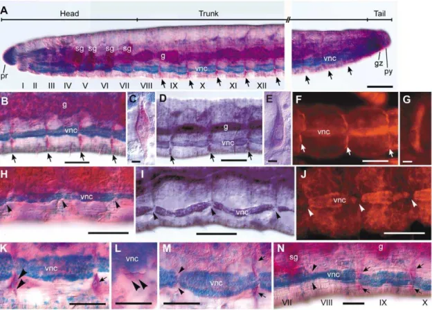
Figure 1. Distribution of neoblasts inE. japonensisand their absence inE. buchholzi.(A–G) IntactE. japonensisworms were stained with methyl green-pyronin (A–C), thionine (D–E), or propidium Iodide (F–G). Neoblast pairs (arrows) are localized on the intersegmental septa along the ventral nerve cord in all segments except for the eight anterior-most segments, i.e. the seven head-specific segments (segment I–VII) and the first trunk segment (segment VIII). (H–J) Neoblasts are absent from the corresponding position (arrowheads) inE. buchholzias revealed by staining with methyl green-pyronin (H), thionine (I), or propidium Iodide (J). (K–N) Behavior of neoblasts (arrows) during regeneration was examined inE. japonensisusing methyl green-pyronin staining. Soon after fragmentation, neoblasts in the segment(s) at the ends of a fragment divide (K, large arrowheads) and both daughter cells migrate together (L, large arrowheads) leaving their site of previous occupancy vacant (M, small arrowheads). The recovery of neoblasts occurs after regeneration has completed, except for in the anterior-most segment, i.e., the new segment VIII in which neoblasts are absent (N, small arrowheads). g, gut; gz, growth zone; pr, prostomium; py, pygidium; sg, septal (pharyngeal) gland; vnc, ventral nerve cord. Scale bar, 100mm for (A–B, D, F, H–J), 5mm for (C, E, G), and 50mm for (K–N).
doi:10.1371/journal.pone.0037319.g001
It has long been observed that polychaete and oligochaete annelids provide excellent model systems for the study of regeneration [3,19]. This is principally due to their high regenerative capacity and metameric body structures that enable quantitative measurements of regeneration activity by counting the number of regenerated segments. However, most studies of annelid regeneration have paid no attention to neoblasts, probably because they have dealt with external morphologies or have employed histological analysis of paraffin-sectioned specimens in which neoblasts are often hard to find. In our present study, neoblasts were distinguished unambiguously in whole-mount specimens of E. japonensis by staining with RNA affinitive dyes such as methyl green-pyronin (MGP), thionine or propidium Iodide (PI). Using this staining approach, the distribution of neoblasts was closely examined in E. japonensis and E. buchholzi
which are similar in size and morphology, belong to the same genus, and can therefore be analyzed using the same methods. An exact comparison of the regeneration patterns of the two species was thus possible.
Although a considerable number of studies have reported on the regeneration ofE. japonensissince our proposal of this species as a new model system for regeneration research in 1999 [4,9– 15,17,21], there has been no report to date on the regeneration of E. buchholzi, or of any other enchytraeid that reproduces only sexually. Out of the several hundred enchytraeid species described to date, only eight have been reported to reproduce asexually by fragmentation and subsequent regeneration (see [4] or [10] and literature cited therein). Regeneration has been studied for three enchytraeid species that reproduce asexually by fragmentation and therefore have high regeneration capacities [4,9–15,18,22] but not for species that reproduce only sexually. Hence, this is the first study report on the regeneration of a non-fragmenting enchy-traeid.
Results
Distribution of neoblasts inE. japonensisand their absence inE. buchholzi
Neoblasts were originally defined as large specialized cells that contribute to mesodermal regeneration in an aquatic oligochaete
Lumbriculus that reproduces asexually by fragmentation and subsequent regeneration [5]. They are large spindle-shaped cells that locate ventro-laterally on the posterior side of the interseg-mental septa along the ventral nerve cord [18,19] and are characterized by an intensely basophilic cytoplasm and a voluminous nucleus with a prominent nucleolus [19,20]. In the present study, it was found that neoblasts could be distinguished unambiguously in whole-mount specimens of E. japonensis by staining with methyl green-pyronin (MGP). By using this staining method, it became evident for the first time that a pair of neoblasts is located in all segments (Fig. 1A–B) except for the eight anterior-most segments (segments I–VIII), i.e. the seven head-specific segments and the first trunk segment (Fig. 1A). The anterior-most neoblasts are located on the posterior side of the intersegmental septa between segments VIII and IX (Fig. 1A,N). By MGP staining, the neoblasts ofE. japonensiswere revealed to have a large nucleus with a prominent nucleolus and cytoplasm that stains intensely red with pyronin (Fig. 1C). Intense labeling of neoblasts was also found inE. japonensisstained with thionine (Fig. 1D,E) and propidium Iodide (PI) (Fig. 1F,G). The intense staining with these three dyes, all of which are known to stain RNA, suggested that the neoblasts are RNA-rich.
In our present study, neoblasts were defined not only by their staining properties but also by their location and morphology. A
cell that fulfilled all the following conditions was recognized as neoblast: (1) large and spindle-shaped, (2) located ventro-laterally on the intersegmental septa along the ventral nerve cord, and (3) intensely stained by MGP, PI or thionine. InE. buchholzi, none of these staining procedures detected any cells that fulfilled these conditions in any segments in intact individuals (Fig. 1H–J) or in regenerating amputees, indicating thatE. buchholzilacks neoblasts in all segments.
It has been reported in studies of other neoblast-bearing oligochaetes that shortly after fragmentation or amputation, neoblasts in the segment(s) at both the anterior and posterior ends of the fragments divide and migrate to the amputated sites, where they take part in blastema formation [5,18,19,23]. In this study, by using MGP staining, it was found that at early stages of
E. japonensisregeneration, both daughter cells of a divided neoblast (Fig. 1K) migrate together (Fig. 1L), leaving behind the site of their previous occupation vacant (Fig. 1M). After regeneration com-pletes, the vacant area is dissolved by the emergence of new neoblasts that are probably derived from the ‘‘neoblast-like cells’’ [15] on intersegmental septa of the segment, but this neoblast recovery did not occur in the anterior-most segment of the stump, i.e., new segment VIII (Fig. 1N). Hence, this segment remains neoblast deficient, as always observed in intact worms (Fig. 1A).
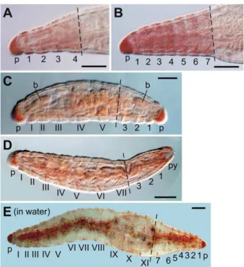
Figure 3. RepresentativeE. japonensisregenerates.(A) Example of a head with four segments regenerated after amputation at the 4th–5th segment. (B) A head with seven segments regenerated after amputation in trunk region. (C) A dicephalic monster with biaxial heads formed after amputation at the 6th segment. A head with three segments was regenerated posteriorly in this case. (D) A normal worm regenerated after amputation at the 7th segment. (E) A long dicephalic monster with biaxial heads formed after amputation at the 11th segment and culture in water instead of agar medium. A complete head with seven segments was regenerated posteriorly in this case. Segments of the original fragments are numbered with Roman numerals, and regenerated segments are numbered using Arabic numerals. The broken lines mark the levels of amputation. The anterior is to the left in each image. p, prostomium; py, pygidium. Scale bars, 100mm.
doi:10.1371/journal.pone.0037319.g003
Regeneration pattern ofE. japonensis
Because spontaneous fragmentation never occurs within the head region of E. japonensis, all of the fragments produced for asexual reproduction contain neoblasts (Fig. 2). These fragments always regenerate a complete head anteriorly and a complete tail posteriorly, irrespective of the region of the body from which they were originally derived, and regenerated worms grow posteriorly via the addition of new segments in the growth zone (Fig. 2) [4]. Hence, no antero-posterior gradient of regeneration ability exists in the trunk region ofE. japonensis. It must be noted, however, that
E. japonensis achieves the regeneration of a complete individual from a body fragment not by a simple restoration of all lost segments, but through a combination of epimorphic recovery of
the head and tail and through the morphallactic transformation of old segments into the appropriate segments [4,13]. This occurs because anterior regeneration inE. japonensisis always limited to the seven head-specific segments, no matter how many segments were originally missing (Fig. 2).
Artificially amputated fragments of E. japonensis generally regenerate into normal worms in the same manner as spontane-ously divided fragments. However, when amputation was carried out within a head region in which neoblasts are absent, the long posterior amputees anteriorly regenerated the missing segments only (Fig. 3A), instead of the seven head-specific segments (Fig. 3B). The number of head segments that regenerated therefore was dependent on the level of amputation along the body axis in the
head region (Fig. 4A, graph). The small anterior amputees, i.e. small head fragments that had been amputated at a site in segments V–VII and thus lacked neoblasts, posteriorly regenerated either a head instead of a tail, resulting in a dicephalic monster (Fig. 3C, Fig. 4B), or a tail, resulting in a normal individual (Fig. 3D, Fig. 4B). This clearly showed that even in the neoblast-bearingE. japonensis, both a head and a tail could be regenerated without neoblasts. The number of regenerated head segments however was never larger than five in the dicephalic monsters produced by amputation within the head region (Fig. 4B, yellow bars), suggesting that neoblasts may be indispensable for the regeneration of a complete head.
When amputation was carried out in the trunk region in which neoblasts are present, the anterior and posterior amputees always regenerated a tail posteriorly and a head anteriorly, resulting in a complete individual (Fig. 4A,B). However, when amputees were cultured in water instead of agar medium, 40% (41 out of 102 examined) of the anterior amputees posteriorly regenerated a head resulting in a long dicephalic monster (Fig. 3E). This occurred probably because ‘‘corrective autotomy’’, which is thought to be important for tail regeneration to occur after artificial amputation, is inhibited in water [21]. In these long dicephalic monsters, a complete head with seven segments was regenerated posteriorly (Fig. 3E), possibly because neoblasts were present in posterior segments of these amputees.
These results suggested that inE. japonensis, each of the head segments that lacks neoblasts has a different positional identity according to its position along the antero-posterior body axis (Fig. 4A,B) and that there is an antero-posterior gradient of regeneration ability in the head region. In contrast, the segments in the trunk region, which contain neoblasts, adopt the same positional identity (of segment VIII) after fragmentation or amputation with respect to their regeneration ability, so that fragments from any body region can regenerate a complete individual (Fig. 2, Fig. 4A,B, Table 1). Moreover, the results of the
experiments dealing with dicephalic monsters suggested that neoblasts may be essential for regeneration of a complete head with seven segments.
Regeneration pattern ofE. buchholzi– anterior direction
In E. buchholzi, head regeneration occurred only when amputation was carried out within or near to the head region (Fig. 4C, Fig. 5A–C). Moreover, the numbers of regenerated head segments decreased with the distance of the amputation site from the original head (Fig. 4C). The range of the numbers of regenerated head segments was 0–4, 0–2, and 0–1 when amputation was carried out in the 4th–14th, 15th–19th, and 21st–30th segments, respectively. In the hypomeric heads, the prostomium and brain were also found to be reduced in size (Fig. 5B).
When amputations were undertaken in the region close to the tail, the small posterior amputees anteriorly regenerated a tail instead of a head, resulting in a bicaudal monster with biaxial tails (Fig. 4C, Fig. 5D). Such a phenomenon never occurs in E. japonensis [4,21] but is known to occur in some other annelids [24,25].
These results suggested that the anterior segments within or near to the head inE. buchholzihave a higher potential for head regeneration, whilst the posterior segments near to the tail have a higher capacity for tail regeneration than the segments of other regions (Fig. 4C, Table 1).
Regeneration pattern ofE. buchholzi– posterior direction
When amputation was carried out within the head region (in the 3rd–7th segments), the small anterior amputees died within seven days without regeneration (none of the 48 examined specimens survived). When amputation was performed immediately posterior to the head segments (in the 8th–11th segments), the amputees generally regenerated a tail, resulting in a normal individual, but on rare occasions (two out of 38 specimens examined), a head Table 1.Comparative summary of the typical regeneration patterns of artificially amputated individuals ofE. japonensisandE. buchholzicultured in agar medium.
Anterior direction
E. japonensis E. buchholzi
Amputation site Regenerate Typical result Regenerate Typical result
Head region (anterior to 7th segment)
Missing segments Normal worm Two to four head segments Worm with a hypomeric
head Anterior trunk region (8th to
,14th segment)
Seven head segments (complete head)
Normal worm One to four head segments Worm with a hypomeric head
Posterior trunk region (posterior to,16th segment)
Seven head segments (complete head)
Normal worm Tail Bicaudal monster
Posterior direction
E. japonensis E. buchholzi
Amputation site Regenerate Typical result Regenerate Typical result
Head region (anterior to 7th segment)
Two to five head segments, or occasionally tail (only when amputated in 6th–7th segment)
Dicephalic monster None - (incapable of surviving)
Anterior trunk region (8th to ,14th segment)
Tail and missing segments Normal worm Tail and missing segments, or rarely an incomplete head
Normal worm (dicephalic monster in rare cases) Posterior trunk region (posterior
to,15th segment)
Tail and missing segments Normal worm Tail and missing segments Normal worm
doi:10.1371/journal.pone.0037319.t001
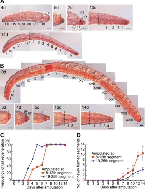
instead of a tail was regenerated, resulting in a dicephalic monster with biaxial heads (Fig. 4D, Fig. 5E). When amputations were carried out in the trunk region, the anterior fragments always regenerated a tail irrespective of the amputation position (Fig. 4D). There was still an antero-posterior gradient of regeneration ability however as tail regeneration occurred more rapidly in the posterior region than in the anterior region, whilst the extent of posterior growth after tail regeneration decreased as the amputa-tion site neared the original tail i.e. if more segments were removed by amputation, more new segments were formed in the growing areas after tail regeneration (Fig. 4D, Fig. 6).
These results suggested that each segment inE. buchholzihas a different positional identity throughout the body with respect to its regenerative ability and that this accords with its position along the antero-posterior body axis. The differing regenerative potentials in different body regions applied to regeneration in both the anterior and posterior direction.
Comparison of cell proliferation activity during the anterior regeneration ofE. japonensisandE. buchholzi
It was found in our analyses that anterior regeneration blastemas of E. buchholziwere smaller than those of E. japonensis
and that regeneration proceeds much more slowly inE. buchholzi
than in E. japonensis (Fig. 7A,B). To examine whether these phenomena are due to lower cell proliferation activity in the blastemas ofE. buchholzicompared with those inE. japonensis, BrdU labeling experiments were performed. InE. japonensis, active cell proliferation began soon after fragmentation and continued at high levels until blastemal segmentation occurred at three days after fragmentation (Fig. 7A). In contrast, in E. buchholzi, cell proliferation activity was very low throughout the regeneration processes except at four days after amputation when the regeneration blastema was formed (Fig. 7B). To assess whether the low proliferation activity observed in E. buchholzi was correlated with the absence of neoblasts, BrdU labeling was monitored in small head fragments ofE. japonensisthat had been artificially amputated within the head region and thus lacked neoblasts. It was found that cell proliferation activity was very low in the regeneration blastemas of these small head amputees (Fig. 7C, arrows) in comparison with those of trunk region amputees in which neoblasts were present (Fig. 7C, arrowheads). These results suggest that neoblasts may contribute to the active proliferations of blastemal cells and to the rapid formation of blastemas with the potential to regenerate a complete head inE. japonensis.
Discussion
As posterior regeneration has been documented in numerous annelid species including those that have been shown to lack neoblasts [19,24,26], it seems obvious that unlike the situation in planarians, neoblasts are dispensable for regeneration in annelids. The analyses in our present study further address the precise functional roles of annelid neoblasts with the aim of providing greater clarity around the differences between the properties of these cells and those of the planarian neoblasts. Self-renewal and pluripotency, the fundamental properties that define a stem cell, have been experimentally determined in planarian neoblasts [2], but not yet in annelid neoblasts. Nevertheless, because they have the same name, annelid neoblasts have sometimes erroneously been regarded as pluripotent somatic stem cells although no experimental evidence has been presented indicating that they have any stem-cell characteristics other than an unspecialized cytological appearance. Indeed, in situ hybridization studies have in fact now shown that the piwi gene, which encodes a key regulator of stem cell self-renewal in various organisms including planarians [8,27], is not expressed inE. japonensisneoblasts [12,15]. Moreover, alkaline phosphatase activity, which is commonly used as a marker for pluripotent stem cells in vertebrates, has also not been detected inE. japonensisneoblasts [10]. These findings suggest that E. japonensis neoblasts may not be pluripotent stem cells. Actually, based on their examination of regeneration of a naid oligochaete, Bilello and Potswald have commented that the neoblasts may simply represent a peritoneal stem cell population that is restricted to regenerating peritoneally derived tissues and should not be considered pluripotent reserve cells [23]. It thus seems more likely that if there are multipotent stem cells in E. japonensis, they would be located in the growth zone from which both germ cells and neoblasts may originate, as shown previously in the polychaete annelid Platynereis dumerilii [28]. Although pluripotent/totipotent adult somatic stem cells are rare, tissue-restricted adult stem cells (such as the neural stem cells and the intestinal stem cells) are common in many animals. It therefore is probable that other tissue-restricted adult stem cells besides neoblasts are present inE. japonensisand inE. buchholzi. Needless
Figure 5. RepresentativeE. buchholziregenerates.(A) Example of a head with three segments regenerated after amputation at the 8th segment. (B) A head with one segment regenerated after amputation at the 12th segment. (C) Undifferentiated blastema of an undeterminable type formed after amputation at around the 20th segment. (D) A bicaudal monster with biaxial tails formed after amputation in the region close to the tail. A tail with three additional segments regenerated anteriorly. (E) A dicephalic monster with biaxial heads formed after amputation at the 11th segments. A head with two segments was regenerated posteriorly in this case. (A–E) Amputees were cultured in 0.6% plain agar for 14 days (A–C), 32 days (E), or 40 days (D), fixed, and then stained with orcein. Segments of the original fragments are numbered with Roman numerals, and regenerated segments are numbered using Arabic numerals. The broken lines mark the levels of amputation. The anterior is to the left and the ventral is down in each image. b, brain; g, gut; m, mouth; p, prostomium; py, pygidium; vnc, ventral nerve cord. Scale bars, 100mm.
to say, much more studies must be carried out to characterize the neoblasts and other stem cells in annelids.
In our present study, comparative analyses of regeneration patterns were carried out between two closely related oligochaete annelids with special regard to the distribution of neoblasts. The results show that the neoblast-bearing species,E. japonensis, which can reproduce asexually by fragmentation, has the ability to regenerate a complete head at any body level where neoblasts are present, whilst the neoblast-lacking species, E. buchholzi, which cannot reproduce asexually, never regenerates a complete head. Moreover, an antero-posterior gradient of regeneration ability is discernible inE. buchholzi, but not inE. japonensis (apart from the
head region). These results suggest that, with respect to regenerative ability, the neoblast-bearing segments ofE. japonensis
adopt the same positional identity (of the 8th segment) after fragmentation or amputation, whereas each neoblast-lacking segment in E. japonensis (i.e. the eight anterior-most segments) and inE. buchholzi(all segments) have a different positional identity that accords with its position along the body axis. Although little is currently known about the cellular and molecular basis of the annelid morphogenetic gradient, it is speculated that the nervous system, which is organized for transmission in an antero-posterior direction, plays an important role in this phenomenon [24].
Figure 6. The rapidity of tail regeneration and the extent of the subsequent posterior growth inE. buchholzi.(A) Tail regeneration and subsequent posterior growth following amputation at the10th segment inE. buchholzi. Regeneration was complete in seven days and nine new segments were formed at 14 days after amputation. (B) Tail regeneration and subsequent posterior growth after amputation at the 24th segment. Regeneration was complete in four days but only four new segments were formed at 14 days after amputation. Segments of the original fragments are numbered with Roman numerals, and regenerated segments are numbered using Arabic numerals. Broken lines mark the levels of amputation. gz, growth zone; py, pygidium. Scale bars, 100mm. (C, D) Graphs showing that regeneration occurs more rapidly in the posterior region than in the anterior region (C), whilst the extent of posterior growth after tail regeneration is larger in the anterior region than in the posterior region (D). The red and blue lines indicate the frequency of tail regeneration (C) and the mean numbers of newly formed posterior segments with standard deviation (D) after amputation at the 8th–12th segment and the 19th–25th segment, respectively. A total of 222 and 152 fragments were examined in (C) and (D), respectively.
doi:10.1371/journal.pone.0037319.g006
The results of our present study also reveal that cell proliferation in the regenerative blastemas begins later and is less active inE. buchholzi than in E. japonensis. This results in the formation of smaller blastemas with a limited regenerative potential in E. buchholzi. Cell proliferation was also found to be less active in the blastemas of E. japonensis small head amputees, which lack neoblasts and never regenerate a complete head. This suggests that neoblasts may contribute to the rapid formation of blastemas with the potential to regenerate a complete head inE. japonensis. This enables any fragment with neoblasts to regenerate a complete individual. In contrast, in neoblast-lacking segments, cell dediffer-entiation seems to occur over a period of time before cell proliferation begins leading to the slow formation of blastemas.
Our results clearly show the correlation between the presence of neoblasts and the absence of morphogenetic gradient. Correlation is also clear between the presence of neoblasts and the active cell proliferation in the regeneration blastemas with high regenerative potential. However, it remains to be accounted for how neoblasts are actually concerned with these phenomena.
Our current data suggest thatE. buchholzi, as well asE. japonensis, can be an effective model system for future regeneration studies. BothE. japonensisandE. buchholziare small terrestrial enchytraeids
that are very easy to culture in the laboratory. Their small size and thin, transparent cuticles make it easier to perform whole-mount observations of morphology and gene expression patterning (e.g. [9,14], present study). In addition, as these two species belong to the same genus, they are phylogenetically quite closely related, similar in size and morphology, and can thus be analyzed using the same methods. However, they have several distinct characteristics (Table 2) that have implications for their usage as an experimental model system. The wide global distribution ofE. buchholzi[29,30] makes it readily accessible, particularly as the international transportation of live animals faces considerable restrictions. Moreover, the ability ofE. buchholzito self-fertilize [31], together with its short generation time (two weeks at 24–25uC), allows for the rapid establishment of pure lines in only a few months [32] (M. Myohara, unpublished data). After repeating selfing six times, the probability of homozygosis at any one locus is.98.4% in each of these lines [32]. The availability of such lines will be advantageous in future genetic and molecular studies of regeneration in annelids. Previously we have isolated 165 genes that were upregulated during regeneration ofE. japonensisby cDNA subtraction cloning [9]. Comparisons of regeneration-upregulated genes between E. Figure 7. Cell proliferation activity during head regeneration inE. japonensisandE. buchholzi.(A) Spontaneous fragments from the trunk region ofE. japonensis. (B) Posterior fragments ofE. buchholzithat were amputated at the 5th–9th segment. (C) Small head fragments ofE. japonensis
that were amputated at the 6th–7th segments (upper three specimens, with arrows indicating weakly-labeled blastemas) and a spontaneous fragment from the trunk region (lower specimen, with arrowheads indicating strongly-labeled blastemas). Fragments were incubated at 23uC, labeled with BrdU for 18 hours, fixed and immunostained for BrdU (yellow dots) and counterstained with propidium Iodide (orange). Chaetae show intense yellow autofluorescence signals. The days after amputation (including BrdU labeling time) are indicated. Broken lines mark the levels of amputation. Scale bars, 100mm.
doi:10.1371/journal.pone.0037319.g007
Table 2.Comparison of the biological and species characteristics ofE. japonensisandE. buchholzi.
E. japonensis E. buchholzi
Size of full-grown worms in laboratory culture
10 mm (50–70 segments) long60.2 mm thick 10 mm (30–40 segments) long60.3–0.4 mm thick
Distribution Reported only from Japan [33] Widely distributed around the world [29,30]
Mode of reproduction Asexually by fragmentation and sexually under certain conditions [4] Only sexually
Self-fertilization Incapable (M. Myohara, unpublished data) Capable [31]
Autotomy Frequent fragmentations for asexual reproduction Capable for detoxification [35]
japonensisand E. buchholzimay lead to the identification of genes that are related to function of annelid neoblasts.
In conclusion, our present results argue for the first time that the presence of neoblasts in annelids correlates with the absence of an antero-posterior gradient of regeneration ability along the body axis, and suggests that the annelid neoblasts are more essential for efficient asexual reproduction than for the regeneration of missing body parts. In addition,Enchytraeus buchholziis proposed as a new model system for future regeneration studies.
Materials and Methods
Worms
Enchytraeus japonensis[33] and Enchytraeus buchholzi [29] worms were provided by Y. Nakamura and have been maintained in our laboratory since 1995 and 1998, respectively. These worms were reared in 1.1% (w/v) plain agar medium in 150615 mm disposable Petri dishes at 23–24uC, and fed with rolled oats. Under these conditions,E. japonensisgrows continuously to about 10 mm in length, consisting of 50–70 segments, and reproduces asexually by fragmentation approximately every two weeks [4]. Under these same conditions, E. buchholzi also grows to about 10 mm in length, consisting of 30–40 segments, but reproduces only sexually. Embryogenesis is completed in both species at 5–6 days after oviposition [10], and juveniles hatched from the cocoon become mature worms and begin to lay eggs at 14 days after oviposition. In both species also, the head comprises seven heteronomous segments that are equipped with specific organs such as the mouth, brain, pharynx, or septal (pharyngeal) glands, and the tail is the pygidium with an anteriorly adjacent growth zone [34].
Whole-Mount Staining
For methyl green-pyronin (MGP) staining, specimens were fixed in 70% ethanol overnight at room temperature and stained in MGP solution (HT70-1; Sigma Diagnostics, St Louis, MO, USA) for 40–60 minutes. For thionine staining, specimens were fixed and stained briefly in 0.5% thionine (Nakarai Chemicals, Kyoto, Japan) in 45% acetic acid for one minute, and then in 0.5% thionine in 70% ethanol for 3–5 minutes, washed in 70% ethanol and then in water. For propidium Iodide (PI) staining, specimens were fixed in 4% paraformaldehyde in phosphate buffered saline (PBS) for 30 minutes at room temperature, washed in PBS for 10 minutes four times, permeabilized with 0.2% Triton X-100 (Sigma Chemical Co, St. Louis, MO, USA) in PBS for 30 minutes, washed in PBS, stained with 2mg/ml PI (P-4170, Sigma) in PBS for 20 minutes at room temperature, and again washed in PBS. For orcein staining, specimens were fixed in freshly prepared AGE fixative (acetic acid: glycerol: ethanol = 4:1:2) for 15–20 minutes, stained in 4% orcein (Merck, Darmstadt, Germany) in AG (acetic acid: glycerol = 4:1) for 20 minutes, and washed with AGE. Stained specimens were whole-mounted in 50% glycerol in water
(for MGP, thionine, and orcein) or in 50% glycerol in PBS (for PI), and examined under a microscope equipped with a differential interference contrast (DIC) (Axiophot 2, Carl Zeiss, Germany). The images were captured using a digital camera system (AxioCam, Carl Zeiss).
Investigation of Artificially Amputated Fragments
Artificial amputations of E. japonensis and E. buchholzi were carried out using needle-sharp tweezers (T-4412, Sigma) and fine-tip dissection scissors (Napox R-12, Natsume Seisakusho Co., Tokyo, Japan), respectively. The amputees were cultured in 0.6% (w/v) plain agar medium in 35610 mm disposable Petri dishes at 23–24uC for 4–5 days in the case ofE. japonensisor for 14–40 days for E. buchholzi. The worms were then fixed and stained with orcein and examined under a DIC-microscope as described above.
BrdU Labeling and Detection
Fragments of E. japonensisand E. buchholzi at five hours to 14 days after amputation were incubated in distilled water containing 20 mM bromodeoxyuridine (BrdU; B-5002, Sigma) for 18 hours at 23uC. To detect BrdU uptake immunohistochemically, speci-mens were fixed in 4% paraformaldehyde in PBS for one hour at room temperature, washed in PBS, treated with 2N HCl for 30 minutes, neutralized with 0.1 M Na2HPO4 (pH 8.5) for 15 minutes twice, washed in PBS, blocked and permeabilized by incubation in PBS containing 1% bovine serum albumin (BSA, B-4287, Sigma) and 0.2% Triton X-100 for 30 minutes. The samples were then labeled with an anti-BrdU monoclonal antibody (MAB3510, Millipore, Jaffrey, NH, USA) diluted 1:20 in 0.2% Triton X-100 in PBS for 2–4 days at 4uC, then washed in PBS. BrdU antigen was visualized by incubation with FITC-labeled anti-mouse IgG (F8521, Sigma) diluted 1:50 in 0.2% Triton X-100 in PBS for four hours at room temperature. The samples were then washed in PBS, counterstained in 2mg/ml PI (P-4170, Sigma) in PBS for 10 minutes at room temperature, washed again in PBS, and whole-mounted in 50% glycerol in PBS, examined under a fluorescence dissection microscope (MZ 16F, Leica Microsystems GmbH, Wetzlar, Germany) and photo-graphed using a digital camera system (VB-7000, Keyence, Osaka, Japan).
Acknowledgments
I am grateful to Y. Nakamura for providingE. japonensisandE. buchholzi
and to M. Okada, M. Hatakeyama, J. M. Lee and C. C. Niva for their helpful comments and encouragement.
Author Contributions
Conceived and designed the experiments: MM. Performed the experi-ments: MM. Analyzed the data: MM. Contributed reagents/materials/ analysis tools: MM. Wrote the paper: MM.
References
1. Poss KD (2010) Advances in understanding tissue regenerative capacity and mechanisms in animals. Nat Rev Genet 11: 710–722.
2. Wagner DE, Wang IE, Reddien PW (2011) Clonogenic neoblasts are pluripotent adult stem cells that underlie planarian regeneration. Science 332: 811–816.
3. Thouveny Y, Tassava RA (1998) Regeneration through phylogenesis. In: Ferretti P, Ge´raudie J, eds. Cellular and Molecular Basis of Regeneration: From Invertebrates to Humans. Chichester, New York: John Wiley & Sons. pp 9–43. 4. Myohara M, Yoshida-Noro C, Kobari F, Tochinai S (1999) Fragmenting oligochaeteEnchytraeus japonensis: a new material for regeneration study. Dev Growth Differ 41: 549–555.
5. Randolph H (1891) The regeneration of the tail inLumbriculus. Zool Anz 14: 154–156.
6. Reddien PW, Alvarado AS (2004) Fundamentals of planarian regeneration. Ann Rev Cell Dev Biol 20: 725–757.
7. Rossi L, Salvetti A, Batistoni R, Deri P, Gremigni V (2008) Planarians, a tale of stem cells. Cell Mol Life Sci 65: 16–23.
8. Aboobaker AA (2011) Planarian stem cells: a simple paradigm for regeneration. Trends Cell Biol 21: 304–311.
9. Myohara M, Niva CC, Lee JM (2006) Molecular approach to annelid regeneration: cDNA subtraction cloning reveals various novel genes that are upregulated during the large-scale regeneration of the oligochaete,Enchytraeus japonensis. Dev Dyn 235: 2051–2070.
10. Myohara M (2004) Differential tissue development during embryogenesis and regeneration in an annelid. Dev Dyn 231: 349–358.
11. Takeo M, Yoshida-Noro C, Tochinai S (2010) Functional analysis ofgrimp, a novel gene required for mesodermal cell proliferation at an initial stage of regeneration inEnchytraeus japonensis(Enchytraeidae, Oligochaete). Int J Dev Biol 54: 151–160.
12. Tadokoro R, Sugio M, Kutsuna J, Tochinai S, Takahashi Y (2006) Early segregation of germ and somatic lineages during gonadal regeneration in the annelidEnchytraeus japonensis. Curr Biol 16: 1012–1017.
13. Takeo M, Yoshida-Noro C, Tochinai S (2008) Morphallactic regeneration as revealed by region-specific gene expression in the digestive tract ofEnchytraeus japonensis(Oligochaeta, Annelida). Dev Dyn 237: 1284–1294.
14. Niva CC, Lee JM, Myohara M (2008) Glutamine synthetase gene expression during the regeneration of the annelidEnchytraeus japonensis. Dev Genes Evol 218: 39–46.
15. Sugio M, Takeuchi K, Kutsuna J, Tadokoro R, Takahashi Y, et al. (2008) Exploration of embryonic origins of germline stem cells and neoblasts in Enchytraeus japonensis(Oligochaeta, Annelida). Gene Expr Patterns 8: 227–236. 16. Weisblat DA (2006) Germline regeneration: the worms’ turn. Curr Biol 16:
R453–R455.
17. Yoshida-Noro C, Tochinai S (2010) Stem cell system in asexual and sexual reproduction ofEnchytraeus japonensis(Oligochaeta, Annelida). Dev Growth Differ 52: 43–55.
18. Christensen B (1964) Regeneration of a new anterior end inEnchytraeus bigeminus (Enchytraeidae, Oligochaeta). Vidensk Medd Dan Natur Foren 127: 259–273. 19. Herlant-Meewis H (1964) Regeneration in annelids. Adv Morphog 4: 155–215. 20. Jamieson BGM (1981) The Ultrastructure of the Oligochaeta. London, New
York: Academic Press.
21. Kawamoto S, Yoshida-Noro C, Tochinai S (2005) Bipolar head regeneration induced by artificial amputation inEnchytraeus japonensis(Annelida, Oligochaeta). J Exp Zoolog A Comp Exp Biol 303: 615–627.
22. Mu¨ller MCM (2004) Nerve development, growth and differentiation during regeneration inEnchytraeus fragmentosusandStylaria lacustris(Oligochaeta). Dev Growth Differ 46: 471–478.
23. Bilello AA, Potswald HE (1974) A cytological and quantitative study of neoblasts in the naidOphidonais sperpentina (Oligochaeta). Wilhelm Roux’ Archiv 174: 234–249.
24. Hyman LH (1940) Aspects of regeneration in annelids. American Naturalist 74: 513–527.
25. Crowell PS (1937) Factors affecting regeneration in the earthworm. J Exp Zool 76: 1–33.
26. Bely AE (2006) Distribution of segment regeneration ability in the Annelida. Integr Comp Biol 46: 508–518.
27. Palakodeti D, Smielewska M, Lu YC, Yeo GW, Graveley BR (2008) The PIWI proteins SMEDWI-2 and SMEDWI-3 are required for stem cell function and piRNA expression in planarians. Rna 14: 1174–1186.
28. Rebscher N, Zelada-Gonza´lez F, Banisch TU, Raible F, Arendt D (2007) Vasa unveils a common origin of germ cells and of somatic stem cells from the posterior growth zone in the polychaetePlatynereis dumerilii. Dev Biol 306: 599–611.
29. Nielsen CO, Christensen B (1959) The Enchytraeidae, critical revision and taxonomy of European species. Natura Jutlandica 8–9: 1–160.
30. Nakamura Y, Christensen B (1978) Enchytraeids in Japan (1). Bulletin of the National Grassland Research Institue (Japan) 12: 32–37.
31. Do´zsa-Farkas K (1995) Self-fertilization: An adaptive strategy in widespread enchytraeids. Eur J Soil Biol 31: 207–215.
32. Hartl DL, Clark AG (1989) Principles of population genetics, 2nd edn. Sunderland (MA): Sinauer Associates. 682 p.
33. Nakamura Y (1993) A new fragmenting enchytraeid species,Enchytraeus japonensis from a cropped Kuroboku soil in Fukushima, Northern Japan (enchytraeids in Japan 5). Edaphologia 50: 37–39.
34. Schmelz RM, Collado R, Myohara M (2000) A taxonomic study ofEnchytraeus japonensis(Enchytraeidae, Oligochaeta): morphological and biochemical com-parisons withE. bigeminus. Zool Sci 17: 505–516.