doi: 10.3389/fmicb.2015.00420
Edited by: Filomena Nazzaro, Consiglio Nazionale delle Ricerche – Istituto di Scienze dell’Alimentazione, Italy
Reviewed by: Efstathios D. Giaouris, University of the Aegean, Greece Taoufik Ghrairi, Faculty of Medicine Ibn El Jazzar of Sousse, Tunisia
*Correspondence: Iqbal Ahmad, Department of Agricultural Microbiology, Aligarh Muslim University, Aligarh 202002-UP, India ahmadiqbal8@yahoo.co.in; Fohad M. Husain, Department of Food Science and Nutrition, College of Food and Agricultural Sciences, King Saud University, Riyadh 11451, Saudi Arabia fahadamu@gmail.com, fhussain@ksu.edu.sa
Specialty section: This article was submitted to Antimicrobials, Resistance and Chemotherapy, a section of the journal Frontiers in Microbiology
Received:11 February 2015 Accepted:21 April 2015 Published:13 May 2015
Citation: Husain FM, Ahmad I, Khan MS, Ahmad E, Tahseen Q, Khan MS and Alshabib NA (2015) Sub-MICs of Mentha piperita essential oil and menthol inhibits AHL mediated quorum sensing and biofilm of Gram-negative bacteria. Front. Microbiol. 6:420. doi: 10.3389/fmicb.2015.00420
Sub-MICs of
Mentha piperita
essential oil and menthol inhibits
AHL mediated quorum sensing and
biofilm of Gram-negative bacteria
Fohad M. Husain1,2*, Iqbal Ahmad1*, Mohammad S. Khan1, Ejaz Ahmad3,Qudisa Tahseen4, Mohd Shahnawaz Khan5and Nasser A. Alshabib2
1Department of Agricultural Microbiology, Aligarh Muslim University, Aligarh, India,2Department of Food Science and
Nutrition, College of Food and Agricultural Sciences, King Saud University, Riyadh, Saudi Arabia,3School of Pharmaceutical
Sciences, Sao Paulo State University, Araraquara, Brazil,4Department of Zoology, Aligarh Muslim University, Aligarh, India, 5Department of Biochemistry, Protein Research Chair, College of Science, King Saud University, Riyadh, Saudi Arabia
Bacterial quorum sensing (QS) is a density dependent communication system that regulates the expression of certain genes including production of virulence factors in many pathogens. Bioactive plant extract/compounds inhibiting QS regulated gene expression may be a potential candidate as antipathogenic drug. In this study anti-QS activity of peppermint (Mentha piperita) oil was first tested using theChromobacterium violaceumCVO26 biosensor. Further, the findings of the present investigation revealed that peppermint oil (PMO) at sub-Minimum Inhibitory Concentrations (sub-MICs) strongly interfered with acyl homoserine lactone (AHL) regulated virulence factors and biofilm formation in Pseudomonas aeruginosa and Aeromonas hydrophila. The result of molecular docking analysis attributed the QS inhibitory activity exhibited by PMO to menthol. Assessment of ability of menthol to interfere with QS systems of various Gram-negative pathogens comprising diverse AHL molecules revealed that it reduced the AHL dependent production of violacein, virulence factors, and biofilm formation indicating broad-spectrum anti-QS activity. Using twoEscherichia colibiosensors, MG4/pKDT17 and pEAL08-2, we also confirmed that menthol inhibited both the las and pqs QS systems. Further, findings of the in vivo studies with menthol on nematode model Caenorhabditis elegans showed significantly enhanced survival of the nematode. Our data identified menthol as a novel broad spectrum QS inhibitor.
Keywords: anti-quorum sensing activity, peppermint oil, menthol, biofilm, molecular docking,C. elegans
Introduction
virtually ineffective (Xavier and Bassler, 2003). It has been found that bacteria living in the biofilm mode of growth are resistant to antibiotic up to 1000 times more than their planktonic coun-terparts (Caraher et al., 2007). Therefore, there is an increased demand for developing alternative strategies to the conventional antibiotic therapy (Zeng et al., 2008). Bacterial quorum sensing (QS) has been identified as a promising anti-infective drug tar-get (Hentzer et al., 2002). It would be possible to repress the expression of QS regulated phenotypes through which the devel-opment of biofilm and virulence are being accomplished in many Gram-negative bacterial pathogens; such interference is expected to be useful in the treatment of bacterial infections (Hentzer and
Givskov, 2003; March and Bentley, 2004). The QS mechanism
enables bacteria to detect their population density through the production, release, and perception of small diffusible molecules called autoinducers and to coordinate gene expression accord-ingly (Williams et al., 2007;Rumbaugh et al., 2012).A wide array of functions in bacteria ranging from bacterial cell motility to complex behaviors such as biofilm formation and production of virulence factors are regulated by QS in pathogenic bacteria (Atkinson et al., 2006;Rumbaugh and Armstrong, 2014). Several Gram-negative pathogens employ N-acyl homoserine lactones (AHLs)-mediated QS systems to coordinate and synchronize spe-cific gene expression of particular phenotypic features between the individual cells (Fuqua et al., 1994).
InPseudomonas aeruginosa, biofilm formation and the pro-duction of various virulence factors are regulated via the action of a hierarchical quorum-sensing system mediated by the two chem-ically different classes of signal molecules, theN-acylhomoserine lactones and the 4-quinolones which comprise of more than 50 compounds and includes the most active signal molecule 2-heptyl-3-hydroxy-4-quinolone which is popularly known as the pseudomonas quinolone signal (PQS;Häussler and Becker, 2008;
Heeb et al., 2011).
Quorum sensing plays an important role during the initial event of infection for the common opportunistic Gram-negative human pathogen P. aeruginosa, which is associated with noso-comial and wound infections, immunocompromised (Obritsch et al., 2005;LaSarre and Federle, 2013) and the genetically inher-ited disease cystic fibrosis (Fothergill et al., 2007). Therefore, compounds that interfere with the QS system to attenuate bac-terial pathogenicity are termed as anti-QS compounds. Such compounds neither kill the bacteria nor stop their growth and are less expected to develop resistance toward antibiotics.
Various compounds both synthetic and natural from algae, fungi, and other organisms have been reported with quorum sensing inhibitory (QSI) activity, but they have little or no ther-apeutic value due to their unstable nature or toxicity associated with them (Rasmussen and Givskov, 2006). Therefore efforts have been directed in search of safe and effective QSI compounds from natural products particularly from medicinal plants, edible vegetables, spices and fruits, marine sponges and seaweeds (Zahin et al., 2010;Husain and Ahmad, 2013;Kalia, 2013).
Previously, we had screened 21 commonly available essen-tial oils for their anti-QS activity using biosensor strains,
Chromobacterium violaceumCV12472 and CVO26. The anti-QS activity of four essential oils namely cinnamon, lavender, clove,
and peppermint was detected. Interestingly, most promising anti-QS activity against bothC. violaceum pigment production and swarming motility of P. aeruginosawas demonstrated by clove oil at sub-Minimum Inhibitory Concentration (sub-MIC;Khan et al., 2009). Similarly, inhibition of QS signals by oils of rose, geranium, rosemary, and lavender has also been reported (Szabo et al., 2010;Yap et al., 2014).
Considering the encouraging results of the primary screen-ing on peppermint oil (PMO; Hi-media, Mumbai, India), we have investigated its anti-pathogenic potential and its major phytoconstituent menthol (Hi-media, Mumbai, India), in three test organisms (C. violaceum, P. aeruginosa, and Aeromonas hydrophila). As above test strains use one or more different types of autoindiceer molecules activity all three organism may provide a strong basis for selection of broad spectrum anti-QS compounds. Both PMO and its major constituent menthol were studied for their efficacyin vitro. Further efficacy of major active component, Menthol was evaluated in Caenorhabditis elegans
model to uncover the therapeutic potential of menthol as effective QS inhibitor.
Materials and Methods
Bacterial Strains and Growth Conditions
Bacterial strains used in this study were Chromobacterium violaceum CV026 (a mini-Tn5 mutant of C. violaceum 31532 that cannot synthesize its own AHL but responds to exoge-nous C4 and C6 AHLs),P. aeruginosaPAO1 (C4 and 3-oxo-C12 HSL producer, McLean et al., 2004), Escherichia coli pEAL08-2 (E. coli DH5α harboring plasmid pEAL08-2, Cugini et al., 2007), andE. coliMG4/pKDT17 (E. coliDH5αharboring plasmid pMG4/pKDT,Pearson et al., 1994). All the bacterial strains were grown in Luria-Bertani (LB) medium at 30◦C for 24 h. When
required, the medium forC. violaceumCV026 was supplemented with hexanoyl homoserine lactone (C6-HSL; Sigma–Aldrich, St Louis, MO, USA).
Determination of Minimum Inhibitory Concentration
Minimum Inhibitory Concentration of the test agents were deter-mined againstC. violaceumCVO26,P. aeruginosaPAO1, andA. hydrophilaWAF38 by broth macrodilution method (Clinical and Laboratory Standards Institute [CLSI], 2007). Sub-MICs were selected for the assessment of anti-virulence and anti-biofilm activity in the above test strains.
Quantitative Estimation of Violacein
centrifuged at 13000 rev/min for 10 min to remove the cells. Two hundred microlitres of the violacein-containing supernatants were added to 96-well flat bottomed microplates (Polylab, India), four wells per each solution, and the absorbance was read with a microplate reader (Thermo Scientific Multiskan Ex, India) at a wavelength of 585 nm. Reduction in the production of pigment in the presence of test agents was measured in terms of % inhibition as, [(OD of control – OD of treated)/OD of control]×100.
Effect on Virulence Factor Production
Effect of sub-MICs of PMO and menthol on virulence factors of P. aeruginosa and A. hyrophila such as LasB elastase, pro-tease, pyocyanin, chitinase, swarming motility, EPS extraction, and quantification was assessed as described previously (Husain et al., 2013).
Assay for Biofilm Inhibition
The effect of PMO and menthol on biofilm formation was measured using the microtitre plate assay (O’Toole and Kolter, 1998). Briefly, 1% overnight cultures (0.4 OD at 600 nm) of test pathogens were added into 1 mL of fresh LB medium in the presence and the absence of sub-MICs of test agents. Bacteria were allowed to adhere and grow without agitation for 24 h at 30◦C. After incubation, microtitre plate was
emp-tied by removing the media along with free-floating plank-tonic cells and the wells were gently rinsed twice with ster-ile water. The surface-attached cells (biofilm) were stained with 200 µL of 0.1% crystal violet (CV; Hi-media, Mumbai,
India) solution. After 15 min, CV solution was discarded com-pletely and wells were filled with 200 µL of 95% ethanol
to solubilize CV from the stained cells. The biofilm biomass was then quantified by measuring the absorbance at OD 470 nm in a microplate reader (Thermo Scientific Multiskan Ex, India).
Scanning Electron Microscopy
Biofilms were grown on glass coverslips, in the treated and untreated cultures of PAO1. After 24 h incubation, the cover slips were rinsed with distilled water to remove planktonic cells and processed for scanning electron microscopy (SEM) examination as described byHusain et al.(2013).
Caenorhabditis elegansSurvival Assay
The method described byMusthafa et al. (2012) was adopted to study the in vivo efficiency of sub-MIC of menthol in
Caenorhabditis eleganshere nematode model. Briefly, the young adult nematodes were infected with PAO1 in the 24-well microtitre plate and incubated at 25◦C for 12 h. After
incu-bation,C. elegans from the wells were washed thrice with M9 buffer to remove surface-bound bacteria. Around 10 infected worms were transferred to the wells of microtitre plate contain-ing 10% LB broth in M9 buffer and incubated at 25◦C with or
without 800 µg/mL menthol treatment. The plate was scored
for live and dead worms every 12 h for 4 days. To assess the toxicity if any of the oil, C. elegans with menthol was main-tained.
GC–MS Analysis
The compositions of the PMO was analyzed by Perkin Elmer GC Autosystem XL and Turbomass with EI source using PE-Wax column (30 m×0.25 mm i.d., film thickness 0.25 mm), carrier
gas was helium with column head pressure 7 psi connected to data station. Temperature programming: 4 min at 60◦C then
using at 4◦C min−1to 200◦C with hold time of 21 min, at 200◦C,
split ratio 1:50. The components were identified by comparing their retention times to those of authentic samples, as well as by comparing their mass spectra with those of Wiley 8 and NIST 05 Libraries described byMasada(1976). Quantitative data were obtained by the peak normalization technique using integrated FID response.
Molecular Docking Analysis
The protein data bank (PDB) structure 2UV0 of LasR was downloaded from Brookhaven Protein Databank for molecu-lar docking of phytoconstituents obtained fromMentha piperita
essential oil to natural autoinducer AHL binding site of LasR. The residues falling within 5 Å of the binding site were extracted and combined to define the binding site residues. From Pubchem database the SDF format for 3D structures of all the phyto-constituents Limonene (I); Menthone (II); Isomenthone (III); 1-Hydroxyoctane (IV); Isopulegol (V); Menthyl acetate (VI); Neoisomenthol (VII); 2-Isopropyl-5-methylcyclohexanol (VIII); Menthol (IX); Lavandulol (X); Piperitone (XI) as well as the nat-ural autoinducer 3-oxo-C12-HSL or AHL (0) with their chemical identifier (CID) 22311; 26447; 6986; 957; 24585; 27867; 19244; 1254; 165675; 5464156; 6987; and 3246941, respectively, were downloaded. The whole LasR molecule was defined as the bind-ing site residues. Molecular dockbind-ing simulation of the phyto-constituents (inhibitors) to LasR was performed with Autodock Vina program (Trott and Olson, 2010). Autodock uses Lamarkian genetic algorithm to calculate the possible conformations of the ligand that binds to the protein. Here the flexible ligands were set to free in the binding clefts of LasR to dock in the most
FIGURE 1 | Quantitative assessment of violacein inhibition in CVO26 by sub-MICs of peppermint oil (PMO).All of the data are presented as mean±SD. Significance at∗
p≤0.05, significance at∗∗
p≤0.005, significance at∗∗∗
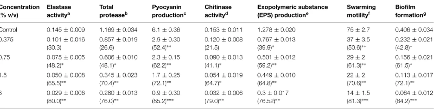
TABLE 1 | Effect of sub-MICs of peppermint oil (PMO) on inhibition of quorum sensing regulated virulence factors inPseudomonas aeruginosaPAO1. Concentration (% v/v) Elastase activitya Total proteaseb Pyocyanin productionc Chitinase activityd Exopolymeric substance (EPS) productione
Swarming motilityf
Biofilm formationg
Control 0.145±0.009 1.169±0.034 6.1±0.36 0.153±0.011 1.278±0.020 75±2.7 0.406±0.034
0.375 0.101±0.016
(30.3)
0.857±0.019 (26.6)
2.9±0.30 (52.4)∗∗
0.120±0.008 (21.5)
0.767±0.013 (39.9)∗
37±3.5 (50.6)∗∗
0.232±0.021 (42.8)∗
0.75 0.075±0.005
(48.2)∗
0.606±0.010 (48.1)∗
2.3±0.15 (62.2)∗∗
0.090±0.013 (41.1)∗
0.501±0.012 (59.2)∗∗
29±2 (61.3)∗∗
0.156±0.021 (61.5)∗
1.5 0.050±0.008
(65.5)∗∗
0.345±0.023 (70.4)∗∗
1.7±0.25 (72.1)∗∗
0.054±0.019 (64.7)∗
0.449±0.010 (64.8)∗∗
22±2 (70.6)∗∗
0.113±0.017 (72.1)∗∗
3 0.029±0.006
(80.0)∗∗
0.280±0.013 (76.0)∗∗
0.9±0.30 (85.2)∗∗∗
0.032±0.006 (79.0)∗∗
0.3±0.017 (76.52)∗∗
14±1.5 (81.3)∗∗∗
0.064±0.012 (84.2)∗∗∗
aElastase activity is expressed as the absorbance at OD 495. bTotal protease activity is expressed as the absorbance at OD
600.
cPyocyanin concentrations were expressed as micrograms of pyocyanin produced per microgram of total protein. dChitinase activity is expressed as the absorbance at OD
570. eEPS production is expressed as absorbance at OD
480. fSwarming motility is expressed as diameter of swarm in mm. gBiofilm formation is expressed as OD
470after incubation with crystal violet.
The data represents mean values of three independent experiments. Significance at∗p≤0.05, significance at∗∗p≤0.005, significance at∗∗∗p≤0.001. Values in the
parentheses indicate percent reduction over control.
TABLE 2 | Effect of sub-MICs of PMO on inhibition of quorum sensing regulated virulence factors inAeromonas hydrophilaWAF-38.
Concentration (% v/v) Total proteasea EPS productionb Biofilm formationc
Control 0.847±0.018 0.834±0.038 0.310±0.024
0.1 0.639±0.034
(24.5)
0.508±0.017 (39.0)
0.187±0.014 (39.6)
0.2 0.413±0.012
(51.2)∗
0.330±0.011 (60.4)∗
0.143±0.015 (53.8)∗
0.4 0.320±0.013
(62.2)∗
0.238±0.015 (71.4)∗∗
0.090±0.021 (70.9)∗∗
0.8 0.245±0.019
(71.0)∗∗
0.184±0.006 (77.9)∗∗∗
0.078±0.013 (74.8)∗∗
aTotal protease activity is expressed as the absorbance at OD 600.
bEPS production is expressed as absorbance at OD 480. cBiofilm formation is expressed as OD
470after incubation with crystal violet.
The data represents mean values of three independent experiments. Significance at∗
p≤0.05, significance at∗∗
p≤0.005, significance at∗∗∗
p≤0.001 Values in the parentheses indicate percent reduction over control.
feasible way. The grid centers of 24.327, 11.9665, and 77.3765 with the grid sizes of 40.368, 46.223, and 56.579 for X, Y, and Z axes, respectively, were used for covering all the binding site residues. For docking simulations the exhaustiveness was set to 8. Best docked structures based on the binding energy scores (G) were chosen for further analyses. The hydrogen bonding and hydrophobic interactions between ligand and protein were calculated by Accelrys DS Visualizer 2.0 (Accelrys Software Inc., 2012) while theFigure 2were generated by PyMol version 0.99 (DeLano, 2002) and Accelrys DS Visualizer 2.0 (Accelrys Software Inc., 2012). To validate the LasR-inhibitor docking experiments, we have compared our docking results with the structures already available at the PDB where the the natural autoinducer AHL has been co-crystallized with LasR, as for example, 2UV0 (BottomLey et al., 2007). Here, all the ligands from the crystal structure have been removed and the AHL was re-docked.
The binding constants (Kb) for protein-ligand interactions were calculated from the obtained free energy changes of docking by using the following equation:
G = −RTlnKb
where R is the gas constant (1.987 cal.mol−1.K−1) and T is 298 K (Ahmad et al., 2011).
Analysis oflasB andpqsA Transcriptional Activity inE. coli
Measurements of β-galactosidase luminescence in E. coli
MG4/pKDT17 andE. colipEAL08-2 was done by the adopting the method described byPearson et al.(1994) andCugini et al.
(2007).
Statistical Analysis
All experiments were performed in triplicates and the data obtained from experiments were presented as mean values and the difference between control and test were analyzed using student’st-test.
Results
Minimum Inhibitory Concentration
Minimum inhibitory concentration of PMO was determined to select the sub-MICs to study the effect on growth and inhi-bition of QS regulated functions. MIC of oil was found to be 0.6% v/v against C. violaceumCVO26, 6.4% v/v against P. aeruginosaPAO1, and 1.6% v/v againstA. hydrophilaWAF38. The MIC of menthol was found to be 1 mg/mL, 800 µg/mL,
FIGURE 2 | (A)Inhibitors obtained from the essential oil of Mentha piperita as well as the natural autoinducer 3-oxo-C12-HSL acyl homoserine lactone (AHL) are docked in the hydrophobic pocket of LasR. (B) Residues involved in the interaction of LasR-inhibitors as revealed from
molecular docking 0, 3-oxo-C12-HSL; I, Limonene; II, Menthone; III, Isomenthone; IV, 1-Hydroxyoctane; V, Isopulegol; VI, Menthyl acetate; VII, Neoisomenthol; VIII, 2-Isopropyl-5-methylcyclohexanol; IX, Menthol; X, Lavandulol; XI, Piperitone.
Effect of Peppermint Oil on Violacein Production
Anti-QS property of M. piperita (peppermint) oil was firstly assessed for pigment inhibition in CVO26. The result of the vio-lacein quantification assay is depicted inFigure 1. The sub-MICs of PMO exhibited concentration-dependent violacein inhibitory activity at all tested concentrations and maximum reduction of 83.3% was recorded. Viable plate count performed on MHA plates at 24 h incubation showed no significant difference in the number of colony-forming units (CFUs) between untreated
C. violaceum CVO26 and C. violaceum CVO26 treated with sub-MICs of the oil (Figure 1).
Effect OF PMO on Production of Virulence Factors and Biofilm Formation
The effect of sub-MICs of PMO in reducing the production of QS-dependent LasB Elastase, protease and chitinase in PAO1 is
presented inTable 1. Significant decrease in LasB elastase activ-ity was observed in the culture supernatant of PAO1 treated with sub-MICs of PMO. A minimum of 30% inhibition was observed when PAO1 was cultured with oil at a concentration of 0.37% v/v and maximum of 80% inhibition was observed at 3% v/v oil con-centration. The impact of PMO in inhibiting the QS-dependent protease activity of PAO1 was also determined and results of the obtained showed 26.6, 48.1, 70.4, and 76% decrease in total pro-tease production when treated with 0.37, 0.75, 1.5, and 3% v/v oil concentrations, respectively. Treatment of PAO1 with sub-MICs of PMO showed significantly reduced chitinase activity, the oil (0.3–3% v/v) demonstrated inhibition in chitinase production to the level of 21–78% (Table 1).
TABLE 3 | Molecular docking results of LasR-inhibitor interactions.
Compounds≈ CID∗ K
b(M−1) G
(kcal.mol−1)
Residues involved
Common residues#
H-bonds
0 3246941 2.39×10⁶ −8.7 20 – 4
I 22311 1.15×10⁵ −6.9 12 8 0
II 26447 1.36×10⁵ −7.0 10 7 1
III 6986 1.61×10⁵ −7.1 14 8 0
IV 957 5.50×10³ −5.1 12 7 1
V 24585 5.25×10⁵ −7.8 13 12 1
VI 27867 4.94×10⁴ −6.4 16 10 1
VII 19244 1.36×10⁵ −7.0 10 7 0
VIII 1254 3.16×10⁵ −7.5 12 8 0
IX 165675 6.22×10⁵ −7.9 14 13 2
X 5464156 3.52×10⁴ −6.2 10 8 0
XI 6987 1.90×10⁵ −7.2 10 7 0
≈
0, 3-oxo-C12-HSL; I, Limonene; II, Menthone; III, Isomenthone; IV, 1-Hydroxyoctane; V, Isopulegol; VI, Menthyl acetate; VII, Neoisomenthol; VIII, 2-Isopropyl-5-methylcyclohexanol; IX, Menthol; X, Lavandulol; XI, Piperitone.
∗
PubChem chemical identifier.
#With respect to autoinducer or natural inhibitor (3-oxo-C12-HSL).
FIGURE 3 | Quantitative assessment of violacein inhibition in CVO26 by sub-MICs of menthol.All of the data are presented as mean±SD. Significance at∗p≤0.05, significance at∗∗p≤0.005, significance at ∗∗∗p≤0.001.
PAO1, at concentrations ranging from 0.3 to 3% v/v as compared to the control. The effect of PMO on swarming motility of PAO1 was also examined. The oil effectively inhibited QS dependent swarming migration in a dose dependent manner inP. aeruginosa
PAO1. The maximum reduction of 81.3% in swarming migration was recorded at highest tested concentration (3% v/v) followed by 70.6, 61.3, and 50.6% at 1.5, 0.75, and 0.37% v/v oil concentration, respectively, (Table 1).
The spectrometric analysis of EPS extracted from oil treated and untreated cultures of PAO1 revealed that the production of EPS decreased with increasing concentration of PMO. The test oil at 3% v/v concentration exhibited 76% decrease in EPS pro-duction inP. aeruginosaPAO1. Anti-biofilm activity of the test
oil shows biofilm inhibition of the pathogen. Addition of 0.37, 0.75, 1.5, and 3% v/v concentration of oil led to a dose dependent
reduction in biofilm formation in the order of 42.7, 61, 73, and 84%, respectively, (Table 1).
Similarly, the oil of peppermint (0.1–0.8% v/v) effectively interfered with the QS system of A. hydrophila WAF38 by significantly reducing the total protease activity to the level of 24.5–71% (p ≤ 0.005) and EPS production by 39–77.9%.
Maximum decrease (74.8%) in biofilm formation at 0.8% v/v concentration of the oil was observed as depicted inTable 2.
Effect of Peppermint Oil onlasSystem
The addition of PMO decreased significantβ-galactosidase activ-ity inE. coliMG4/pKDT17 by up to 41.9 and 54.5% at 1.5 and 3% v/v (Figure 5), demonstrating that PMO reduces AHL levels significantly to inhibit las-controlled transcription.
GC-MS Analysis of Peppermint Oil
Major ingredient of PMO as revealed by GC-MS analy-sis was menthol (36.87%), and other constituents identi-fied were menthone (16.44%), neoisomenthol (11.33%), iso-menthone (10.47%), menthyl acetate (7.47%), 2-isopropyl-5-methylcyclohexanol (2.74%), piperitone (2.17%), and limonene (0.53%) as given in Supplementary Table S1 and depicted in Supplementary Figure S1.
Molecular Docking Analysis
For the better understanding of LasR-phytoconstituents binding the complementary applications of molecular docking of com-pounds on LasR has been performed with Autodock simulation analyses. The best energy ranked results are shown inFigure 2
TABLE 4 | Effect of sub-MICs of menthol on inhibition of quorum sensing regulated virulence factors inP. aeruginosaPAO1.
Concentration (µg ml−1)
Elastase activitya
Total proteaseb
Pyocyanin productionc
Chitinase activityd
EPS productione
Swarming motilityf
Biofilm formationg
Control 0.141±0.024 1.010±0.027 5.47±0.1 0.128±0.015 0.997±0.032 55±2.7 0.677±0.050
100 0.092±0.015
(34.7)
0.661±0.033 (34.5)
2.2±0.43 (59.7)∗
0.109±0.015 (14.8)
0.612±0.038 (38.6)
41±3.5 (25)
0.577±0.022 (14.7)
200 0.062±0.019
(56)∗∗
0.351±0.029 (65.2)∗∗
2.0±0.1 (63.4)∗
0.084±0.022 (34.3)∗
0.581±0.016 (41.7)
27±4.0 (51)∗
0.384±0.041 (43.2)∗
400 0.034±0.012
(75.8)∗∗∗
0.199±0.019 (80.2)∗∗∗
1.4±0.32 (74.4)∗∗
0.071±0.010 (44.5)∗
0.515±0.014 (48.3)∗
18±2.0 (67)∗∗
0.311±0.035 (54)∗∗
800 0.030±0.009
(78.7)∗∗
0.159±0.019 (84.2)∗∗∗
0.9±0.038 (83.5)∗∗∗
0.058±0.014 (54.6)∗
0.421±0.009 (57.7)∗
12±3.5 (78)∗∗
0.207±0.009 (69.4)∗∗
aElastase activity is expressed as the absorbance at OD 495. bTotal protease activity is expressed as the absorbance at OD
600.
cPyocyanin concentrations were expressed as micrograms of pyocyanin produced per microgram of total protein. dChitinase activity is expressed as the absorbance at OD
570. eEPS production is expressed as absorbance at OD
480. fSwarming motility is expressed as diameter of swarm in mm. gBiofilm formation is expressed as OD
470after incubation with crystal violet.
The data represents mean values of three independent experiments. Significance at *p≤0.05, significance at **p≤0.005, significance at∗∗∗p≤0.001.
Values in the parentheses indicate percent reduction over control.
FIGURE 4 | Scanning electron microscopic (SEM) images for inhibition of biofilm ofPseudomonas aeruginosaPA01 at sub-MICs. (A)Control;(B)PMO (3% v/v);(C)Menthol (800µg mL−1);(D)Aeromonas hydrophilaWAF38control;(E)PMO (0.8% v/v );(F)menthol (200µg mL−1).
as in the ligand-complexed crystal structure. This docked struc-ture was further evaluated by superimposing with the LasR-AHL bound crystal structure and was found to be quite significant. From the above interacted residues Y47, A50, Y56, W60, Y64, D73, V76, and S129 are the main residues responsible for the acti-vation of LasR. In case of all the 11 phytocompounds, most of the residues involved in the interactions are common with the natural ligand AHL (Table 3). Flexible ligands were set to free in the LasR to dock in the most feasible orientation according to the least free energy change (G) and the interactions between the LasR and
the inhibitors were exclusively hydrophobic in nature as reflected by several non-polar residues at binding site. These residues were in the proximity distance of 5 Å of the bound ligands. The essential oil constituents were docked in the active site with binding affinities ranging from−5.1 to−7.9 kcal/mol as
com-pared to the binding affinity of AHL (−8.7 kcal/mol) and from
TABLE 5 | Effect of sub-MICs of menthol on inhibition of quorum sensing regulated virulence factors inA. hydrophilaWAF-38.
Concentration (µg ml−1)
Total proteasea
EPS productionb
Biofilm formationc
Control 0.854±0.043 1.06±0.038 0.301±0.016
25 0.754±0.024
(11.7)
0.698±0.024 (34.1)
0.217±0.031 (27.9)
50 0.636±0.023
(25.5)
0.442±0.010 (58.3)∗
0.166±0.012 (44.8)∗
100 0.512±0.034
(40.0)
0..369±0.026 (65.1)∗
0.082±0.007 (72.7)∗∗
200 0.405±0.02
(52.5)∗
0.353±0.018 (66.6)∗
0.06±0.003 (80.0)∗∗∗
aTotal protease activity is expressed as the absorbance at OD 600. bEPS production is expressed as absorbance at OD
480. cBiofilm formation is expressed as OD
470after incubation with crystal violet.
The data represents mean values of three independent experiments. Significance at∗
p≤0.05, significance at **p≤0.005, significance at∗∗∗
p≤0.001. Values in the parentheses indicate percent reduction over control.
FIGURE 5 | Effect of PMO onlassystems.β-Galactosidase activity was measured in theEscherichia coliMG4/pKDT17 with and without sub-MICs of PMO. All of the data are presented as mean±SD. Significance at∗
p≤0.05.
most of the residues of LasR interacted hydrophobically with all the 11 tested compounds. Highest affinities are shown by men-thol followed by isopulegol and 1-Hydroxyoctane as given in
Table 3.
Effect of Menthol on Violacein Production
Menthol exhibited a concentration dependent decrease in QS regulated violacein production. Maximum reduction of 85% was recorded at 400µg/mLconcentration while lowest of 26%
decrease over control was observed at 50µg/mLmenthol concen-tration (Figure 3).
Effect of Menthol on Virulence Factor Production
Effect of menthol on QS regulated virulence factors ofP. aerug-inosa PAO1 revealed a concentration dependent decrease in
all the functions. Highest reduction in all the virulence factors was observed at 800µg/mLin PAO1. Decrease in total protease
activity was highest (84.2%) followed by pyocyanin production (83.5%), elastase activity (78.7%), swarming motility (78%), EPS production (57.7%), and least in chitinase activity (54.6%) as presented inTable 4.
In A. hydrophila WAF38, menthol inhibited total protease significantly (52.5%) at 200 µg mL−1 while at lower
concen-trations reduction observed was not statistically significant. EPS produced by untreatedA. hydrophilaWAF38 was lowered signif-icantly (58.3–66.6%) at sub-MICs (50–200µg/mL) as depicted in
Table 5.
Effect of Menthol on Biofilm Formation
A significant decrease in biofilm formation was observed in test bacterial strains when grown in the presence of menthol. Highest reduction (69.4%) in biofilm formation was observed at 800 µg/mLconcentration followed by 54 and 43.2% reduction at 400 and 200µg/mL, respectively. To ascertain the structures
visualized by light microscopy that exhibited antibiofilm activ-ity, we used SEM to elucidate the potential of menthol against biofilm formation (Figure 4).The results of electron microscopic analysis revealed that the control slides showed well developed dense biofilm growth of PAO1, whereas, PAO1 treated with the menthol developed poor biofilm growth compared to that of the control sample (Figure 4). In A. hydrophila WAF38, biofilm formation was also reduced considerably ranging from 27.9–80% over untreated control at sub-MICs of menthol tested (Table 5).
Effect of Menthol onlasandpqsSystems
To exclude the influence of other QS systems in P. aerug-inosa, we used E. coli MG4/pKDT17 that produces LasR and contains the lasB promoter fused to lacZ. The addition of menthol decreased β-galactosidase luminescence in E. coli
MG4/pKDT17 up to 60% at 800 µg/mL (Figure 6A), which shows that menthol directly inhibits las-controlled transcrip-tion.
Pyocyanin production which is mainly regulated by the pqs system, therefore a heterologous strain E. coli pEAL08-2 that produces PqsR and contains thepqsA promoter fused to lacZ
was used to determine whether the inhibition of pyocyanin pro-duction was directly due to the effects of pqs system (Cugini et al., 2007). The addition of menthol reduced theβ-galactosidase luminescence in E. coli pEAL08-2 up to 55% at 800 µg/mL (Figure 6B), which proved that menthol directly inhibits PQS-stimulated transcription.
In VivoAssessment withC. elegans
The anti-infection potential of the sub-MIC of menthol was assessed using a liquid killing assay ofC. elegansanimal model by PAO1 in a 24-well microtitre plate. Complete mortality of the P. aeruginosa PAO1 preinfected C. elegans was observed within 72 h. However, C. elegans preinfected with PAO1 fur-ther treated with menthol (800 µg/mL) displayed enhanced
survival upto 58% (Figure 7). However, menthol alone
FIGURE 6 | Effect of menthol onlasandpqssystems. (A)β-Galactosidase activity was measured in theE. coliMG4/pKDT17 with and without sub-MICs of menthol.(B)β-Galactosidase activity was measured in theE. colipEAL08-2 with and without menthol. All of the data are presented as mean±SD. Significance at
∗p≤0.05, significance at∗∗p≤0.005.
Discussion
In the present investigation, we showed the QS inhibitory poten-tial of PMO and menthol based on its ability to inhibit AHL-dependent violacein production inC. violaceum and virulence factors such as elastase, protease, pyocyanin, EPS production, and biofilm formation in PAO1. It has previously been confirmed that in C. violaceum, the CviIR-dependent QS system coordi-nates the production of violacein pigment, and the compound, which has the ability to inhibit the violacein production without any antibacterial activity, is considered to be the promising QS inhibitor (Choo et al., 2006;Zhu and Sun, 2008). In this study, a dose dependent decrease of AHL mediated violacein production
FIGURE 7 | Anti-infection potential of sub-MIC of menthol (800µg/mL) in increasing the survival ofCaenorhabditis eleganspreinfected with P. aeruginosaPAO1.Means values of triplicate independent experiments and SDs are shown.
was recorded inC. violaceumCVO26. The concentration depen-dent response of anti-QS potential as found in our study is also supported by the findings on clove oil (Khan et al., 2009), mar-joram (Kerekes et al., 2013), and three isothiocyanates (Borges et al., 2014) that demonstrated reduction in AHL dependent vio-lacein production inC. violaceumCVO26 without inhibition of growth.
The LasIR-encoded protease and elastase play a key role in the pathogenesis of PAO1 (Kessler et al., 1993). These enzymes degrade the structural components of the infected tissue and enhance the growth and invasiveness of the organism. In this present investigation, the oil of peppermint and menthol demon-strated a concentration-dependent inhibition of virulence factor production such as total protease, elastase in PAO1, as shown in
Table 1. This result is in agreement with the study ofAdonizio et al. (2008), who demonstrated significant inhibition of LasB activity by medicinal plants from Florida. In addition to this recently, flavanones (Vandeputte et al., 2011), Lagerstroemia speciosafruit extract (Singh et al., 2012) andSclerocarya birrea
bark extract (Sarkar et al., 2014) have shown to inhibit elas-tase activity to substantial levels. Our data shows that menthol decreases both the elastase activity of PAO1 and the transcrip-tional activation oflasBinE. coli, which indicates that menthol inhibits thelassystem. Pyocyanin metabolite causes severe toxic effects by damaging the neutrophil-mediated host defense in patients with cystic fibrosis (Fothergill et al., 2007). Similarly, menthol reduced both the pyocyanin production of PAO1 and the transcriptional activation ofpqsAinE. coli, which indicates that menthol inhibits thepqssystem. Considering thelasandpqs
the production of pyocyanin significantly. QS-regulated flagellar and pili dependent swarming motility is considered as one of the virulence factors because of its involvement in biofilm formation (Niu and Gilbert, 2004). In the present study, the oil of pepermint and menthol reduced the swarming migration of target organ-ism. Our findings are in agreement with the results ofKhan et al.
(2009) in which sub-MICs of clove oil decreased the swarming behavior in PAO1 by 78%. Recently, studies onCapparis spinosa,
Cuminum cyminum,andS. birrea(Packiavathy et al., 2011, 2012;
Sarkar et al., 2014) have shown significant reduction in swarming motility ofP. aeruginosaPAO1.
Exopolymeric substance plays a defining role in maintain-ing the biofilm architecture. Therefore, decrease in production of EPS will render the biofilm structure weak and suscepti-ble (Bomchil et al., 2003). The observed results in the present study indicated the potential PMO to inhibit EPS production in PAO1 at sub-MIC values. This is probably the first report on the inhibition of EPS byM. piperitaoil inP. aeruginosaPAO1. Formation of biofilm by PAO1 is QS regulated and this mode of growth confers increased drug resistance and infection caused is quite severe in patients suffering from cystic fibrosis. AHL medi-ated cell–cell signaling plays a crucial role in the initiation and maturation of biofilms in PAO1 (Rasmussen et al., 2005). The results of our present investigation revealed the biofilm inhibit-ing potential of PMO and its major phytoconstituent menthol against PAO1 biofilms in a concentration dependent manner, as shown in Figure 3. Recent studies on herbal extract ofM. piperitahave demonstrated 57% reduction in adhesion property ofP.aeruginosa(Sandasi et al., 2011); our findings on the biofilm inhibitory property ofM. piperitaoil finds support from above work. Further, the results obtained are comparable to the decrease in biofilm formation reported in studies for methyl eugenol (Packiavathy et al., 2012), marjoram (Kerekes et al., 2013), and
Rosa rugosatea extract (Zhang et al., 2014).
The mortality of the nematode by PAO1 is caused by the cyanide asphyxiation and paralysis (Gallagher and Manoil, 2001). We demonstrated an enhanced survival of pre-infected nema-tode model maintained with menthol (800µg/mL) as depicted
in the Figure 7. The data obtained clearly indicates that the
menthol interferes with the QS of PAO1 leading to reduced mor-tality of theC. elegans. The findings are similar to the previous observations ofHusain et al.(2013), on enhanced survival of the nematode (C. elegans) after treatment with sub-MICs of clove oil.
Conclusion
In many pathogens, virulence potential and biofilm formation is under the regulation of QS. Therefore, instead of bactericidal or bacteriostatic strategies, attenuation of virulence via QS inhibi-tion approach could be more effective strategy to combat bacterial infection caused by drug resistant bacteria.. In the present study oil of M. piperita and menthol showed QS inhibitory proper-ties, resulting in decreased biofilm formation capabilities and the attenuation ofP. aeruginosavirulencein vitroandin vivo. Findings of this study also suggest that essential oil ofM. piperita
and menthol can inhibit QS, biofilm and its related virulence processes in pathogenic bacteria, and can be exploited by the pharmaceutical industry for the development of new safe broad spectrum antibiofilm and anti-QS drugs with reduced toxicity and antibiotic resistance.
Acknowledgments
The authors extend their appreciation to the Deanship of Scientific Research at KSU for funding this work through research group project number RGP-215.
Supplementary Material
The Supplementary Material for this article can be found online at: http://journal.frontiersin.org/article/10.3389/fmicb. 2015.00420/abstract
References
Accelrys Software Inc. (2012).Discovery Studio Modeling Environment, Release 3. 5, San Diego: Accelrys Software Inc.
Adonizio, A. L., Kong, K. F., and Mathee, K. (2008). Inhibition of Quorum sensing-controlled virulence factor production inPseudomonas aeruginosaby south Florida plant extracts.Antimicrob. Agents Chemother.52, 198–203. doi: 10.1128/AAC.00612-07
Ahmad, E., Rabbani, G., Zaidi, N., Singh, S., and Rehan, M. (2011). Stereo-selectivity of human serum albumin to enantiomeric and isoelectronic pol-lutants dissected by spectroscopy, calorimetry and bioinformatics.PLoS ONE 6:e26186. doi: 10.1371/journal.pone.0026186
Atkinson, S., Chang, C. Y., Sockett, R. E., Cámara, M., and Williams, P. (2006). Quorum sensing inYersinia enterocoliticacontrols swimming and swarming motility.J. Bacteriol.188, 1451–1461. doi: 10.1128/JB.188.4.1451-14 61.2006
Blosser, R. S., and Gray, K. M. (2000). Extraction of violacein from Chromobacterium violaceum provides a new quantitative bioassay for N-acyl homoserine lactone autoinducers.J. Microbiol. Methods40, 47–55. doi: 10.1016/S0167-7012(99)00136-0
Bomchil, N., Watnick, P., and Kolter, R. (2003). Identification and charac-terization of a Vibrio cholerae gene, mbaA, involved in maintenance of biofilm architecture.J. Bacteriol.185, 1384–1390. doi: 10.1128/JB.185.4.1384-13 90.2003
Borges, A., Serra, S., Abreu, A. C., Saavedra, M. J., Salgado, A., and Simoes, M. (2014). Evaluation of the effects of selected phytochemicals on quorum sensing inhibition and in vitro cytotoxicity. Biofouling 30, 183–195. doi: 10.1080/08927014.2013.852542
BottomLey, M. J.,Muraglia, E.,Bazzo, R., and Carfì, A. (2007). Molecular insights into quorum sensing in the human pathogenPseudomonas aeruginosafrom the structure of the virulence regulator LasR bound to its autoinducer.J. Biol. Chem. 282, 13592–13600. doi: 10.1074/jbc.M700556200
Caraher, E., Reynolds, G., Murphy, P., McClean, S., and Callaghan, M. (2007). Comparison of antibiotic susceptibility of Burkholderia cepacia complex organisms when grown planktonically or as biofilm in vitro. Eur. J. Clin. Microbiol. Infect. Dis.26, 213–221. doi: 10.1007/s10096-007-0256-x
Clinical and Laboratory Standards Institute [CLSI]. (2007).Performance Standards for Antimicrobial Susceptibility Testing: Seventeenth Informational Supplement, Vol. 27. M100-S17. Wayne: CLSI.
Costerton, J. W., Lewandowski, Z., Caldwell, D. E., Korber, D. R., and Lappin-Scott, H. M. (1995). Microbial biofilms.Annu. Rev. Microbiol.49, 711–745. doi: 10.1146/annurev.mi.49.100195.003431
Cugini, C., Calfee, M. W., Farrow, J. M. III, Morales, D. K., Pesci, E. C., and Hogan, D. A. (2007). Farnesol, a common sesquiterpene, inhibits PQS production inPseudomonasaeruginosa.Mol. Microbiol.65, 896–906. doi: 10.1111/j.1365-2958.2007.05840.x
Davies, D. (2003). Understanding biofilm resistance to antibacterial agents.Nat. Rev. Drug Discov.2, 114–122. doi: 10.1038/nrd1008
DeLano, W. L. (2002).The PyMOL Molecular Graphics System, DeLano Scientific, CA, USA: San Carlos.
Fothergill, J. L., Panagea, S., Hart, C. A., Walshaw, M. J., Pitt, T. L., and Winstanley, C. (2007). Widespread pyocyanin overproduction among isolates of a cystic fibrosis epidemic strain.BMC Microbiol.7:45. doi: 10.1186/1471-2180-7-45
Fuqua, W. C., Winans, S. C., and Greenberg, E. P. (1994). Quorum sensing in bac-teria: the LuxR-LuxI family of cell density-responsive transcriptional regulators. J. Bacteriol.176, 269–275.
Gallagher, L. A., and Manoil, C. (2001).Pseudomonas aeruginosa PAO1 kills Caenorhabditis elegansby cyanide poisoning.J. Bacteriol.183, 6207–6214. doi: 10.1128/JB.183.21.6207-6214.2001
Häussler, S., and Becker, T. (2008). ThePseudomonasquinolone signal (PQS) balances life and death inPseudomonas aeruginosapopulations.PLoS Pathog. 4:e1000166. doi: 10.1371/journal.ppat.1000166
Heeb, S., Fletcher, M. P., Chhabra, S. R., Diggle, S. P., Williams, P., and Camara, M. (2011). Quinolones: from antibiotics to autoinducers.FEMS Microbiol. Rev.35, 247–274. doi: 10.1111/j.1574-6976.2010.00247.x
Hentzer, M., and Givskov, M. (2003). Pharmacological inhibition of quorum sensing for the treatment of chronic bacterial infections.J. Clin. Invest.112, 1300–1307. doi: 10.1172/JCI20074
Hentzer, M., Givskov, M., and Parsek, M. R. (2002). Targeting quorum sensing for treatment of chronic bacterial biofilm infections.Lab. Med. 33, 295–306. doi: 10.1309/EYEV-WT6T-GKHE-C8LM
Husain, F. M., and Ahmad, I. (2013). Quorum sensing inhibitors from natural products as potential novel anti-infective drug.Drugs Fut.38, 691–706. doi: 10.1358/dof.2013.038.10.2025393
Husain, F. M., Ahmad, I., Asif, M., and Tahseen, Q. (2013). Influence of clove oil on certain quorum sensing regulated functions and biofilm ofPseudomonas aeruginosaandAeromonas hydrophila.J. Biosci.38, 1–10. doi: 10.1007/s12038-013-9385-9
Kalia, V. C. (2013). Quorum sensing inhibitors: an overview.Biotechnol. Adv.31, 224–245. doi: 10.1016/j.biotechadv.2012.10.004
Kerekes, E. B., Deák, É., Takó, M., Tserennadmid, R., Petkovits, T., Vágvölgyi, C., et al. (2013). Anti-biofilm forming and anti-quorum sensing activity of selected essential oils and their main components on food-related micro-organisms. J. Appl. Microbiol.115, 933–942. doi: 10.1111/jam.12289
Kessler, E., Safrin, M., Olson, J. C., and Ohman, D. E. (1993). Secreted LasA ofPseudomonas aeruginosais a staphylolytic protease.J. Biol. Chem. 268, 7503–7508.
Khan, M. S. A., Zahin, M., Hasan, S., Husain, F. M., and Ahmad, I. (2009). Inhibition of quorum sensing regulated bacterial functions by plant essential oils with special reference to clove oil.Lett. Appl. Microbiol.49, 354–360. doi: 10.1111/j.1472-765X.2009.02666.x
LaSarre, B., and Federle, M. J. (2013). Exploiting quorum sensing to con-fuse bacterial pathogens. Microbiol. Mol. Biol. Rev. 77, 73–111. doi: 10.1128/MMBR.00046-12
March, J. C., and Bentley, W. E. (2004). Quorum sensing and bacte-rial cross-talk in biotechnology.Curr. Opin. Biotechnol. 15, 495–502. doi: 10.1016/j.copbio.2004.08.013
Masada, Y. (1976).Analysis of essential oils by gas chromatography and mass spectrometry. New York, NY: John Wiley & Sons.
McLean, R. J. C., Pierson, L. S. III, and Fuqua, C. (2004). A sim-ple screening protocol or the identification of quorum sensing signal antagonists.J. Microbiol. Methods 58, 351–360. doi: 10.1016/j.mimet.2004. 04.016
Musthafa, K. S., Balamurugan, K., Pandian, S. K., and Ravi, A. V. (2012). 2,5-Piperazinedione inhibits quorum sensing-dependent factor produc-tion in Pseudomonas aeruginosa PAO1. J. Basic Microbiol. 52, 1–8. doi: 10.1002/jobm.201100292
Niu, C., and Gilbert, E. S. (2004). Colorimetric method for identifying plant essen-tial oil components that affect biofilm formation and structure.Appl. Environ. Microbiol.70, 6951–6956. doi: 10.1128/AEM.70.12.6951-6956.2004
O’Toole, G. A., and Kolter, R. (1998). Initiation of biofilm formation in Pseudomonas fluorescensWCS365 proceeds via multiple, convergent signaling pathways: a genetic analysis.Mol. Microbiol.28, 449–461. doi: 10.1046/j.1365-2958.1998.00797.x
Obritsch, M. D., Fish, D. N., MacLaren, R., and Jung, R. (2005). Nosocomial infections due to multidrug-resistant Pseudomonas aeruginosa: epi-demiology and treatment options. Pharmacotherapy 25, 1353–1364. doi: 10.1592/phco.2005.25.10.1353
Packiavathy, I. A. S. V., Agilandeswari, P., Musthafa, K. S., Pandian, S. K., and Ravi, A. V. (2012). Antibiofilm and quorum sensing inhibitory potential ofCuminum cyminumand its secondary metabolite methyl eugenol against Gram negative bacterial pathogens.Food Res. Int.45, 85–92. doi: 10.1016/j.foodres.2011.10.022 Packiavathy, I. A. S. V., Agilandeswari, P., Ramaswamy, B. R., Pandian, S. K., and Ravi, A. V. (2011). Antiquorum sensing and antibiofilm potential ofCapparis spinosa.Arch. Med. Res. 42, 658–668. doi: 10.1016/j.arcmed.2011.12.002 Pearson, J. P., Gray, K. M., Passador, L.,Tucker, K. D., Eberhard, A., Iglewski,
B. H., et al. (1994). Structure of the autoinducer required for expression of Pseudomonas aeruginosa virulence genes.Proc. Natl. Acad. Sci. U.S.A. 91, 197–201. doi: 10.1073/pnas.91.1.197
Rasmussen, T. B., Bjarnsholt, T., Skindersoe, M. E., Hentzer, M., Kristoffersen, P., Kote, M., et al. 2005). Screening for quorum-sensing inhibitors (QSI) by use of a novel genetic system, the QSI selector.J. Bacteriol.187, 1799–1814. doi: 10.1128/JB.187.5.1799-1814.2005
Rasmussen, T. B., and Givskov, M. (2006). Quorum sensing inhibitors: a bargain of effects.Microbiology152, 895–904. doi: 10.1099/mic.0.28601-0
Rumbaugh, K. P., and Armstrong, A. (eds). (2014). “The role of quorum sensing in biofilm development,” inAntibiofilm Agents, Springer Series on Biofilm,Vol. 8, From Diagnosis to Treatment and Prevention(Berlin: Springer), 97–113. Rumbaugh, K. P., Trivedi, U., Watters, C., Burton-Chellew, M. N., Diggle, S. P., and
West, S. A. (2012). Kin selection, quorum sensing and virulence in pathogenic bacteria.Proc. R. Soc. B279, 3584–3588. doi: 10.1098/rspb.2012.0843 Sandasi, M., Leonard, C. M., Van Vuuren, S. F., and Viljoen, A. M. (2011).
Peppermint (Mentha piperita) inhibits microbial biofilms in vitro.S. Afr. J. Bot. 77, 80–85. doi: 10.1016/j.sajb.2010.05.011
Sarkar, R., Chaudhary, S. K., Sharma, A., Yadav, K. Y., Nema, N. K., Sekhoacha, M., et al. (2014). Anti-biofilm activity of Marula- A study with standard-ized bark extract.J. Ethnopharmacol.154, 170–175. doi: 10.1016/j.jep.2014. 03.067
Singh, B. N., Singh, H. B., Singh, A., Singh, B. R., Mishra, A., and Nautiyal, C. S. (2012).Lagerstroemia speciosafruit extract modulates quorum sensing-controlled virulence factor production and biofilm formation inPseudomonas aeruginosa.Microbiology158, 529–538. doi: 10.1099/mic.0.052985-0 Szabo, M. A., Varga, G. Z., Hohmann, J., Schelz, Z., and Szegedi, E. (2010).
Inhibition of quorum-sensing signals by essential oils.Phytother. Res. 24, 782–786.
Trott, O., and Olson, A. J. (2010). AutoDock vina: improving the speed and accuracy of docking with a new scoring function, efficient optimization and multithreading.J. Comp. Chem.31, 455–461.
Vandeputte, O. M., Kiendrebeogo, M., Rasamiravaka, T., Stevigny, C., Duez, P., Rajaonson, S., et al. (2011). The flavanone naringenin reduces the production of quorum sensing-controlled virulence factors inPseudomonas aeruginosaPAO1. Microbiology157, 2120–2132. doi: 10.1099/mic.0.049338-0
Williams, P., Winzer, K., Chan, W. C., and Camara, M. (2007). Look who’s talking: communication and quorum sensing in the bacterial world.Philos. Trans. R. Soc. Lond. B Biol. Sci.362, 1119–1134. doi: 10.1098/rstb.2007.2039
Xavier, K. B., and Bassler, B. L. (2003). LuxS quorum sensing: more than just a numbers game. Curr. Opin. Microbiol.6, 191–197. doi: 10.1016/S1369-5274(03)00028-6
with antibiotic against plasmid-conferred multi-drug-resistantEscherichia coli. J. Appl. Microbiol.116, 1119–1128. doi: 10.1111/jam.12444
Zahin, M., Hasan, S., Aqil, F., Khan, M. S. A., Husain, F. M., and Ahmad, I. (2010). Screening of Indian medicinal plants for their anti-quorum sensing activity. Indian J. Exp. Biol.48, 1219–1224.
Zeng, Z., Qian, L., Cao, L., Tan, H., Huang, Y., Xue, X., et al. (2008). Virtual screening for novel quorum sensing inhibitors to eradicate biofilm formation of Pseudomonas aeruginosa.Appl. Microbiol. Biotechnol. 79, 119–126. doi: 10.1007/s00253-008-1406-5
Zhang, J., Rui, X., Wang, L., Guan, Y., Sun, G., and Dong, M. (2014). Polyphenolic extract fromRosa rugosatea inhibits bacterial quorum sensing and biofilm formation.Food Control42, 125–131. doi: 10.1016/j.foodcont.2014.02.001 Zhou, L., Zheng, H.,Tang, Y.,Wengong, Y. U., and Qianhong, G. (2013).Eugenol
inhibits quorum sensing at sub-inhibitory concentrations.Biotechnol. Lett.35, 631–637. doi: 10.1007/s10529-012-1126-x
Zhu, H., and Sun, S. J. (2008). Inhibition of bacterial quorum sensing-regulated behaviors byTremella fuciformisextract.Curr. Microbiol. 57, 418–422. doi: 10.1007/s00284-008-9215-8
Conflict of Interest Statement:The authors declare that the research was con-ducted in the absence of any commercial or financial relationships that could be construed as a potential conflict of interest.