Ellagic Acid Derivatives from
Terminalia chebula
Retz.
Downregulate the Expression of Quorum Sensing Genes
to Attenuate
Pseudomonas aeruginosa
PAO1 Virulence
Sajal Sarabhai1, Prince Sharma1, Neena Capalash2*
1Department of Microbiology, Panjab University, Chandigarh, India,2Department of Biotechnology, Panjab University, Chandigarh, India
Abstract
Background: Burgeoning antibiotic resistance in Pseudomonas aeruginosa has necessitated the development of anti pathogenic agents that can quench acylhomoserine lactone (AHL) mediated QS with least risk of resistance. This study explores the anti quorum sensing potential ofT. chebulaRetz. and identification of probable compounds(s) showing anti QS activity and the mechanism of attenuation ofP. aeruginosaPAO1 virulence factors.
Methods and Results:Methanol extract ofT. chebulaRetz. fruit showed anti QS activity usingAgrobacterium tumefaciens
A136. Bioactive fraction (F7), obtained by fractionation of methanol extract using Sephadex LH20, showed significant reduction (p,0.001) in QS regulated production of extracellular virulence factors inP. aeruginosaPAO1. Biofilm formation and alginate were significantly (p,0.05) reduced with enhanced (20%) susceptibility to tobramycin. Real Time PCR of F7 treatedP. aeruginosashowed down regulation of autoinducer synthase (lasIandrhlI) and their cognate receptor (lasRand
rhlR) genes by 89, 90, 90 and 93%, respectively. Electrospray Ionization Mass Spectrometry also showed 90 and 64% reduction in the production of 3-oxo-C12HSL and C4HSL after treatment. Decrease in AHLs as one of the mechanisms of quorum quenching by F7 was supported by the reversal of inhibited swarming motility in F7-treatedP. aeruginosaPAO1 on addition of C4HSL. F7 also showed antagonistic activity against 3-oxo-C12HSL-dependent QS inE. colibioreporter.C. elegans fed on F7-treatedP. aeruginosashowed enhanced survival with LT50 increasing from 24 to 72 h. LC-ESI-MS of F7 revealed the presence of ellagic acid derivatives responsible for anti QS activity inT. chebulaextract.
Conclusions:This is the first report on anti QS activity ofT. chebulafruit linked to EADs which down regulate the expression oflasIRandrhlIRgenes with concomitant decrease in AHLs inP. aeruginosaPAO1 causing attenuation of its virulence factors and enhanced sensitivity of its biofilm towards tobramycin.
Citation:Sarabhai S, Sharma P, Capalash N (2013) Ellagic Acid Derivatives fromTerminalia chebulaRetz. Downregulate the Expression of Quorum Sensing Genes to AttenuatePseudomonas aeruginosaPAO1 Virulence. PLoS ONE 8(1): e53441. doi:10.1371/journal.pone.0053441
Editor:Vasu D. Appanna, Laurentian University, Canada
ReceivedSeptember 5, 2012;AcceptedNovember 28, 2012;PublishedJanuary 8, 2013
Copyright:ß2013 Sarabhai et al. This is an open-access article distributed under the terms of the Creative Commons Attribution License, which permits unrestricted use, distribution, and reproduction in any medium, provided the original author and source are credited.
Funding:Funding in the form of a fellowship to Sajal Sarabhai from the Council of Scientific and Industrial Research (CSIR) is acknowledged [Grant no:09/ 135(0544)/2008-EMR-I]. The funders had no role in study design, data collection and analysis, decision to publish, or preparation of the manuscript.
Competing Interests:The authors have declared that no competing interests exist.
* E-mail: [email protected]
Introduction
P. aeruginosa is the major cause of secondary infections in immunocompromised patients with cystic fibrosis, burn wound and HIV causing maximum morbidity and mortality [1]. It is a clinically important opportunistic pathogen responsible for 57% of total nosocomial infections [2]. To facilitate the establishment of infection,P. aeruginosaproduces both cell-associated and extracel-lular virulence factors globally regulated by well defined quorum sensing systems arranged in hierarchical manner withlassystem at the top, positively controlling the activity ofrhlsystem [3]. Thelas system utilizes N-(3-oxododecanoyl)-L-homoserine lactone (3-oxo-C12HSL) whereasrhlsystem functions by means of N-butanoyl-L-homoserine lactone (C4HSL) as the signal molecules [4]. Intermediate between the two is the quinolone system which utilizes 2-heptyl-3-hydroxy-4-quinolone as the signal molecule [5]. Pyocyanin and rhamnolipids production is controlled by rhlIR system whereas elastase and proteolytic activities bylasIRsystem [6]. P. aeruginosa also adopts biofilm mode of growth that is
regulated jointly by lasIR and rhlIR system [7] making it recalcitrant to various antimicrobial treatments.
reduces the cellular concentration of transcriptional regulatorluxR ofVibrio fischericloned inE. coli[11]. Curcumin, fromCurcuma longa [12], Ajoene from Allium sativum [13], Iberin from Armoracia rusticana[14] attenuate P. aeruginosa virulence by downregulating the expression of QS genes. Bioreporter strains with promoters of QS genes fused withlacZshowed downregulation of QS genes on treatment with aqueous extracts ofConocarpus erectus,Bucida buceras and Callistemon viminalis [15]. Plant polyphenols like epigalloca-techin, ellagic acid, tannic acid [16] and chemically synthesized 4-Nitro-pyridine–N-oxide [17], S-adenosylhomocysteine [18], iso-thiocyanate [19] were capable of antagonizing AHL dependent QS in bioreporter strains. However, their toxicity in mammalian cells limits their use as drugs [20].
T. chebula Retz. belongs to Combretaceae family [21] and is commonly known as harad or black myroblans. A very well known ayurvedic formulation in India known as ‘‘Triphala’’ contains equal parts ofT. chebula,T. bellericaandEmbilica officinalisand has been scientifically proven to promote immunity, health and longevity [22]. Organic and aqueous extracts ofT. chebulaexhibit antioxidant [23], antimicrobial [24], antianaphylactic [25], antidiabetic [26], antimutagenic [27], anticancerous [28], apo-ptotic [29], anticaries [30], antifungal [31] and antiviral [32] activities.T. chebulafruit extract is effective antimicrobial against methicillin resistantStaphylococcus aureusand trimethoprim-sulpha-methoxazole resistant uropathogenicE. colistrain [33]. To the best of our knowledge,Terminaliaspecies have not been explored for anti QS activity. However, tannin rich fraction ofT. cattappahas been shown to inhibit QS regulated violacein production in Chromobacterium violaceum JCM1249 and QS controlled biofilm maturation and LasA staphylolytic activity inP. aeruginosaATCC 10145 [34]. The broad spectrum of activities in T. chebula is attributed to the presence of different types of phytochemicals where hydrolysable tannins contributed 40% of the total content that includes simple gallate esters, ellagic acid derivatives and glycosides, and various ellagitannins [35]. This study explores the anti QS potential ofT. chebula fruit for attenuation of virulence factors ofP. aeruginosaPAO1 and identification of compounds (s) responsible for the activity. Mechanism of anti QS activity has also been elucidated.
Materials and Methods
Bacterial strains, plasmids and culture conditions are described in Table 1.
Preparation of Plant Extract
Dried fruit of T. chebula, confirmed as T. chebula Retz. by National Institute of Scientific Communication And Information Resource (NISCAIR), New Delhi, India, was ground to fine powder and extracted with water and methanol in Soxhlet apparatus for 10–12 h, separately. The extracts were air dried and reconstituted in water and methanol, respectively.
Disc Diffusion Assay for Anti QS Activity
A136 was used as biosensor strain for disc diffusion assay [43]. 10ml of aqueous and methanol extracts ofT. chebulawere used in the assay with curcumin (3mg/ml) [12], methanol and water as controls.
The integrity of AHLs in the presence ofT. chebulaextract was also checked using biosensor A136. AHLs were extracted from 100 ml of cell free supernatant of P. aeruginosa PAO1 using acidified ethyl acetate [44] and dissolved in 100ml of DMSO. 20ml of AHLs were incubated with 0.5 mg/ml ofT. chebulaextract for overnight at 37uC. AHLs were re-extracted after incubation and AHL mediatedb-galactosidase activity in biosensor A136 was estimated [45].
Separation of Bioactive Fraction from the Methanol Extract ofT. chebula
4.0 mg of methanol extract, dissolved in ethanol, was loaded onto the Sephadex LH 20 column (GE healthcare) (3064 cm with
i.d. 2 cm) and fractionation was conducted by successive elution of sample with increasing concentration of methanol (0, 5, 10, 20, 30, 50 and 100%) in ethanol (100 ml of each solvent). Individual fractions (F1–F7) were dried at 30uC and re-suspended in 1 ml of methanol. Phytochemical analysis of the fractions was done for the presence of terpenoides, flavonoids, saponins, tannins and alkaloids [46]. The fraction that inhibited the production of violacein pigment in biosensor CVO26 [47] was used for further work.
Table 1.Bacterial strains and plasmids used in this study.
Strain Genotype or phenotype Growth conditions Reference or source
P. aeruginosa
PAO1 Wild type LB or bactopeptone at 37uC 36
PAOJP2 lasI2rhlI2derivative of PAO1;TetrHgr LB or bactopeptone at 37
uC; Tetracycline 50mg/ml,HgCl215mg/ml
37
GFP tagged PAO1 pSM2472withgfp LB at 37uC 38
A.tumefaciensA136 pCF372withtraI::lacZandpCF218 withtraR;Spectr,Tetr
LB at 30uC;Spectinomycin 50mg/ml, Tetracycline 5mg/ml
39
C. violaceumO26 Mini Tn5 mutant of CV31532;Kmr LB at 30uC;Kanamycin 30mg/ml 40
E.coli
OP50 Wild type food forC. elegans LB or Nematode Growth Medium at 37uC CGC centre,
University of Minnesota, Twin cities, USA
DH5a(pSC11+pJN105L) PlasI::lacZ;lasRin pJN105;Gmrampr LB at 37
uC;Ampicllin 100mg/ml, Gentamycin 15mg/ml
41, 42
Quantitation of Extracellular Virulence Factors ofP. aeruginosa PAO1
Overnight grown culture of P. aeruginosa PAO1 was diluted with fresh 2% bactopeptone (1:100) and incubated at 37uC for 16 h at 150 rpm. PAOJP2, autoinducer mutant (lasI2rhlI2) was
taken as negative control. Cell free supernatant was used for quantification of virulence factors. Pyocyanin pigment was extracted from culture supernatant (5 ml) using chloroform in the ratio of 3:2 and re-extracted with 1.0 ml of 0.2 M HCl and absorbance was read at 540 nm [1]. 250ml elastin congo red
solution (5 mg/ml in 0.1 M Tris-HCl pH 8;1 mM CaCl2) was incubated with 750ml cell free supernatant at 37uC for 16 h at
200 rpm. The mixture was centrifuged at 3000 g for 10 min and absorbance was read at 490 nm to estimate elastase activity [47]. Rhamnolipids were quantitated by adjusting the pH of the culture supernatant to 2 with HCl and the resultant suspension was centrifuged at 8000 g for 10 min. Absorbance was read at 570 nm [48]. Protease activity was determined using 2% azocasein solution prepared in 50 mM phosphate buffer saline (PBS), pH 7. The substrate and culture supernatant were incubated at 37uC in 1:1 ratio for 1 h in a reaction volume of 400ml. The reaction was stopped by the addition of 500ml
of 10% trichloroacetic acid and centrifuged at 8000 g for 5 min to remove residual azocasein. The absorbance of supernatant was read at 400 nm [15]. All absorbance values were reported as OD of virulence factors per growth OD600 nm to normalize the effect of bioactive fraction on bacterial growth.
Alginate Production
P. aeruginosaPAO1 was grown in 10 ml LB for 3 days at 37uC in 35 mm petri plate under static conditions to form biofilm. The exhausted medium in the plate were collected and loosely adhered bacterial cells were removed by repeated washing with 0.85% saline. Alginate in culture supernatant was precipitated by 2% cetylpyridinium chloride and quantified by carbazole reagent [49].
Biofilm Formation
Biofilms were developed in 96 well polystyrene microtiter plate. 200ml ofP. aeruginosa(OD600 nm,1) culture in LB broth with 1% glycerol was incubated for 24 h at 37uC under static conditions. The supernatant surrounding the biofilm was collected and planktonic cells were quantitated by serial dilution method. Thereafter, biofilm was given three washings with PBS (50 mM, pH 7) to wash off loosely adhered planktonic bacterial cells. Subsequently, the biofilm was fixed with 200ml of methanol for
15 min, air dried and stained with 200ml of 0.5% (w/v) crystal violet for 15 min. The plate was washed with PBS (50 mM, pH 7) three times to remove excessive stain. 200ml of 95% (v/v) ethanol
was added to extract bound crystal violet and Biofilm Index was tabulated as OD570 nm/600 nm[50].
For visualizing biofilm, sterilized glass cover slips were immersed in LB broth containing 1% glycerol, inoculated with 1% of overnight grown GFP taggedP. aeruginosa[38] in a 35 mm petri plate and incubated at 37uC under static conditions. The medium was changed after every 24 h for 3 days. The coverslips were washed with PBS (50 mM, pH 7) and stained with 20mM
Propidium Iodide. Confocal Laser Scanning Microscope (CLSM) Figure 1. Schematic representation of bioassay guided fractionation ofT. chebulafruit extract.
doi:10.1371/journal.pone.0053441.g001
images of biofilm were observed under 63X magnification and analyzed with Neiss viewer image analysis software.
C. elegansKilling Assay
The wild typeC. elegans(Bristol) N2 hermaphrodite strain was used as in vivo model system. Worms were synchronized by hypochlorite treatment of gravid adults. Synchronized worms Figure 2. Effect of different fractions (F1–F7) on the production of virulence factors byP. aeruginosaPAO1 A)Pyocyanin B)Elastase C)Rhamnolipids D) Protease (* p,0.05, ¥ p,0.001).Bars indicate standard deviations for triplicate sets of experiments.
doi:10.1371/journal.pone.0053441.g002
Figure 4. CLSM images of biofilm formed byP. aeruginosaPAO1 (63X magnification) A)Untreated B) Treated with 1 mg/ml F7.
doi:10.1371/journal.pone.0053441.g004
Figure 5.C. elegans-P. aeruginosakilling assay A) LT50ofC. elegansincreased from 24 to 72 h when fed onP. aeruginosaPAO1 treated with 0.5 mg/ml of F7.B) Microscopic images ofC. elegans(100X) fed on 1)P. aeruginosaPAO1 2)P. aeruginosa+0.5 mg/ml F7 3)E. coli
OP50+0.5 mg/ml F7.
doi:10.1371/journal.pone.0053441.g005
were grown to L4 or young adult stage by incubating them at 25uC in Nematode Growth Medium (NGM) for killing assays. BHI agar plates were seeded with 10ml of overnight culture of E. coli OP50 or P. aeruginosa PAO1 or PAOJP2 (lasI2rhlI2) and
incubated at 37uC for 24 h to form lawn of bacteria [51]. Nematodes were washed off from stock plates and suspended in a minimal volume of M9 buffer (pH 6.5). 20 adult nematodes were picked and placed onto the bacterial lawn, incubated at 25uC and were observed for killing after every 24 h for 7 days. The number of worms that survived was tabulated to observe change in LT50 value (time required to kill 50% of worms). Experiments were performed in triplicates. Killing curves represent the mean of three separate experiments. Bacterial population inside nematode gut was determined by the method described by Rudrappa and Bias [12].
Mechanism of Anti QS Activity of Bioactive Fraction
Expression of QS genes. P. aeruginosa PAO1 was treated with bioactive fraction and total RNA was extracted by TRIZOL reagent (Sigma). First strand cDNA synthesis was done as per the manufacturers protocol (Fermentas). qRT PCR was done using SYBR green mastermix (Fermentas). In 10ml reaction mixture, 5ml of SYBR green mastermix, 100 ng of cDNA, 5mM target gene primers (lasI,lasR,rhlIorrhlR) and 1mM 16s rRNA primers (internal housekeeping gene) were used [52]. The qRT PCR was done using Eppendorf real plex system with two step PCR programme: 95uC for 10 min, (denaturation at 95uC for 15s and annealing at 60uC for 1 min) X40 cycles. Relative expression of gene (RQ) was calculated by 22DDct
and percent reduction was calculated as (1-RQ) X 100.
AHLs production. AHLs were extracted from 20 ml of cell free supernatantP. aeruginosaPAO1 treated with bioactive fraction [44] and subjected to Electrospray Ionization Mass Spectrometry (ESI-MS) to determine 3-oxo-C12HSL and C4HSL content. Sample was directly injected into a Finnegan Navigator with the nebulizer tip at 250uC and 4.52 kV. The cone voltage was 5 kV. The scans were averaged over 0.5–1.0 min (15–30 scans). Mass spectrum was observed for various m/z peaks of AHLs [53] and for change in their relative peak intensity.
Reversal of swarming motility. 2ml overnight grown culture ofP. aeruginosa(OD600 nm,1) was inoculated on swarming
plates (0.5% LB agar) containing bioactive fraction alone and in combination with 2mM C4HSL. The plates were incubated at 37uC for 16 h to observe swarming motility [50].
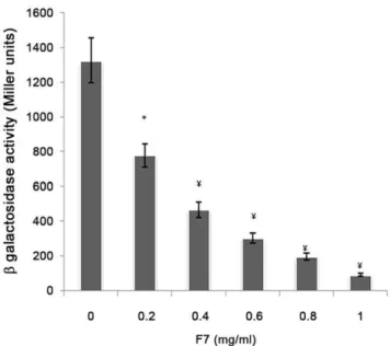
Antagonistic activity. E. coli DH5a, harboring pSC11 (PlasI::lacZ) was electroporated with pJN105L (containing lasR gene) and used as bioreporter to check antagonistic activity of bioactive fraction. Overnight grown culture was diluted 1:10 with fresh LB, incubated at 37uC and 150 rpm till OD600 nmreached 0.3 [19]. Expression oflas Rwas induced by addition of 4 mg/ml of arabinose. 10mM 3-oxo-C12HSL was added along with a concentration gradient of bioactive fraction (0.2–1.0 mg/ml) and incubated further under similar conditions. b-Galactosidase activity was measured in the bioreporter strain as described [44].
Purification and Characterization of Putative Active Compound(s)
50ml of bioactive fraction was loaded on forward Silica Gel60F254 and separated into distinct spots using ethyl acetate, methanol and water (8:1:1) solvent system. The individual spots were scraped out and compounds were re-extracted from bound silica with a mixture of ethyl acetate: methanol (9:1). The solvent was evaporated and residue was re-suspended in minimal volume of methanol. Each spot was then observed for QS inhibition using CVO26 [53].
The spot with anti QS activity was further analyzed using by LC ESI MS using Agilent 1100HPLC (HP 1101; Agilent technologies, Waldbronn, Germany). 20ml of sample in methanol was injected into reverse phase C18column (25064 mm with i.d. 5mm). The mobile phase consisted of 2% acetic acid in water (solvent A) and acetonitrile (solvent B) with following gradient profile: initially 95% A for 10 min; to 90% A for 1 min; to 80% A for 10 min; to 60% A for 10 min to 0% A for 5 min and continuing at 0% A until completion of the run. Mass spectra was obtained in the negative ion mode using 100 V fragmenter voltage and mass range of 100–1500D, Drying gas temperature was 350uC, capillary voltage 2500 Vand nebulizer pressure was 30 psi. Compounds were identified by comparing the standard masses and fragmentation peaks obtained with those available in literature for fruit ofTerminaliaspecies [35].
Statistical Analysis
All the statistical analyses were performed using student t test and p,0.05 was considered significant.
Results
Anti QS Activity ofT. chebulaExtract
In disc diffusion assay, both aqueous (5 mg/ml) and methanol (1 mg/ml) extracts exhibited anti QS activity as shown by viable white colonies of A136 around the disc in a background of blue colonies (Fig. S1). As methanol extract showed anti QS activity at lower concentration, further experiments were done with it. Methanol extract did not bind or brought any structural change in AHL molecules as there was no significant difference (p = 0.125) in
b-galactosidase activity of biosensor A136 when incubated with the extract-treated and untreated AHLs.
Bioassay Guided Fractionation ofT. chebulaMethanol Extract
In order to separate the anti QS component(s), methanol extract fractionation scheme was followed as illustrated in Fig. 1. Fractions F6 and F7 showed anti QS activity indicated by 66 and 83% (p,0.001) reduction in violacein pigment production by biosensor Figure 6. Relative expression of lasIR and rhlIR genes of P.
aeruginosa PAO1 in the presence of 0.5 mg/ml F7 as
deter-mined by qRT PCR.
CVO26, respectively (Fig. S2). However, anti QS components were probably more concentrated in F7 as it showed anti QS activity even at 0.1 mg/ml (data not shown). Phytochemical analysis of different fractions revealed the presence of terpenoides in F1 and F2, flavonoids in F6 and hydrolysable tannins in all seven fractions. However, protein precipitation assay confirmed the presence of higher amount of hydrolysable tannins in F4–F7 (Data not shown).
Effect of F7 on the Production of Virulence Factors inP. aeruginosa PAO1
Significant reduction (p,0.001) in pyocyanin (60%), elastase (50%), rhamnolipids (58%) and protease (55%) production in the presence of 0.3 mg/ml of F7 was observed while F6 showed reduction (p,0.001) at 0.4 mg/ml. F4 and F5 did not affect the production of pyocyanin and elastase. However, there was reduction (p,0.05) in case of rhamnolipids and protease whereas F1-3 did not show any effect (Fig. 2). The virulence factors were reduced by the bioactive fraction to the level equivalent to that in PAOJP2. Alginate, an important component of extracellular polysaccharides ofP. aeruginosa biofilm matrix and known to be
regulated by QS was also significantly (p,0.05) reduced by 50% at 5 mg/ml (Fig. S3).
Effect of F7 on Biofilm Formation byP. aeruginosaPAO1 Prophylactic efficiency of F7 was shown by significant reduction (65%, p,0.05) in biofilm formation in 24 h at 1 mg/ml (Fig. 3) that was increased to 85% at 5 mg/ml (p,0.001) (data not shown) with concomitant increase in planktonic bacterial cells by 1.2 log folds (from 2.7560.786106 to 6.760.346107CFU/ml). CLSM images of biofilm formed by GFP tagged P. aeruginosa in the presence of 1 mg/ml of F7 and stained with PI showed bacterial cells scattered singly on the adherent surface. Z stack analysis of the images showed 1–1.5mm thickness that corresponds to size of single bacterial cell. However, in untreated sample, bacterial aggregation and formation of bacterial microcolonies was observed that measured to 10mm. Non significant signal of PI indicated the absence of antibacterial effect of F7 and inhibition of biofilm formation was thus due to its anti QS activity (Fig. 4). Minimum biofilm eradication concentration (MBEC) of tobra-mycin was found to be 100mg/ml for P. aeruginosa PAO1.
Tobramycin at sub inhibitory 20mg/ml reduced biofilm
forma-tion by 60% (p,0.05) after 24 h. However, when tobramycin Figure 7. Swarming motility ofP. aeruginosaPAO1 a) Untreated b) Treated with 0.5 mg/ml of F7 c) reversal of inhibited swarming motility by the addition of exogenous C4HSL(2mM).
doi:10.1371/journal.pone.0053441.g007
(20mg/ml) was used along with F7 (1 mg/ml) biofilm was reduced
by 80% (p,0.001) showing enhanced susceptibility to tobramycin (Fig. 3).
C. elegansKilling Assay
C. elegans N2 fed on P. aeruginosa PAO1 showed decrease in motility within 12 h of incubation that subsequently led to death of 50% worm (LT50) within 24 h. However, LT50 increased to 72 h when worms were allowed to feed on F7 (0.5 mg/ml) treated P. aeruginosa PAO1 and an increase was also seen with PAOJP2 an autoinducer deficient mutant (LT50144 h) (Fig. 5A). Microscopic examination of P. aeruginosa fed C. elegans showed distention of gut as possible indication of infection like process. This distention was also present in worms fed on F7 treated P. aeruginosa but exhibited prolonged survival rates (Fig. 5B1and 5B2). Analysis of bacterial load in the worm gut after 24 h of feeding onP. aeruginosaPAO1 did not show significant difference (p = 0.198) (560.316106CFU/ml/worm in untreated and
860.586106CFU/ml/worm in treated groups) indicating the
attenuation of virulence of P. aeruginosa PAO1 colonizing worm gut, without affecting its viability.C. elegans fed onE. coliOP50 in the presence of F7 (0.5 mg/ml) had normal physiology with proper egg laying,life cycle (2.5 days at 25uC), motility and intact gut morphology (Fig. 5B3) showing absence of F7 toxicity.
Mechanism of Anti QS Activity of F7
Real time PCR showed 89, 90, 90 and 93% reduction in the expression oflasI,lasR,rhlIandrhlR,respectively with 0.5 mg/ml of F7(Fig. 6). This was supported by the reduction of peak intensity of 3-oxo-C12HSL by 64% (peaks corresponding to 3-oxo-C12HSL at m/z 316 ammonium and 595 dimer adduct) and 90% reduction in C4-HSL (peaks corresponding to C4HSL at m/z 159) on ESI MS analysis of AHLs after F7 exposure (Fig. S4). The reduction in both AHLs was consistence with the reduction in virulence factors controlled by 3-oxo-C12HSL (elastase and protease) and C4HSL (rhamnolipids and pyocyanin) in P. aeruginosa PAO1(Fig. 2). Growth ofP. aeruginosaPAO1 was monitored in the presence of different concentrations of F7 (0–1.25 mg/ml) and it showed insignificant change in growth (p = 0.132) at 0.5 mg/ml (Fig. S5) indicating quorum sensing inhibition as the mechanism for the reduction in AHLs and not the killing of cells. Restoration of inhibited swarming motility on addition of 2mM C4HSL further supported the observation (Fig. 7). F7 at 1 mg/ml also reducedb -galactosidase activity by 93% inE. colibioreporter strain showing its antagonistic activity towards transcriptional regulator lasR (Fig. 8).
Identification of Bioactive Constituents
TLC of F7 using silica gel 60 F254resolved into four different spots (S1, S2, S3 and S4) with Rf value of 0.07, 0.38, 0.61 and 0.72. Anti QS activity was found in S2 and S3 using CVO26 (data not shown). LC-ESI-MS analysis of S2 showed three peaks at RT of 1.26, 13.36, 34.63 min. On comparing the peaks with the reported MS fragmentation data of polyphenols fromTerminalia species (Table S1) [35], peak obtained at RT 1.26 min may be of 3-O-methyl-4-O-(b–D-xylopyranosyl) ellagic acid, that is a glyco-sylated derivative of ellagic acid with m/z peaks of 447,315,126,217 representing C20H16O12 (mol wt. 448). RT 13.36 min peak probably corresponded to ellagic acid molecule with molecular formula of C14H6O2(mol wt. 302) and [m/z+H2] of 283,255,243,200,173 whereas peak at RT 34.63 min could be the methylated derivative of (S)-flavogallonic acid C22H12O13(mol Figure 8. Antagonistic activity of F7 against 3-oxo-C12HSL
mediated QS inE. colibioreporter strain harboring pSC11(P la-sI)::lacZ) and lasR expression vector pJN105L.
doi:10.1371/journal.pone.0053441.g008
Table 2.Putative anti QS compounds as shown by LC-ESI-MS fragmentation data for the bioactive fraction.
Proposed compounds
Molecular Formula
Molecular Weight
Retention Time
(min) (M+H)2
MS-MS fragmentation Peaks
Spot S2
3-O-methyl-4-O-(b-D-xylo pyranosyl)ellagic acid
C20H16O12 448 1.26 447 447,315, 217,126
Ellagic acid C14H6O2 302 13.36 301 255,283,243,200,173
Methyl S-flavogallonic acid C22H12O13 485 34.63 484 323,255,227,200
Spot S3:
S flavogallonic acid C21H10O13 470 0.74 469 305,217, 145,126
3,4,8,9,10-pentahydroxyldibenzo(b.d) Pyran-6-one
C20H16O12 276 13.63 275 276,255,227,201,173
Unknown Unknown Unknown 34.63 Unknown 106,265,293,393
wt.485) with [m/z+H2] of 323, 255,227,200 (Table 2). In S3 fraction, three RT peaks were identified at 0.74,13.63, 34.63 min. Peak at RT 0.74 min may be of (S)-flavogallonic acid C21H10O13 (mol wt.470) that gave [m/z+H2] peak of 469. (S)-flavogallonic
acid possesses both ellagic acid and gallic acid moieties. Therefore, [m/z+H2] peaks of both gallic acid at 126 and ellagic acid at [m/ z+H2] 305, 217,145 were observed. Peak at RT 13.63 min
corresponded to the molecular formula of C20H16O12 (mol wt. 276) that is 3,4,8,9,10–pentahydroxyldibenzo(b,d)pyran-6-one molecule and showed m/z of 276,255,227,201,173 that corre-sponded to the standard MS-MS fragmentation data of this molecule. Peaks at RT 34.63 min showed [m/z+H2] of 393, 293,
265, 106. The nucleus of this compound consists of C3H603as the major [m/z+H2] peak observed was at 106. However, exact chemical formula could not be elucidated.
Discussion
Plant products have been used traditionally for the treatment of various ailments and this is attributed to the wide range of phytochemicals present in them. In the present study, both aqueous and methanol extract showed anti QS activity. However, methanol extract exhibited greater anti QS activity indicating that effective phytochemical compound(s) have been partitioned more in organic phase. In most of the studies, organic extracts of plants have showed anti QS activity as reported in toluene extracts of Allium sativum [54], ethanol extracts of C. arbiflorum leaves [1], Mangifera indicaandPuncia granatum[55].
Methanol extract ofT. chebulaon partial purification led to the separation of hydrolysable tannins as a group of phytochemicals responsible for the anti QS activity. Tannins in plants protect them from predators and also play role in plant growth regulation [56]. Initially tannins were regarded as anti nutritional but due to their antioxidant and antimicrobial properties, they are now being used in various medicinal formulations [57]. Ellagic acid, gallic acid, corilagen, chebulagic acid and punicalagin are some of the known polyphenolics isolated fromT. chebulafruit [21]. Anti QS activity has been linked only with ellagic acid which reduced swarming motility and biofilm formation in soil isolate, P. putida [16]. However, ellagic acid has not been shown to attenuateP. aeruginosa virulence. In the present study, LC-ESI-MS analysis of tannin-rich bioactive fraction showed the presence of ellagic acid derivatives (EADs) as major compounds. EADs constitute the polyphenolic
compounds that possess ellagic acid as the core molecule. Bioactive fraction, on mass identification, revealed the presence of glycosides derivatives of ellagic acid. EADs extracted fromRubus ulmifoliusalong with some sapogenin related compounds have been shown to reduce biofilm formation in Staphylococcus aureus with enhancement to antibiotics (Clindamycin, Daptomycin, Oxacillin) susceptibility [58]. To the best of our knowledge, anti QS activity of ellagic acid derivatives fromT. chebulaand its use to controlP. aeruginosavirulence has not been studied before.
Pyocyanin, elastase, protease and rhamnolipids are regarded as indicators of the optimal operation of QS regulon inP. aeruginosa. Reduction in their production level indicates the anti QS potential of the tested compound(s). Elastase and protease form important determinants in colonizing the host tissues [59] whereas pyocyanin chelates the bound iron from transferrin for optimal virulence expression [60]. Rhamnolipids constitute an important surfactant that assists in surface motility ofP. aeruginosarequired for biofilm initiation [61]. Bioactive fraction was able to reduce all of them to the level comparable inlasI2rhlI2mutant PAOJP2, indicating the
effectiveness of EADs in attenuatingP. aeruginosavirulence factors. Alginate is one of the important virulence determinants in P. aeruginosaand is present as a constituent of exopolysacchrides (EPS) in biofilms. [62]. Tannic acids have been reported to cause massive decrease in EPS production inStreptococcus species [63] which may explain the decrease in alginate content inP. aeruginosa biofilm after treatment with bioactive fraction that contains hydrolysable tannins. This also increased the planktonic bacterial cell count in the medium surrounding biofilm making them vulnerable to the action of tobramycin.
In vitroattenuation of virulence factors correlated well with the in vivostudy. Slow killing ofC. elegansoccurs due to colonization and proliferation of bacteria in the worm gut [64]. CFU analysis of worm gut fed on treated and untreatedP. aeruginosashowed that bioactive fraction was able to attenuate the virulence of colonizing bacteria indicating therapeutic potential of the fraction. Phena-zines, produced byP. aeruginosacauses lethal paralysis of muscular tissues inC. elegansleading to asphyxia and death of worms within 4–24 h. F7 was found to inhibit the pyocyanin (phenazine) pigment production that may be the reason for increase in LT50 of worms fed on treatedP. aeruginosa.
QS can be inhibited in various ways. Halogenated furanones or synthetic analogs act as signal mimics resulting in a decrease in QS gene expression [20]. Lactonases and acylases from Gram positive bacteria cause enzymatic degradation of AHLs resulting in inhibition of QS in Gram negative bacteria [65]. QscR a negative transcriptional regulator homolog oflasRandrhlRinP. aeruginosa also led to similar inhibition of QS [41]. Lactonolysis, a pH meditated degradation of AHLs inhibits QS [66]. As pH ofT. chebula extract was neutral hence there was no spontaneous inactivation of AHLs. EADs-rich bioactive fraction inhibited QS by reducing AHLs production as shown in the present study. In the previous studies, anti biofilm activity of tannin rich fraction fromT. cattapahas been reported but the underlying mechanism has not been explained [34]. Similarly, proanthocyanidins (condensed tannins from cranberries) has been reported to inhibit swarming motility in P. aeruginosa by multiple mechanisms that include binding with lipopolysacchrides, flagellin subunits, and reduction in rhamnolipids production [67] but their effect onlasIR andrhlIRremained unexplored. Plants extracts ofConocarpus erectus, Callistemon viminalis, Bucida buceras and Combretum arbiflorum have been reported to downregulate the expression oflasIRand rhlIR using bioreporter strains whereas in the current study, qRT PCR analysis provides more specific analysis of QS gene expression inP. aeruginosatreated with bioactive fraction ofT. chebula. Antagonist Figure 9. Comparative effect of F7 and ellagic acid on the
production of virulence factors at 0.5 mg/ml.Elastase OD490 nm/ 600 nm0.654,Pyocyanin OD540 nm/600 nm1.08, Rhamnolipids OD570 nm/
600 nm0.456 and Protease OD400 nm/600 nm0.876 were taken as 100% in
untreatedP. aeruginosaPAO1. doi:10.1371/journal.pone.0053441.g009
activity of synthetic QS inhibitor viz. isothiocyanate and haloacetamides has been reported as these compounds displace 3-oxo-C12HSL from the ligand binding domain of lasR [19]. Antagonist activity was also displayed by the bioactive fraction of T. chebula.
Since ellagic acid formed the core molecule in F7, effect of ellagic acid was checked onP. aeruginosavirulence factors. There was only 10–15% reduction in virulence factors production at concentration of 0.5 mg/ml (Fig. 9). In contrast, F7 was able to show more than 50% reduction in virulence factors production in P. aeruginosa PAO1 at the same concentration suggesting the importance of ellagic acid derivatives in the inhibition of QS inP. aeruginosaPAO1. Further studies are required to find out which EADs have antagonistic activity that act by down regulating the lasIR and rhlIRsystem. The combination of EADs needs to be worked out to develop an effective anti QS formulation to control AHL mediated virulence in pathogens.
Supporting Information
Figure S1 Disc diffusion assay for anti quorum sensing activity of T. chebula using A. tumefaciens A136 as biosensor. 1. Aqueous extract (5 mg/ml) 2. Methanol extract (1 mg/ml) 3.Curcumin (3mg/ml ) as positive control 4.Methanol and 5.Water as negative controls.
(TIF)
Figure S2 Anti quorum sensing activity of different fractions at 0.5 mg/ml shown as reduction in violacein production by CV026 in the presence of 50 nM C6HSL
(*,p,0.05, ¥,p,0.001 ). Bars indicates standard deviations for triplicate sets of experiments.
(TIF)
Figure S3 Effect of F7(1–10 mg/ml) on alginate content in biofilms ofP. aeruginosaPAO1.
(TIF)
Figure S4 ESI-MS analysis of AHLs extracted fromP. aeruginosa PAO1 A) untreated B) treated with 0.5 mg/ml of F7.
(TIF)
Figure S5 Effect of F7 (0–1.25 mg/ml) on growth of P. aeruginosaPAO1.
(TIF)
Table S1 Compound(s) isolated fromT. chebulafruit [35]. (TIF)
Acknowledgments
We thank Dr. K.H. McClean for providing biosensor strainsA. tumefaciens A136 andC. violaceum026, Dr. K.P. Rambaugh for PAOJP2, Dr. J.A. Haagensen for GFP tagged PAO1, Dr. E.P. Greenberg for plasmids pSC11 and pJN105L and CGC centre, University of Minnesota, USA for C. elegansN2 andE. coliOP50.
Author Contributions
N/A. Conceived and designed the experiments: NC PS. Performed the experiments: SS. Analyzed the data: SS PS NC. Contributed reagents/ materials/analysis tools: PS NC. Wrote the paper: SS PS NC.
References
1. Vandeputte OM, Kiendrebeogo M, Rajaonson S, Diallo B, Mol A, et al. (2010) Identification of catechin as one of the flavonoids fromCombretum albiflorumbark extract that reduces the production of quorum sensing controlled virulence factors inPseudomonas aeruginosaPAO1. Appl Environ Microbiol 76: 243–253. 2. Oncul O, Ulkur E, Acar A (2009) Prospective analysis of nosocomial infections
in a burn care unit, Turkey. Indian J Med Res 130: 758–764.
3. Jimenez PN, Koch G, Thompson JA, Xavier KB, Cool RH, et al. (2012) The multiple signaling systems regulating virulence in Pseudomonas aeruginosa. Microbiol Mol Biol Rev 76: 46–65.
4. Rasmussen TB, Givskov M (2006) Quorum sensing inhibitors: a bargain of effects. Microbiology 152: 895–904.
5. Dekimpe V, Deziel E (2009) Revisiting the quorum-sensing hierarchy in Pseudomonas aeruginosathe transcriptional regulator RhlR regulates LasR-specific factors. Microbiology 155: 712–723.
6. Senturk S, Ulusoy S, Bosgelmez-Tinaz G, Yagci A (2012) Quorum sensing and virulence ofPseudomonas aeruginosaduring urinary tract infections. J Infect Dev Ctries 6: 501–507.
7. Klausen M, Heydorn A, Ragas P, Lambersten L, Molin S (2003) Biofilm formation byPseudomonas aeruginosawild type, flagella and type IV mutants. Mol Microbiol 48: 1511–1524.
8. Doring G, Conway SP, Heijerman HGM, Hodson ME, et al. (2000) Antibiotic therapy againstPseudomonas aeruginosain cystic fibrosis: A European consensus. Eur Respir J 16: 749–767.
9. Hoiby N, Bjarnsholt T, Givskov M, Molin S, Ciofu O (2010) Antibiotic resistance of bacterial biofilms. Int J Antimicrob Agents 35: 322–332. 10. Sadlon AE, Lamson DW (2010) Immune-modifying and antimicrobial effects of
Eucalyptus oil and simple inhalation devices. Altern Med Rev 15: 33–47. 11. Manefield M, Rasmussen TB, Henzter M, Andersen JB, Steinberg P, et al.
(2002) Halogenated furanones inhibit quorum sensing through accelerated LuxR turnover. Microbiology 148: 1119–1127.
12. Rudrappa T, Bais HP (2008) Curcumin a known phenolic fromCurcuma longa attenuates the virulence ofPseudomonas aeruginosa PAO1 in whole plant and animal pathogenicity models. J Agric Food Chem 56: 1955–1962.
13. Jakobsen TH, Van Gennip M, Phipps RK, Shanmugham MS, Christensen LD, et al. (2012) Ajoene, a sulfur-rich molecule from garlic, inhibits genes controlled by quorum sensing. Antimicrob Agents Chemother 56: 2314–2325. 14. Jakobsen TH, Bragason SK, Phipps RK, Christensen LD, Gennip M, et al.
(2012) Food as a source for quorum sensing inhibitors: Iberin from horseradish revealed as a quorum sensing inhibitor ofPseudomonas aeruginosa. Appl Environ Microbiol 8: 2410–2421.
15. Adonizio A, Kong KF, Mathee K (2008) Inhibition of quorum sensing controlled virulence factor production inPseudomonas aeruginosaby south florida plant extracts. Antimicrob Agents Chemother 52: 198–203.
16. Huber B, Eberl L, Feucht W, Polster J (2003) Influence of polyphenols on bacterial biofilm formation and quorum sensing. Z Naturforsch 58: 879–884. 17. Kociolek MG (2009) Quorum sensing inhibitors and Biofilms. Anti infect Agents
8: 315–326.
18. Hoang TT, Schweizer HP (1999) Characterization ofP. aeruginosaenoyl acyl carrier protein reductase a target for antimicrobial triclosan and the role in AHL mediated QS inSerratia marcescens. Mol Microbiol 45: 1655–1671.
19. Amara N, Mashiach D, Amar P, Krief AH, Stephane J, et al. (2009) Covalent Inhibition of bacterial quorum sensing. J Am Chem Soc 131: 10610–10619. 20. Ni N, Li M, Wang J, Wang B (2009) Inhibitors and antagonists of bacterial
quorum sensing. Med Res Rev 29: 65–124.
21. Juang LJ, Sheu SJ, Lin TC (2004) Determination of hydrolysable tannins in the fruit ofTerminalia chebulaRetz. by high-performance liquid chromatography and capillary electrophoresis. J Sep Sci 27: 718–724.
22. Srikumar R, Jeya PN, Sheela DR (2005) Immunomodulatory activity of triphala on neutrophil functions. Biol Pharm Bull 28: 1398–1403.
23. Chalise JP, Acharya K, Gurung N, Bhusal RP, Gurung R, et al. (2010) Antioxidant activity and polyphenol content in edible wild fruits from Nepal. Int J Food Sci Nutr 61: 425–432.
24. Kannan P, Ramadevi SR, Hopper W (2009) Antibacterial activity ofTerminalia chebulafruit extract. Afr J Microbiol Res 3: 180–184.
25. Shin TY, Jeong HJ, Kim DK (2001) Inhibitory action of water soluble fraction of T. chebulaon systemic and local anaphylaxis. J Ethnopharmacol 74: 133–140. 26. Gao H, Huanf YN, Gao B, Kawabata J (2008) Chebulagic acid is a potent
alphaglucosidase inhibitor. Biosc Biotechnol Biochem 72: 601–603.
27. Kaur K, Arora S, Kumar S, Nagpal A (2002) Modulatory effect of phenolic fractions fromT. arjunaon the mutagenicity in Ames test. J Environ Pathol Toxicol Oncol 21: 45–56.
28. Saleem A, Husheem M, Harkonen P, Pihlaja K (2002) Inhibition of cancer cell growth by crude extract and the phenolics ofTerminalia chebulaRetz. Fruit. J Ethnopharmacol 81: 327–335.
29. Reddy DB, Reddy TCM, Jyotsna G, Sharan S, Priya N, et al. (2009) Chebulagic acid, a COX-LOX dual inhibitor isolated from the fruits ofTerminalia chebula Retz., induces apoptosis in COLO-205 cell line. J Ethnopharmacol 124: 506– 512.
31. Agarwal V, Lal P, Pruthi V (2008) Prevention ofCandida albicansbiofilm by plant oils. Mycopathol 165: 3–19.
32. Cheng HY, Lin CC, Lin TC (2002) Anti herpes simplex virus type 2 activity of casuarinin from the bark ofTerminalia arjunaLinn. Antiviral Res 55: 447–455. 33. Bag A, Bhattacharyya SK, Bharati P, Pal NK, Chattopadhyay RR (2009)
Evaluation of antibacterial properties of Chebulic myrobalan (fruit ofTerminalia chebula Retz.) extracts against methicillin resistant Staphylococcus aureus and trimethoprim sulphamethoxazole resistant uropathogenic Escherichia coli. Afr J Plant Sci 3: 25–29.
34. Taganna JC, Quanico JP, Perono RM, Amor EC, Rivera WL (2011) Tannin-rich fraction from Terminalia catappa inhibits quorum sensing (QS) in Chromobacterium violaceumand the QS-controlled biofilm maturation and LasA staphylolytic activity inPseudomonas aeruginosa. J Ethnopharmacol 134: 865–871. 35. Pfundstein B, Desouky SKE, Hull WE, Haunber R, Erben G, et al. (2010) Polyphenolic compounds in the fruit of Egyptian medicinal plants (Terminalia bellerica, Terminalia chebulaandTerminalia horrida): Characterization, quantitation and determination of antioxidant capacities. Phytochemistry 71: 1132–1148. 36. Iglewski BH, Pesci EC, Pearson JP, Seed PC (1997) Regulation oflasandrhl
Quorum Sensing inPseudomonas aeruginosa. J Bacteriol 79: 3127–3132. 37. Rambaugh KP, Beale E, Tann MW, Guigen Li (2006)C. eleganssenses bacterial
autoinducers. Appl Environ Microbiol 72: 5135–5137.
38. Haagensen JA, Klausen M, Ernst RK, Miller SI, Folkesson A, et al. (2007) Differentiation and distribution of colistin and sodium dodecylsulphate tolerant cells inP. aeruginosabiofilms. J Bacteriol 189: 28–37.
39. Fuqua C, Winans SC (1996) Conserved cis-acting promoter elements are required for density-dependent transcription ofAgrobacterium tumefaciensconjugal transfer genes. J Bacteriol 78: 435–440.
40. McClean KH, Winson MK, Fish L, Taylor A, Chhabra SR, et al. (1997) Quorum sensing and Chromobacterium violaceum: exploitation of violacein production and inhibition for the detection of N-acylhomoserine lactones. Microbiology 143: 3703–3711.
41. Chugani SA, Whiteley M, Lee KM, Argenio D, Manoil C, et al. (2000) QscR, a modulator of quorum-sensing signal synthesis and virulence inPseudomonas aeruginosa. Proc Natl Acad Sci USA 98: 2752–2757.
42. Lee JH, Lequette Y, Greenberg (2006) Activity of purified QscR, aPseudomonas aeruginosaorphan quorum-sensing transcription factor. Mol Microbiol 59: 602– 609.
43. Adonizio A, Kong KF, Mathee K (2006) Anti-quorum sensing activity of medicinal plants in Southern Florida. J Ethnopharmacol 105: 27–35. 44. Shaw PD, Ping G, Daly SL, Cha C, Cronan JE, et al.(1997) Detecting and
characterizing N-acyl-homoserine lactone signal molecules by thin-layer chromatography. Proc Natl Acad Sci USA 94: 6036–6041.
45. Miller JH (1972) Experiments in molecular genetics; Cold Spring Harbor Laboratory Press: Woodbury, NY: 352–355.
46. Harborne J (1973) Phytochemicals Methods: A guide to modern techniques of plant analysis. Chapman and Hall, London.
47. Zhu H, Thuruthyl SJ, Willcoxd DP (2001) Determination of quorum-sensing signal molecules and virulence factors ofPseudomonas aeruginosaisolates from contact lens-induced microbial keratitis. J Med Microbiol 51: 1063–1070. 48. McClure CD, Schiller NL (1992) Effects ofPseudomonas aeruginosarhamnolipids
on human monocyte-derived macrophages. J Leukocyte Biol 51: 97–102.
49. Franklin JM, Ohman FM (1993) Identification of algF in the Alginate Biosynthetic Gene Cluster of Pseudomonas aeruginosa which is required for Alginate acetylation. J Bacteriol 175: 5057–5065.
50. Babic F, Venturi V, Vlahoviek GM (2010) Tobramycin at subinhibitory concentration inhibits the RhlI/R quorum sensing system in a Pseudomonas aeruginosaenvironmental isolate. BMC Infect Dis 10: 148–160.
51. Gallagher LA, Manoil C (2001)P. aeruginosakillsC. elegansby cyanide poisoning. J Bacteriol 183: 6207–6214.
52. Wang EW, Jung JY, Pashia ME, Nason R (2005) OtopathogenicPseudomonas aeruginosastrains as competent biofilm formers. Arch Otolaryngol Head Neck Surg 131: 983–989.
53. Makemson J, Eberhard A, Mathee K (2006) Simple electrospray mass spectrometry detection of acylhomoserine lactones. Luminescence 21: 1–6. 54. Bjarnsholt T, Jensen P, Rasmussen TB, Christophersen L, Calum H, et al.
(2005) Garlic blocks quorum sensing and promotes rapid clearing of pulmonary Pseudomonas aeruginosainfections. Microbiology 151: 3873–3880.
55. Zahin M, Hasan S, Aqil F, Khan MS, Husain FM, et al. (2010) Screening of certain medicinal plants from India for their anti-quorum sensing activity. Indian J Exp Biol 48: 1219–1224.
56. Drewnowski A, Carneros CG (2000) Bitter taste, Phytonutrients and consumers; a review Am J Clin Nutr 72: 1424–1435.
57. Vattem DA, Ghaedian R, Shetty K (2005) Enhancing health benefits of berries through phenolic antioxidant enrichment: focus on cranberry. Asia Pac J Clin Nutr 14: 120–130.
58. Quave CL, Carmona ME, Compare CM, Hobby G, Hendrikson H, et al. (2012) Ellagic acid derivatives fromRubus ulmifoliusinhibitStaphylococcus aureusbiofilm formation and improve response to antibiotics PLoS ONE 7: e28737–38745. 59. Stehling EG, da Silveira WA, da Silva Leite D (2008) Study of biological
characteristics ofPseudomonas aeruginosastrains isolated from patients with cystic fibrosis and from patients with extra-pulmonary infections. Braz J Infect Dis 12 : 86–88.
60. Lau GW, Hassett DJ, Ran H, Kong F (2004)The role of pyocyanin in Pseudomonas aeruginosainfection. Trends Mol Med 10: 599–606.
61. May CO, Tufenkji N (2011) The swarming motility ofP. aeruginosais blocked by cranberry proanthocyanidins and other tannin containing materials. Appl Environ Microbiol 77: 3061–3067.
62. Balasubramanian D, Kong KF, Jayawardena SR, Leal SM, Sautter RT, et al. (2011) Co-regulation of {beta}-lactam resistance, alginate production and quorum sensing inPseudomonas aeruginosa.J Med Microbiol 60: 147–156. 63. O’Donovan L,Brooker JD (2001) Effect of hydrolysable and condensed tannins
on growth, morphology and metabolism ofStreptococcus gallolyticus(S. caprinus) and Streptococcus bovis. Microbiology 147: 1025–1033.
64. Tan MW, Rahme LG, Sternberg JA, Tompkins RG, Ausubel FM (1999) Pseudomonas aeruginosakilling ofCaenorhabditis elegansused to identifyP. aeruginosa virulence factors. Proc Natl Acad Sci USA 96: 2408–2413.
65. Hentzer M, Eberl L, Nielsen J, Givskov M (2003) Quorum sensing : a novel target for the treatment of biofilm infections. BioDrugs 17: 241–250. 66. Yates EA, Philipp B, Buckley C, Atkinson S, Chhabra SR, et al. (2002)
N-Acylhomoserine lactones undergo lactonolysis in a pH, temperature and Acyl Chain Length-Dependent Manner during Growth ofYersinia pseudotuberculosisand Pseudomonas aeruginosa.Infect Immun 70: 5636–5646.