Streptococcus mutans
Biofilm Formation and Adherence
to Oral Epithelial Cells
Telma Blanca Lombardo Bedran1, Louis Grignon2, Denise Palomari Spolidorio3, Daniel Grenier2* 1Department of Oral Diagnosis and Surgery, Araraquara Dental School, State University of Sa˜o Paulo, Sa˜o Paulo, Brazil,2Oral Ecology Research Group, Faculty of Dentistry, Universite´ Laval, Quebec City, Quebec, Canada,3Department of Physiology and Pathology, Araraquara Dental School, State University of Sa˜o Paulo, Sa˜o Paulo, Brazil
Abstract
Triclosan is a general membrane-active agent with a broad-spectrum antimicrobial activity that is commonly used in oral care products. In this study, we investigated the effect of sub-minimum inhibitory concentrations (MICs) of triclosan on the capacity of the cariogenic bacteriumStreptococcus mutansto form biofilm and adhere to oral epithelial cells. As quantified by crystal violet staining, biofilm formation by two reference strains ofS. mutanswas dose-dependently promoted, in the range of 2.2- to 6.2-fold, by 1/2 and 1/4 MIC of triclosan. Observations by scanning electron microscopy revealed the presence of a dense biofilm attached to the polystyrene surface. Growth ofS. mutansin the presence of triclosan at sub-MICs also increased its capacity to adhere to a monolayer of gingival epithelial cells. The expression of several genes involved in adherence and biofilm formation inS. mutanswas investigated by quantitative RT-PCR. It was found that sub-MICs of triclosan significantly increased the expression ofcomD,gtfC, andluxS, and to a lesser extent ofgtfBandatlAgenes. These findings stress the importance of maintaining effective bactericidal concentrations of therapeutic triclosan since
sub-MICs may promote colonization of the oral cavity byS. mutans.
Citation:Bedran TBL, Grignon L, Spolidorio DP, Grenier D (2014) Subinhibitory Concentrations of Triclosan PromoteStreptococcus mutansBiofilm Formation and Adherence to Oral Epithelial Cells. PLoS ONE 9(2): e89059. doi:10.1371/journal.pone.0089059
Editor:Jens Kreth, University of Oklahoma Health Sciences Center, United States of America
ReceivedDecember 4, 2013;AcceptedJanuary 13, 2014;PublishedFebruary 13, 2014
Copyright:ß2014 Bedran et al. This is an open-access article distributed under the terms of the Creative Commons Attribution License, which permits unrestricted use, distribution, and reproduction in any medium, provided the original author and source are credited.
Funding:This study was supported by the Canadian Institutes of Health Research (#95335). The funders had no role in study design, data collection and analysis, decision to publish, or preparation of the manuscript.
Competing Interests:The authors have declared that no competing interests exist.
* E-mail: [email protected]
Introduction
Triclosan (2,4,49-trichloro-29-hydroxydiphenyl ether) is a non-ionic molecule with broad-spectrum antimicrobial activities against both bacteria (Gram positive and negative) and fungi [1]. More specifically, it is a general membrane-active agent that causes structural perturbations resulting in a loss of permeability-barrier functions [2]. Triclosan has also been reported to possess an anti-inflammatory activity since it reduces cytokine secretion by host cells such as gingival fibroblasts [3,4]. Given this interesting dual action associated to triclosan and the fact that its high substantivity can be associated to a long-lasting effect, it has been used in oral care products (mouthwash, toothpaste) as an active agent for the reduction of dental plaque accumulation and the control of inflammatory gingivitis. The systematic review per-formed by Davies et al. [5] supports the benefits of triclosan-containing toothpaste for reduction of dental plaque and gingivitis. Being a general anti-plaque agent, triclosan may also be effective for the management of dental caries, a chronic infectious disease associated with a progressive destruction of the hard tooth structures (enamel, dentine, cementum) by the action of acido-genic/aciduric bacteria, mainly Streptococcus mutans, embedded in the dental biofilm [6,7]. To support that, it has been reported that triclosan-containing toothpastes enhance the anti-caries potential of fluoride in dentifrices [8]. Phan and Marquis [9] brought evidence that the ability of triclosan to inhibit glycolysis ofS. mutans
in biofilms may be, at least in part, responsible for its anti-caries effect.
The effective therapeutic results of antimicrobials are optimal when the concentration is above the minimum inhibitory concentration (MIC). Several studies have reported that antimi-crobials at sub-MICs may modulate biological characteristics of bacteria, including their ability to colonize the host [10–15]. In this study, we investigated the effect of triclosan sub-MICs onS. mutans
biofilm formation and adherence to oral epithelial cells. In addition, the effect of triclosan at sub-MICs on expression of several genes involved in adherence and biofilm formation was investigated.
Materials and Methods Bacteria and Growth Conditions
S. mutans ATCC 25175 (serotype c) and ATCC 35668 (unknown) were used in this study. Bacteria were grown aerobically at 37uC in Todd-Hewitt broth (BBL Microbiology Systems, Cockeysville, MD, USA) supplemented with hemin (10mg/ml) and vitamin K (10mg/ml) (THB-HK).
Determination of MIC of Triclosan
to 16108colony-forming units [CFU]/ml). Samples (100ml) were
added to the wells of a 96-well tissue culture plate containing serial dilutions (125 to 0.50mg/ml) of triclosan (Sigma-Aldrich Canada
Ltd., Oakville, ON, Canada) in culture medium (100ml). Control
bacteria were cultivated in the absence of triclosan. After incubation for 24 h at 37uC, bacterial growth was monitored by recording the OD660 using a microplate reader. The MIC was
defined as the lowest concentration of triclosan that completely inhibitsS. mutansgrowth (OD660#0.05).
Biofilm Assay
S. mutanswas grown (24 h) in a flat-bottomed 96-well microplate as above in the absence (control) and presence of triclosan at 1/2, 1/4, or 1/8 MIC. Similar assays were also performed using THB-HK supplemented with 0.25% sucrose. The medium, free-floating bacteria, and loosely-bound biofilm were then removed by aspiration, and the wells were washed three times with 50 mM phosphate-buffered saline (pH 7.2; PBS). The biofilms were stained with 0.04% crystal violet (100ml) for 10 min. The wells were washed three times with PBS to remove unbound crystal violet dye and dried for 2 h at 37uC. After adding 100ml of 95% (v/v) ethanol to each well, the plate was shaken for 10 min to release the dye from the biofilms and the absorbance at 550 nm (A550) was recorded.
Scanning Electron Microscopy
TheS. mutans(ATCC 25175) biofilm was examined by scanning electron microscopy. One ml ofS. mutansresuspended to an OD660
of 0.1 in culture THB-HK6 triclosan at 1/2 or 1/4 MIC was added into wells of a 6-well plate containing a 13 mm-diameter plastic coverslip. After 24 h incubation, medium and free-floating bacteria were removed. The biofilms were incubated overnight in fixation buffer (4% (w/v) paraformaldehyde, 2.5% (w/v) glutar-aldehyde, 2 mM CaCl2 in 0.2 M cacodylate buffer, pH 7.2),
washed with 0.1 M cacodylate buffer pH 7.0 (3620 min) and post-fixed for 90 min at room temperature in 1% (w/v) osmic acid containing 2 mM potassium ferrocyanide and 6% (w/v) sucrose in cacodylate buffer. Samples were dehydrated through a graded series of ethanol (50, 70, 95 and 100%), critical point dried, gold sputtered and examined using a JEOL JSM6360LV scanning electron microscope operating at 30 kV.
Assay for Adherence to Oral Epithelial Cells
S. mutansATCC 25175 cells cultivated in the absence (control) or presence of triclosan at 1/2, 1/4 or 1/8 MIC were labeled with fluorescein isothyocyanate (FITC) as previously reported [16]. The immortalized human gingival epithelial cell line OBA-9 used in this study, kindly provided by Dr. Marcia Mayer (Departamento de Microbiologia, Institute of Biomedical Sciences, Universidade de Sa˜o Paulo, Sa˜o Paulo, Brazil), was initially described by Kusumotoet al. [17]. The epithelial cells were cultured (96-well
Table 1.Primers used for the quantitative RT-PCR analysis.
Genes Primer sequences Product size (bp)
16S rRNA Sense: 5-CCATGTGTAGCGGTGAAATGC-39 144
Antisense: 59-TCATCGTTTACGGCGTGGAC-39
atlA Sense: 59-TCCAATTGCAGCAAACACAGGA-39 139
Antisense: 59-AGTACTTGCCTGAGACGGAACTGTT-39
comD Sense: 59-TTCCTGCAAACTCGATCATATAGG-39 113
Antisense: 59-TGCCAGTTCTGACTTGTTTAGGC-39
gtfB Sense: 59-AGCCGAAAGTTGGTATCGTCC-39 123
Antisense: 59-TGACGCTGTGTTTCTTGGCTC-39
gtfC Sense: 59-TTCCGTCCCTTATTGATGACATG-39 122
Antisense: 59-AATTGAAGCGGACTGGTTGCT-39
luxS Sense: 59-CCAGGGACATCTTTCCATGAGAT-39 147
Antisense: 59-ACGGGATGATTGACTGTTCCC-39
doi:10.1371/journal.pone.0089059.t001
Table 2.Effect of triclosan sub-MICs on biofilm formation and planktonic growth by S. mutans.
Strain
Presence of
sucrose Biofilm formation (A550) Planktonic growth (OD660)
Control 1/2 MIC 1/4 MIC 1/8 MIC Control 1/2 MIC 1/4 MIC 1/8 MIC
ATCC 25175 2 0.2760.04 1.6860.04* 1.3360.04* 0.3660.11 0.4560.12 0.2860.06* 0.3160.05 0.3460.08
+ 1.2660.07 1.7360.20* 1.4160.18 1.1960.13 0.5360.08 0.3460.12* 0.4460.09 0.5960.14
ATCC 35668 2 0.3160.06 0.9460.12* 0.6760.15* 0.3160.09 0.5760.09 0.3960.04* 0.4260.14 0.6460.11
+ 0.7460.05 0.9660.09* 0.8260.14 0.8560.11 0.6560.12 0.3860.10* 0.5360.15 0.6460.07
Data are expressed as means6standard deviations. Controls refer to the absence of triclosan. *Significantly different atp,0.01 compared to control.
microplate) in Keratinocyte-Serum Free Medium (K-SFM, Life Technologies Inc., Burlington, ON, Canada) containing insulin, epidermal growth factor, and fibroblast growth factor, and supplemented with 100mg/ml of penicillin G/streptomycin at 37uC in a 5% CO2atmosphere until they reached confluence. The
adherence assay ofS. mutansto epithelial cells was carried out as described in a previous study [16]. After removing unbound bacteria and washing wells, the relative fluorescence units (RUF; excitation wavelength 495 nm; emission wavelength 525 nm) corresponding to the degree of bacterial adherence were determined using a microplate reader.
Determination of Cell Surface Hydrophobicity
The relative cell surface hydrophobicity of S. mutans ATCC 29175 grown in THB-HK6triclosan at 1/2, 1/4, or 1/8 MIC was determined by measuring their absorption to n-hexadecane according to the procedure described by Rosenberget al.[18].
RNA Isolation and Quantitative RT-PCR
To investigate the effect of sub-MICs of triclosan on expression of several genes involved in adherence and biofilm formation,S. mutansATCC 25175 was grown to mid-log phase (OD660= 0.45)
and then triclosan was added at 1/2, 1/4 or 1/8 MIC prior to
further incubate at 37uC for 2 h. Control cells were incubated in the absence of triclosan. Bacteria were collected by centrifugation (7,0006g for 5 min) and treated with an RNAprotect bacterial
reagent (Qiagen Canada Inc., Montreal, QC, Canada). Bacterial cells were then lysed and RNA was isolated and purified using the RNeasy minikit (Qiagen Canada Inc.). The amounts of mRNA were quantified with the ExperionTMsystem (Bio-Rad Laborato-ries, Mississauga, ON, Canada). The reverse transcription-polymerase chain reaction (RT-PCR) analysis was performed as follows. RNA from each sample (100 ng/ml) was reverse-transcribed using Maloney murine leukemia virus reverse tran-scriptase and random hexamers in a Bio-Rad MyCyclerTM thermal cycler (Bio-Rad Laboratories). Reverse transcription conditions were 5 min at 70uC, 10 min at 25uC, 50 min at 37uC, and 15 min at 70uC. Real-time PCR was used for quantification ofatlA,comD,gtfB,gtfC, andluxSmRNA expression. 16S rRNA gene was used as an internal control for data normalization. The primers used for the quantitative RT-PCR were purchased from Life Technologies Inc. (Burlington, ON, Canada) and are listed in Table 1. The sequences of primers were obtained from a previous study [19] while the primers for atlA
were designed in this study. Triplicate reactions were prepared with 25ml of PCR mixture containing 12.5ml of IQ SYBR Green
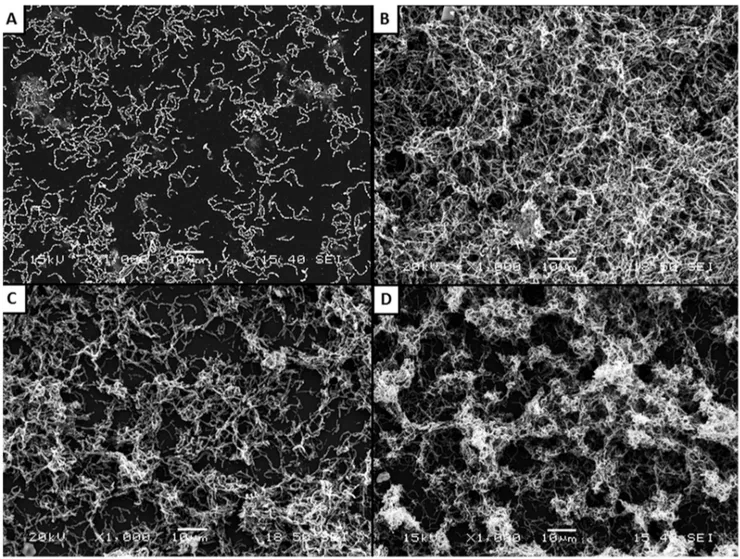
Figure 1. Scanning electron micrographs ofS. mutansATCC 25175 biofilm following growth in THB-HK (Panel A) supplemented with 1/2 MIC of triclosan (Panel B), or 1/4 MIC of triclosan (Panel C) or sucrose used as a positive control (Panel D).
Supermix, 5ml of cDNA, 1ml of gene-specific primer, and 6.5ml
of RNase- and DNase-free water. The samples were amplified using a Bio-Rad MyCyclerTMthermal cycler (Bio-Rad Laborato-ries). The amplification conditions for atlA, comD, gtfC, luxSand 16S rRNA were 95uC for 3 min followed by 30 cycles at 95uC for 45 s, 60uC for 45 s and 72uC for 30 s, while that ofgtfBwas 95uC for 3 min followed by 40 cycles at 95uC for 45 s, 55uC for 45 s and 72uC for 30 s. To validate the specificity of each primer pair, temperature curve analyses were performed.
Statistical Analysis
Unless specified otherwise, assays were run in triplicate and the means6standard deviations were calculated. Data were analyzed using the Studentt-test.
Results
Using a microdilution broth method, the MIC of triclosan forS. mutans ATCC 25175 and ATCC 35668 was 7.8mg/ml.
There-after, biofilm formation by S. mutans was investigated following growth in culture medium6triclosan at 1/2, 1/4, or 1/8 MIC. As reported in Table 2, biofilm formation by both strains of S. mutanswas dose-dependently induced by sub-MICs of triclosan, as determined by crystal violet staining. At 1/2 and 1/4 MIC of triclosan, the biofilm ofS. mutansATCC 25175 was increased by 6.2- and 5-fold, while that of strain ATCC 35668 was increased by 3- and 2.2-fold, respectively. The biofilm was not significantly affected following growth in the presence of triclosan at 1/8 MIC. The effect of adding 0.25% sucrose to THB-HK on biofilm formation induced by sub-MICs of triclosan was also tested. As shown in Table 2, in the absence of triclosan, an important biofilm
was formed by both strains ofS. mutans. The triclosan sub-MICs-inducing effect on biofilm formation was much less significant in the presence of sucrose. Planktonic cells, estimated by recording the OD660of the bacterial suspensions surrounding the biofilm,
were significantly decreased following growth in the presence of 1/ 2 MIC of triclosan in the presence or not of sucrose (Table 2). Given that the triclosan-induced biofilm formation was optimal for
S. mutansATCC 25175 grown in the absence of sucrose, this strain and condition were selected for further analyses.
Scanning electron microscopy analysis was performed to observe the triclosan sub-MICs-induced biofilm formation byS. mutans ATCC 25175. As shown in Figure 1A, individual short chains of S. mutans were observed attached to the polystyrene surface when growth was carried out in THB. However, when the culture medium was supplemented with 1/2 and 1/4 MIC of triclosan (Figures 1B and 1C), a thick biofilm made of aggregates and microcolonies of S. mutans almost completely covered the surface of the polystyrene support. Sucrose, a well-known biofilm-promoting agent used as positive control, also induced the formation of biofilm (Figure 1D).
Thereafter, we further investigated the impact of triclosan at sub-MICs on the host colonization properties ofS. mutansATCC 25175 by evaluating the effect on adherence to gingival epithelial cells. As reported in Figure 2, triclosan at 1/2 and 1/4 MIC promoted the adherence of FITC-labeledS. mutansto a monolayer of gingival epithelial cells. More specifically, at 1/2 MIC of triclosan, the adherence of S. mutans to epithelial cells was increased by 42.5%.
We then attempted to identify the mechanism by which triclosan at sub-MICs may increase the capacity of S. mutans to form biofilm and adhere to epithelial cells. Since the hydrophobic
Figure 2. Effect of sub-MICs of triclosan on adherence ofS. mutansATCC 25175 to gingival epithelial cells.RFU: Relative Fluorescence Units. Data are expressed as means6standard deviations. Significant increase (*,p,0.01) compared to control bacteria grown in the absence of triclosan.
properties of the bacterial cell surface may be involved in adherence and biofilm formation, we tested the effect of growing
S. mutansin the presence of triclosan at sub-MICs on cell surface hydrophobicity. No significant modifications in cell surface hydrophobicity were observed (data not shown).
The expression profile of five genes related to adherence and biofilm formation inS. mutanswas determined following incubation (2 h) ofS. mutansin the absence and presence of triclosan at 1/2 and 1/4 MIC. As reported in Figure 3, the genes gtfC
(glucosyltransferase C), comD (histidine kinase sensor protein), and luxS (autoinducer 2 synthase) were those for which the expression was the most upregulated. More specifically, triclosan at 1/2 MIC, increasedgtfC,comD, andluxSexpression by 3.6-, 3.1-, and 4-fold, respectively. Although the upregulation of atlA
(autolysin) and gtfB (glucosyltransferase B) expression was less pronounced, it was significantly increased following incubation of
S. mutanswith triclosan at sub-MICs.
Discussion
Triclosan is a broad spectrum antimicrobial agent used in oral care products to control dental plaque [20]. Although numerous studies investigated the antibacterial properties of triclosan towards oral bacteria [20,21], there are no data in the literature on the effects of this compound at sub-MICs. Since there are a number ofin vivocircumstances where concentrations of triclosan may be at subinhibitory levels, we investigated the effects of sub-MICs of this antimicrobial agent on the cariogenic bacteriumS. mutansin regard to its capacity to colonize the host.
Previous studies have shown that antimicrobial agents at subMICs can either increase or decrease biofilm formation by bacterial pathogens [10,11,13,14]. Our study brought clear evidence that triclosan at sub-MICs significantly increases the biofilm formation capability of S. mutans. To the best of our knowledge, this is the first report on the effect of sub-MICs of triclosan on bacterial adherence properties. Prior to our study, only one research group reported on the effect of an antimicrobial agent on biofilm formation byS. mutans. More specifically, Dong
et al.[19] recently showed that sub-MICs of chlorhexidine appear to solidify and strengthenS. mutansbiofilm. The ability of nicotine to enhanceS. mutansbiofilm formation has also been reported [22]. Although the primary natural location ofS. mutansis the dental biofilm, we showed that growing S. mutans in the presence of triclosan at sub-MICs increased its capacity of adherence to epithelial cells. If the epithelial barriers are breached, adheredS. mutansmay invade tissue, enter the bloodstream, and ultimately induce infective endocarditis. Since S. mutans is an important causative agent of subacute infective endocarditis in particular in subjects with predisposing cardiac conditions [23], further studies should investigate the effects of sub-MICs of triclosan on adherence to endothelial cells.
We then attempted to identify the mechanism by which triclosan at sub-MICs may increaseS. mutans biofilm formation and adherence to epithelial cells. The cell surface hydrophobicity of bacteria is known to contribute to their adherence properties [24]. Wuet al.[25] reported that sub-MICs of specific antibiotics can increase the surface hydrophobicity of another important cariogenic bacterium,Streptococcus sobrinus, a phenomenon that may
Figure 3. Effect of sub-MICs of triclosan on mRNA expression of specific genes involved in adherence and biofilm formation inS. mutansATCC 25175.Data are expressed as means6standard deviations. The expression was normalized to 16S rRNA. Significant increase (*,p, 0.05; **,p,0.01; ***,p,0.001) compared to untreated control bacteria.
increase their adherence property. In the present study, triclosan at sub-MICs had no effect on the surface hydrophobicity of S. mutans, a result that ruled out the involvement of this mechanism in the increased adherence properties ofS. mutans.
S. mutanscan use sucrose to synthesize extracellular polysaccha-rides via glucosyltransferases, more specifically GtfB and GtfC [26]. In this study, although the expression ofgtfB and gtfC was increased inS. mutansexposed to triclosan at sub-MICs, it is likely not responsible for the increased biofilm formation observed since sucrose was not used in the culture medium.
Biofilm formation is largely influenced by bacterial communi-cation via quorum-sensing signaling system [27]. More specifically, in S. mutans, the comD gene product, an histidine kinase sensor protein for the competence-stimulating peptide (CSP), is known to play a critical role in biofilm formation [27]. Moreover, LuxS is produced by many Gram positive bacteria, including S. mutans, and is involved in the production of autoinducer 2, another signaling molecule playing a role in biofilm formation [28]. Our study showed that both comC and luxS genes were significantly upregulated whenS. mutanswas cultivated in the presence of sub-MICs of triclosan. This is likely contributing to the increased biofilm formed under this condition. Dong et al. [19] also reported on the capacity of sub-MICs of antimicrobial agents, more specifically sodium fluoride and tea polyphenols, to increase the mRNA expression ofcomDandluxS.
The autolysin AtlA (also known as Smu0630) ofS. mutanshas been reported to play a critical role in biofilm formation regardless of the carbohydrate source. [29]. Interestingly, AtlA has been identified as a fibronectin-binding protein that contributes to bacterial survival in the bloodstream and consequently as a virulence factor for infective endocarditis [30]. This cell surface adhesion whose expression was found to be upregulated in S. mutans exposed to sub-MICs of triclosan may contribute to the increased adherence to epithelial cells.
Conclusions
Our study showed that sub-MICs of triclosan can enhance biofilm formation and epithelial cell adherence ofS. mutans. We also brought evidence that this may be modulated by an increased expression of specific genes coding for cell surface adhesins or involved in quorum-sensing. Collectively, our data stress the importance of maintaining MIC of therapeutic triclosan to efficiently prevent colonization of the oral cavity byS. mutans.
Author Contributions
Conceived and designed the experiments: DG DPS. Performed the experiments: TBLB LG. Analyzed the data: TBLB DG DPS. Contributed reagents/materials/analysis tools: DG. Wrote the paper: DG DPS TBLB.
References
1. Schweizer HP (2001) Triclosan: a widely used biocide and its link to antibiotics. FEMS Microbiol Lett 202: 1–7.
2. Villalain J, Mateo CR, Aranda FJ, Shapiro S, Micol V (2001) Membranotropic effects of the antibacterial agent triclosan. Arch Biochem Biophys 390: 128–136. 3. Mustafa M, Wondimu B, Ibrahim M, Modeer T (1998) Effect of triclosan on interleukin-1bproduction in human gingival fibroblasts challenged with tumor necrosis factora. Eur J Oral Sci 106: 637–643.
4. Modeer T, Bengtsson A, Rolla F (1996) Triclosan reduces prostaglandin biosynthesis in human gingival fibroblasts challenged with interleukin-1in vitro. J Clin Periodontol 23: 927–933.
5. Davies RM, Ellwood RP, Davies GM (2004) The effectiveness of a toothpaste containing triclosan and polyvinyl-methyl ether maleic acid copolymer in improving plaque control and gingival health: a systematic review. J Clin Periodontol 31: 1029–1033.
6. Takahashi N, Nyvad B (2008) Caries ecology revisited: microbial dynamics and the caries process. Caries Res 42: 409–418.
7. Takahashi N, Nyvad B (2011) The role of bacteria in the caries process: ecological perspectives. J Dent Res 90: 294–303.
8. Mann J, Vered Y, Babayof I, Sintes J, Petrone ME, et al. (2001) The comparative anticaries efficacy of a dentifrice containing 0.3% triclosan and 2.0% copolymer in a 0.243% sodium fluoride/silica base and a dentifrice containing 0.243% sodium fluoride/silica base: a two-year coronal caries clinical trial on adults in Israel. J Clin Dent 12: 71–76.
9. Phan TN, Marquis RE (2006) Triclosan inhibition of membrane enzymes and glycolysis ofStreptococcus mutansin suspensions and biofilms. Can J Microbiol 52: 977–983.
10. Majta´n J, Majta´nova´ L, Xu M, Majta´n V (2008)In vitroeffect of subinhibitory concentrations of antibiotics on biofilm formation by clinical strains ofSalmonella entericaserovarTyphimuriumisolated in Slovakia. J Appl Microbiol 104: 1294– 1301.
11. Starner TD, Shrout JD, Parsek MR, Appelbaum PC, Kim G (2008) Subinhibitory concentrations of azithromycin decrease nontypeableHaemophilus influenzae biofilm formation and diminish established biofilms. Antimicrob Agents Chemother 52: 137–145.
12. Wojnicz D, Jankowski S (2007) Effects of subinhibitory concentrations of amikacin and ciprofloxacin on the hydrophobicity and adherence to epithelial cells of uropathogenicEscherichia colistrains. Int J Antimicrob Agents 29: 700– 704.
13. Hoffman LR, D’Argenio DA, MacCoss MJ, Zhang Z, Jones RA, et al. (2005) Aminoglycoside antibiotics induce bacterial biofilm formation. Nature 436: 1171–1175.
14. Rachid S, Ohlsen K, Witte W, Hacker J, Ziebuhr W (2000) Effect of subinhibitory antibiotic concentrations on polysaccharide intercellular adhesin expression in biofilm-forming Staphylococcus epidermidis. Antimicrob Agents Chemother 44: 3357–3363.
15. Erdeljan P, MacDonald KW, Goneau LW, Bevan T, Carriveau R, et al. (2012) Effects of subinhibitory concentrations of ciprofloxacin on Staphylococcus
saprophyticusadherence and virulence in urinary tract infections. J Endourol 26: 32–37.
16. Marquis A, Genovese S, Epifano F, Grenier D (2012) The plant coumarins auraptene and lacinartin as potential multifunctional therapeutic agents for treating periodontal disease. BMC Complement Altern Med 12: 80. 17. Kusumoto Y, Hirano H, Saitoh K, Yamada S, Takedachi M, et al. (2004)
Human gingival epithelial cells produce chemotactic factors interleukin-8 and monocyte chemoattractant protein-1 after stimulation with Porphyromonas gingivalisvia Toll-like receptor 2. J Periodontol 75: 370–379.
18. Rosenberg M, Gutnick D, Rosenberg E (1980) Adherence of bacteria to hydrocarbons: a simple method for measuring cell-surface hydrophobicity. FEMS Microbiol Lett 9: 29–33.
19. Dong L, Tong Z, Linghu D, Lin Y, Tao R, et al. (2012) Effects of sub-minimum inhibitory concentrations of antimicrobial agents onStreptococcus mutansbiofilm formation. Int J Antimicrob Agents 39: 390–395.
20. Wade WG, Addy M (1992) Antibacterial activity of some triclosan-containing toothpastes and their ingredients. J Periodontol 63: 280–282.
21. Haraszthy VI, Reynolds HS, Sreenivasan PK, Subramanyam R, Cummins D, et al. (2006) Media- and method-dependent variations in minimal inhibitory concentrations of antiplaque agents on oral bacteria. Lett Appl Microbiol 43: 256–261.
22. Huang R, Li M, Gregory RL (2012) Effect of nicotine on growth and metabolism ofStreptococcus mutans. Eur J Oral Sci 120: 319–325.
23. Mylonakis E, Calderwood SB (2001) Infective endocarditis in adults. N Engl J Med 345: 1318–1330.
24. Doyle RJ, Rosenberg M, Drake D (1990) Hydrophobicity of oral bacteria. In: Doyle RJ and Rosenberg M, editors. Microbial cell surface hydrophobicity. American Society for Microbiology, Washington, D.C. 387–419.
25. Wu Q, Wang Q, Taylor KG, Doyle RJ (1995) Subinhibitory concentrations of antibiotics affect cell surface properties ofStreptococcus sobrinus. J Bacteriol 177: 1399–1401.
26. Koo H, Xiao J, Klein MI (2009) Extracellular polysaccharides matrix – an often forgotten virulence factor in oral biofilm research. Int J Oral Sci 1: 229–234. 27. Senadheera D, Cvitkovitch DG (2008) Quorum sensing and biofilm formation
by Streptococcus mutans. In: Utsumi R, editor. Bacterial signal transduction: networks and drug targets. Landes Bioscience and Springer Science+Business Media. 178–188.
28. Nobbs AH, Lamont RJ, Jenkinson HF (2009) Streptococcus adherence and colonization. Microbiol Mol Biol Rev 73: 407–450.
29. Brown TA, Ahn SJ, Frank RN, Chen YY, Lemos JA, et al. (2005) A hypothetical protein ofStreptococcus mutansis critical for biofilm formation. Infect Immun 2005, 73: 3147–3151.