Recebido em 07.07.2000. / Received in July, 07thof 2000.
Aprovado pelo Conselho Consultivo e aceito para publicação em 04.06.2002. / Approved by the Consultive Council and accepted for publication in June, 04stof 2002. * Trabalho realizado no Serviço de Dermatologia da Faculdade de Ciências Médicas de Pernambuco. / Work done at the "Serviço de Dermatologia da Faculdade de Ciências Médicas de
Pernambuco."
1Médica dermatologista / Dermatologist.
2Preceptora de Dermatologia. / Dermatology preceptor 3Preceptora de Dermatopatologia/ Dermatopathology preceptor
4Livre-Docente e Doutor em Dermatologia. Professor Adjunto e Chefe do Serviço. / Ph.D. in dermatology, and lecturer. Adjunct Professor and Head of the Service
©2003by Anais Brasileiros de Dermatologia
Síndrome de reiter: relato de caso
*
Reiter's syndrome: a case report
*
Ana Elisabete Simões de Sousa
1Aldejane Gurgel
2Juliana Albuquerque de Sousa
1Eliane Alencar
3Márcia Maria Ribeiro Costa
1Emmanuel Rodrigues de França
4Resumo:Relato de um caso de síndrome de Reiter em paciente jovem, do sexo masculino, com lesões der-matológicas típicas e achado positivo para o antígeno do complexo de histocompatibilidade HLA-B27. O quadro surgiu após infecção intestinal por Salmonella enteritidis, evoluindo com melhora após utilização de tetraciclina, prednisona e indometacina. Episódio recidivante foi tratado com metotrexato. É feita uma revisão da literatura, abordando os aspectos clínicos, laboratoriais, etiológicos e fisiopatogênicos dessa sín -drome.
Palavras-chave: Doença de Reiter; Salmonella enteritidis.
Summary:A case of Reiter's disease is reported in a young male showing typical dermatological
lesions and positive finding for the HLA-B27 antigen histocompatibility complex. The condition arose
following enteric infection with Salmonella enteritidis. Remission followed treatment with
tetracy-cline, prednisone and indomethacin. The relapse of disease was treated with methotrexate. A review of clinical, physiopathologic and laboratory findings of Reiter's syndrome are presented.
Key words: Reiter Disease; Salmonella enteritidis.
Caso Clínico /
Case Report
INTRODUÇÃO
A síndrome de Reiter é condição rara, de distribuição universal. Acomete preferencialmente indivíduos adultos do sexo masculino. Caracteriza-se por poliartrite periférica soronegativa, com duração mais longa do que um mês, manifestando-se geralmente após quadro infeccioso disenté-rico ou urogenital.1Acometimentos mucocutâneo, ungueal e
ocular são comuns. Os autores acompanharam um paciente com síndrome de Reiter com manifestações clínicas e labo-ratoriais típicas. Neste trabalho, relatam o caso, consideran-do o possível envolvimento infeccioso, genético e imunoló-gico em sua etiopatogenia, ainda obscura.
RELATO DO CASO
INTRODUCTION
Reiter's syndrome is a rare globally distributed condition, occurring most commonly in men. It is charac-terized by seronegative peripheral polyarthralgia, lasts for over a month, and is generally manifested subsequent to an infectious dysenteric or urogenitial condition.1
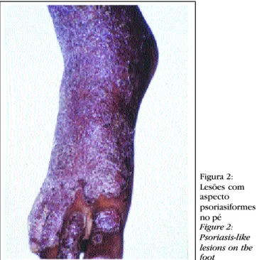
Paciente de 16 anos, do sexo masculino, natural e procedente de Recife, PE, procurou o ambulatório do Hospital Universitário Oswaldo Cruz - Faculdade de Ciências Médicas, apresentando poliartralgia periférica e lesões cutâneo-mucosas disseminadas, de aspecto psoriasiforme. Relatava episódio prévio de infecção intestinal há cerca de três semanas. No momento da consulta foram observados edema, dor à mobilização e rigidez das articulações dos punhos, cotovelos, joelhos e tornozelos, com limitação dos movimentos. Ao exame dermatológico, apresentava placas eritêmato-escamo-crostosas, localizadas no tronco (Figura 1), membros superiores e inferiores (Figura 2); lesões eritêmato-es-camativas ceratósicas nas regiões plantares (Figura 3); e lesões eritêmato-escamosas de aspecto circinado na glande (Figura 4).
O estudo histopatológico de uma lesão cutânea do tronco demonstrou hiperceratose compacta e focos de paraceratose, acantose, alongamento das papilas dérmi -cas e infiltrado celular predominantemente linfocitário intersticial e perivascular. O hemograma evidenciou ane-mia com discreta leucocitose, VSH elevado (62mm/h) e
PCRqualitativo elevado (+++). A pesquisa do fator reu-matóide foi negativa pela reação do látex, e a do antíge-no do complexo de histocompatibilidade HLA-B27, posi-tiva. As reações sorológicas para sífilis (VDRL e FTA-ABS) foram negativas, bem como a pesquisa de anticor-pos anti-HIV, feita pelo método Elisa. O sumário de urina evidenciou numerosos piócitos por campo. A urocultura foi positiva para Staphylococcus aureus, e a
coprocultu-CASE REPORT
A 16-year-old male patient, born and raised in Recife, Pernambuco State (PE), sought care at the outpatient clinic of the Hospital Universitário Oswaldo Cruz -Faculdade de Ciências Médicas (Hospital Universitário Oswaldo Cruz Faculty of Medical Sciences). He showed peripheral polyarthralgia and disseminated mucocuta-neous lesions with psoriatic-like features. The patient reported having a previous episode of intestinal infection roughly three weeks earlier. At the moment of consultation, edema was observed, with pain and stiffness in the joints when moving the wrist, elbow, knee and ankle, including limitation of movement. The dermatological examination showed erythematous, crusty and scaly plaques located on the trunk (Figure 1), and upper and lower limbs (Figure 2); erythematous and erosive keratotic plaques on the plantar regions (Figure 3); and erythematous squamous circinate-like lesions on the glans penis (Figure 4).
The histopathologic study of a cutaneous lesion on the trunk showed compact hyperkeratosis and parakerato-sis clusters, acanthoparakerato-sis, lengthening of the dermal papilla and predominately interstitial and perivascular lymphocy-te cellular infiltralymphocy-te. The hemogram evidenced anemia with discreet leukocytosis, high VSH (62 mm/h) and high quali-tative PCT (+++). The study of the rheumatic factor was negative for latex reaction, and the study of the HLA-B27
antigen histocompatibility complex was positive. Serological reactions for the diagnosis of syphilis (VDRL
and FTA-ABS) were negative, as was the anti-HIVantibody
test, done by Elisa method. The urine summary evidenced
Figura 1: Lesões eritêmato-escamativas no tronco
Figure 1: Erythematous scaly lesions on the trunk
Figura 2: Lesões com aspecto psoriasiformes no pé
Figura 4: Balanite circinada
Figure 3: Keratoderma blennorrhagica
numerous pyocytes per field. Urine culture was positive for
Staphylococcus aureus, and copro-culture was positive for Salmonella enteritidis. Radiographic examina-tion of the knee joint only showed signs of effusion. Analysis of arti-cular fluid evidenced neutrophil and hyperprotein with negative cultures. Electrocardiogram and ecocardiogram were normal. Ophthalmologic evaluation did not show changes.
Treatment was initiated with tetracycline, 500 mg every six hours over 10 days, associated with 40 mg prednisone daily and 25 mg indometacin daily, due to severe articular involvement. After four weeks, the patient showed significant clinical improvement of the arthralgia and cutaneous condition. The corticoid dose (5 mg weekly) was tapered next to an increase in the indome-tacine dose (25 mg weekly). The patient was released from hospital with doses of 20 mg prednisone daily and 75 mg daily indometacin. He was kept under outpatient follow-up, with later withdrawal of the drugs.
Three months after being released from hospital, the patient showed relapsing of the dermatological and articu-lar condition, after which he was interned again. He then initiated treatment with methotrexate in a weekly dose of 25 mg IM. Five weeks later he showed regression of the condition, and was granted asymptomatic hospital release. Two months later, in outpatient control, the patient was asymptomatic without taking medication.
DISCUSSION
Reiter's syndrome belongs to a group of disea-ses called seronegative s p o n d y l o a r t h r o p a t h i e s , which exhibit a hereditary predisposition associated with the presence of the
anti-ra mostrou-se positiva paanti-ra
Salmonella enteritidis. O exame radiográfico da articulação do joe-lho demonstrou apenas sinais de efusão. A análise do fluido articu-lar evidenciou neutrofilia e hiper-proteinemia com culturas negati-vas. O eletrocardiograma e o eco-cardiograma foram normais. A avaliação oftalmológica não mos-trou alterações.
Foi instituído tratamento com tetraciclina, 500mg a cada seis horas, durante 10 dias, associada à prednisona 40mg/dia e indometaci-na 25mg/dia, devido ao grave
envolvimento articular. Após quatro semanas, o paciente apresentou melhora clínica significativa da artralgia e do quadro cutâneo, sendo iniciada a diminuição da dose do corticóide (5mg por semana) com aumento na dose da indometacina (25mg por semana), recebendo alta hospita-lar em uso de 20mg/dia de prednisona e 75mg/dia de indometacina. Manteve-se em acompanhamento ambula-torial, com posterior retirada das drogas.
Três meses após a alta hospitalar, o paciente apre-sentou recidiva do quadro dermatológico e articular, sendo novamente internado; iniciou, então, tratamento com meto-trexato em dose semanal de 25mg IM. Após cinco semanas apresentou regressão do quadro, recebendo alta hospitalar assintomático.
Dois meses após, o paciente encontrava-se em contro-le ambulatorial, assintomático e sem uso de medicação.
DISCUSSÃO
A síndrome de Reiter faz parte de um grupo de doenças denominadas espon-diloartropatias soronegativas, que exibem predisposição hereditária associada à pre-sença do antígeno do comple-xo de histocompatibilidade
Figura 3: Queratoderma
blenorrágico
Figure 4:
maior (HLA-B27) e têm em comum graus variáveis de mani-festações artrocutâneas e oculares.
Sua etiopatogenia não está completamente com-preendida, porém a consistente associação com o HLA-B27
e sua ocorrência em vários membros de uma mesma famí-lia falam fortemente a favor de uma predisposição consti-tucional geneticamente determinada.2Esses pacientes
res-pondem de formas variadas a diversos agentes infecciosos, indicando que fatores imunorreguladores também sejam importantes em sua patogênese.3O HLA-B27, positivo no
paciente em estudo, é o maior determinante de susceptibi-lidade para a síndrome de Reiter, sendo encontrado em per-centual que varia de 70 a 90% dos pacientes.4O risco rela
-tivo de os indivíduos B27positivos desenvolverem a doen-ça é 25 vezes maior em comparação com os B27negativos.5
Entretanto os casos B27negativos demonstram que o gene simplesmente não é essencial. O mecanismo da doença parece envolver uma resposta imunológica específica do tipo celular, centrada pelos linfócitos T CD8, que reconhe-cem os antígenos do microorganismo apresentado pelas moléculas da classe I do complexo de histocompatibilida -de maior (MHC) e iniciam a resposta inflamatória.6A
ocorrência da síndrome de Reiter na presença de uma imunode -ficiência acentuada, como nos pacientes HIV positivos, sugere que as células T helper não estão envolvidas em sua patogênese.3 O papel do vírus
HIV na patogênese dessa doença merece investigações mais amplas. O próprio vírus pode causar artrite diretamente ou ser responsável por maior susceptibilidade para infecções por microorganis-mos artritogênicos, uma vez que esses indivíduos estão mais expostos a vários organismos oportunistas.7,8Nenhum
agente infeccioso isoladamente pode ser responsabilizado pelo desenvolvimento da doença.
Há duas formas epidemiológicas distintas: uma endê-mica ou venérea, iniciada por uretrite inespecífica, cujo agen-te infeccioso mais importanagen-te é a Chlamydia trachomatis, sendo também implicados o Ureaplasma e o Mycoplasma
como possíveis agentes;7outra epidêmica ou pós-disentérica,
na qual se inclui o paciente em questão, em seguimento após uma infecção intestinal por microorganismos Gram negati-vos chamados artritogênicos, incluindo os gêneros Shigella,
Salmonella, Yersinia, Campylobacter eClostridium.8,9,10
Embora possa ocorrer em qualquer faixa etária, a sín-drome de Reiter acomete preferencialmente o indivíduo adulto do sexo masculino, sendo considerada a causa mais comum de artrite em pacientes jovens.11,12Clinicamente manifesta-se com
envolvimento do sistema articular, cutâneo-mucoso e ocular. A doença articular caracteriza-se por acometimento poliarticular, envolvendo predominantemente as articulações dos membros inferiores e sacroilíaca, apresentando entesopa-tia e artrite.13Os sítios mais envolvidos são o tornozelo, o pé
gen of the major histocompatibility complex (HLA-B27). They share common variable degrees of arthrocutaneous and ocular manifestations.
Its etiopathogenesis is not completely understood. However the consistent association with HLA-B27and its occurrence in various members of a single family speak strongly in favor of a genetically determined constitutional predisposition.2These patients respond variably to diverse
infectious agents, indicating that immunoregulatory fac-tors are also important in its pathogenesis.3HLA-B27,
posi-tive in the patient being studied, is the greatest determining factor of vulnerability to Reiter's syndrome, encountered in percentages varying from 70 to 90% of patients.4The
rela-tive risk of B27 posirela-tive individuals developing the disease is 25 times higher in comparison with B27 negatives.5
Nonetheless, B27 negative cases show that the gene is simply not essential. The disease mechanism seems to involve a specific cell-like immunologic response centered around T CD8 lymphocytes that recognize the microorga-nism antigens demonstrated by the class I molecules of the major histocompatibility complex (MHC) and initiate the inflammatory response.6The occurrence of Reiter's
syndro-me in the presence of accentuated immunodeficiency, as in
HIV-positive patients, suggests that the T helper cells are not involved in its pathogenesis.3The role of the HIVvirus
in the pathogenesis of this disease deserves broader inves-tigation. The virus itself may cause arthritis directly or be responsible for higher vulnerability to infections by arthri-togenic microorganisms, once these individuals are more exposed to various opportunistic organisms.7,8 No
infec-tious agent alone may be held responsible for the develop-ment of the disease.
There are two distinct epidemiological forms: an endemic or venereal one, initiated by non-specific urethri-tis, whose most important infectious agent is Chlamydia trachomatis, with Ureaplasmaand Mycoplasmaalso impli-cated as possible agents;7 the other form is epidemic or
post-dysenteric, which includes the patient being studied, subsequent to an intestinal infection by Gram negative microorganisms called arthritogenic, including the
Shigella, Salmonella, Yersinia, Campylobacter and
Clostridium generae.8,9,10
Although Reiter's syndrome is not age specific, it most commonly affects male adults, as well as being consi-dered the most common cause of arthritis in young patients.11,12It is clinically manifested with involvement of
the joint, mucocutaneous and ocular systems.
The articular disease characterized by polyarticu-lar affliction predominantly involving the lower limbs and sacroiliac joints, showed enthesopathy and arthritis.13The
Characteristic symptoms include pain and rigidity after periods of rest, improving with exercise. The episodes tend to be self-limited, but they may recur for years or decades. Roughly a quarter of patients with chronic Reiter's syndro-me develop sacroiliatis or ankylosing spondylitis.15In the
patient studied in this paper, articular dysfunction was sig-nificant. In line with the literature findings, the involve-ment of the wrist, elbow, knee and foot joints was observed, which involved pain and rigidity in the joints.
The mucocutaneous disease shows variable inci-dence, with lesions present in proportions varying from 8 to 31% of cases. In spite of the literature referring to their lower frequency in post-dysenteric cases,16,17cutaneous and
florida mucose manifestations, like circinate balanitis, keratoderma blennorrhagica and ungual involvement, were present in the patient studied. Circinate balanitis is the most common finding, seen in 36% of patients with cutaneous condition.18It is characterized by surface ulcers
located in the mucosa of the penis, which may coalesce, taking on the circinate distribution. There may also be involvement of the penis base and scrotal sac. Keratoderma blennorrhagica most frequently affects the palmoplantar regions. Although it may be a typical mani-festation of Reiter's syndrome, it is not a frequent finding, found only in 15% of patients.19 Beginning with
erythema-tous macules, it progresses rapidly into papules or pseudo-vessels that may coalesce. The final appearance of the lesion is hyperkeratotic plaques, with patches of eroding skin and color varying from erythematous to yellow-oran-ge. At times, keratoderma blennorrhagica may progress with erythrodermia, especially in cases associated with
HIV infection.19 Ungual and periungual involvement is
common and generally severe. It is characterized by thic-kening of the nail plate, which becomes yellow and fragile, with possible occurrence of real onycholysis with nail loss. Pitting of the nails, characteristic of psoriasis, is not seen.20
Ocular involvement, which may be present in roughly half of cases,21was not observed in the patient
stu-died. Conjunctivitis is the most common manifestation wit-nessed, though irritation, uveitis and keratitis may also occur.22
Other manifestations could be present. During the acute phase, constitutional sign and symptoms are prominent. Additional findings include: erythema nodosum, car -diac valvular disease, pulmonary parenchymal inflamma-tion, peripheral neuropathy and nephropathies.23
From the laboratorial point of view, there is no spe-cific test to confirm the diagnosis of Reiter's syndrome. The hemogram is normal or there is variable leukocytosis, and the reagents of the acute phase (VSHand PCR) are
gene-e o jogene-elho.14Os sintomas característicos incluem dor e rigidez
após períodos de repouso, com melhora após exercícios. Os episódios tendem a ser autolimitados, mas podem recorrer por anos ou décadas. Aproximadamente um quarto dos pacientes com síndrome de Reiter crônica desenvolve sacroi-leíte ou espondilite anquilosante.15No paciente em estudo o
comprometimento articular foi significativo, e, em acordo com os achados da literatura, observou-se envolvimento das articulações dos punhos, cotovelos, joelhos e pé, com dor e rigidez nas articulações envolvidas.
A doença mucocutânea apresenta incidência variável, com lesões presentes em proporção que varia de oito a 31% dos casos. Apesar de a literatura referir sua menor freqüência nos casos pós-disentéricos,16,17 manifestações cutâneas e mucosas
floridas, como a balanite circinada, o queratoderma blenorrági-co e envolvimento ungueal, estavam presentes no paciente estu-dado. A balanite circinada é o achado mais comum, visto em cerca de 36% dos pacientes com envolvimento cutâneo.18
Caracteriza-se por úlceras superficiais úmidas localizadas na mucosa do pênis, que podem coalescer, assumindo distribuição circinada. Pode haver também envolvimento da base do pênis e da bolsa escrotal. O queratoderma blenorrágico acomete mais freqüentemente as regiões palmoplantares. Embora seja uma manifestação típica da síndrome de Reiter, não é achado fre-qüente, sendo encontrado em apenas 15% dos pacientes.19
Inicia-se por máculas eritematosas evoluindo rapidamente para pápulas ou pseudovesículas que podem coalescer. A aparência final da lesão é de placa hiperceratósica, com colarete descama-tivo e coloração que varia do eritema ao amarelo-alaranjado. Ocasionalmente, o queratoderma blenorrágico pode evoluir com eritrodermia, sobretudo nos casos associados à infecção pelo HIV.19O envolvimento ungueal e periungueal é comum e
em geral grave. Caracteriza-se por espessamento da lâmina ungueal, que se encontra amarelada e frágil, podendo ocorrer onicólise franca com perda ungueal. Pittingungueal, caracterís-tico da psoríase, não é visto.20
O envolvimento ocular, que pode estar presente em aproximadamente metade dos casos,21não foi observado no
paciente em estudo. A conjuntivite é a manifestação mais comum, podendo também ocorrer irite, uveíte anterior e queratite.22
Outras manifestações podem estar presentes. Durante a fase aguda, sinais e sintomas constitucionais são proeminentes. Achados adicionais incluem: eritema nodo-so, doença valvular cardíaca, inflamação parenquimatosa pulmonar, neuropatia periférica e nefropatias.23
sensí-vel e específico da atividade da doença do que o VSH.24
Níveis de PCRsão significativamente mais altos em pacien-tes com doença ativa, enquanto o VSHpode não ser estatis-ticamente diferente na doença ativa e na inativa.24
Imunocomplexos podem ser detectados no soro, porém a pesquisa do fator reumatóide deve ser negativa.25A
positivi-dade para o antígeno do complexo de histocompatibilipositivi-dade
HLA-B27 é suporte diagnóstico, estando relacionado com
doença mais grave e protraída.26Tendo em vista o aumento
de casos da síndrome de Reiter em pacientes com Aids, tes-tes sorológicos para HIVdevem ser realizados em todos os pacientes.27Sumário de urina é de pouca utilidade, porém o
exame direto e cultura da secreção uretral são importantes na investigação. Urocultura e coprocultura devem ser realiza-das para pesquisa de microorganismos. No caso do paciente objeto deste estudo, a coprocultura demonstrou crescimento da Salmonella enteritidis, que foi responsabilizada pelo desencadeamento do quadro, no paciente com predisposição genética definida pela presença do HLA-B27. O exame radio-gráfico das articulações acometidas pode, em casos de artri-te crônica, deartri-tectar sinais de efusão, artriartri-te franca, erosões, osteoporose periarticular e entesopatia.15 O fluido articular
aspirado pode ser turvo ou amarelado, porém geralmente estéril. Apesar do grave envolvimento clínico articular observado no paciente, o estudo radiológico das articulações envolvidas evidenciou apenas derrame articular. Mesmo na ausência de sintomas oculares justifica-se a avaliação oftal-mológica pelo freqüente comprometimento ocular. Eletrocardiograma e ecocardiograma devem ser realizados para exclusão de alterações cardíacas. As alterações histopa-tológicas das lesões mucocutâneas não são diagnósticas. Nos cortes histopatológicos observam-se alterações indistin-guíveis da psoríase, quando a mesma está completamente desenvolvida, sendo, portanto, um diagnóstico de compati-bilidade com a clínica.
No diagnóstico diferencial devem ser consideradas outras formas de espondiloartropatias (espondilite anquilosan-te e artrianquilosan-te psoriática), artrianquilosan-te gonocóccica, doença de Lyme, gota, artrite reumatóide, síndrome de Behçet, febre reumática, psoríase, candidíase, eritema polimorfo e líquen plano.
Não há cura para a síndrome de Reiter. Aproximadamente dois terços dos pacientes apresentam remissão do quadro em seis meses.28Recorrências são fre
-qüentes, com 40% dos pacientes apresentando um segundo episódio,29 como observado no paciente em questão. Na
fase aguda, repouso e antiinflamatório não hormonal podem ser úteis. Fisioterapia passiva e ativa devem ser recomendadas para evitar seqüelas, manter a mobilidade e prevenir a fibrose articular. Nos casos graves e crônicos o tratamento é difícil. Antibióticos são utilizados com o obje -tivo de encurtar a evolução e impedir o início da artrite
rea-rally high. Reactive protein C is a more sensitive and spe-cific indicator of the disease activity than is VSH.24
PCR
levels are significantly higher in patients with active disea-se, while the VSHmay be statistically different in the acti-ve and inactiacti-ve diseases.24 Immune complexes may be
detected in the serum, however research into the rheuma-toid factor should be negative.25 Positivity for the antigen
of the histocompatibility complex HLA-B27is a diagnostic support, being related to the most severe and protracted disease.26 Bearing in mind the increase in cases of Reiter's
syndrome among AIDS patients, serological tests for HIV
have to be performed in all patients.27Urine summary is of
little use. However, direct examination and a culture of urethral secretion are important in the investigation. Uroculture and copro-culture have to be performed in the search for microorganisms. Regarding the patient being studied in this paper, copro-culture showed an increase of
Salmonella enteritidis, held responsible for triggering the condition, in a patient with a genetic disposition defined by the presence of HLA-B27. The radiographic test of the affected joints can, in cases of chronic arthritis, detect signs of effusion, frank arthritis, erosion, periarticular osteoporosis and enthesopathy.15 The aspired articular
fluid can be muddy or yellow, though generally sterile. In spite of the severe articular clinical involvement observed in the patient, the radiological study of the joints involved evidenced only articular apoplexy. Even in the absence of ocular symptoms the ophthalmologic evaluation is justified due to frequent ocular involvement. Electrocardiogram and ecocardiogram have to be performed to exclude car-diac alterations. Histopathologic alterations of mucocuta-neous lesions were not been diagnosed. In histopathologi-cal cuts, changes indistinguishable from psoriasis were observed, as in the completely developed form of the latter, yet with the diagnosis being compatible with the clinical condition.
The differential diagnosis has to consider other forms of spondyloarthropathy (ankylosing spondylitis and psoriatic arthritis), gonococcic arthritis, Lyme disease, gout, rheumatoid arthritis, Behcet's syndrome, rheumatic fever, psoriasis, candidiasis, polymorphic erythema and lichen planus.
There is no cure for Reiter's syndrome. Roughly two thirds of patients show remission of the condition within six months.28 Recurrences are frequent with 40%
of patients having a second episode,29as observed in the
12. Cron RQ, Sherry DD. Reiter's syndrome associated with cryptosporidial gastroenteritis. J Rheumatol 1995;22:1962. 13. Pacheco Tena; Burgos Vargas; Vazquez Mellado; Cazarin J; Perez Diaz. A proposal for the classification of patients for clini-cal and experimental studies on reative arthritis. J Rheumatol; 26(6):1338-46, 1999 Jun.
14. Azouz EM, Duffy CM. Juvenile spondyloarthropathies: clinical manifestations and medical imaging . Skeletal Radiol 1995;24:399.
15. McEwen C et al. Ankylosing spondylitis and spondylitis
accompanying ulcerative colitis, rgional enteritis, psoriasis and Reiter's disease. Arthritis Rheum 1971;14:291.
16. Calin A. Keratoderma blenorrhagicum and mucocutaneous manifestations of Reiter's syndrome. Ann Rheum Dis 1979;38 (Suppl.): 68-72.
17. Paronen I. Reiter's disease: a study of 344 cases observed in Finland. Acta Med Scand 1948; 131 (Suppl. 212): 1-112.
18. Aho K et al. HLA B27 in reactive arthritis following infection.
Ann Rheum Dis 1975;34:29.
19. Hancock JAH. Surface manifestations of Reiter's disease in the male. Br J Venereal Dis 1960; 36: 36.
20. Fitzpatrick Thomas B et al. Dermatology in general medicine.
5th ed; 1999;182:2085-94.
21. Lee DA, Barker SM, Su WP et al. The clinical diagnosis of
Reiter's syndrome. Ophthalmic and non-ophthalmic aspects. Ophthalmology 1986;93:350-6.
22. Saari KM, Vilppula A, Lassus A et al. Ocular inflammation in
Reiter's disease after Salmonella enteritis. Am J Ophthalmol 1980; 90:63-8.
23. Satko SG, Iskandar SS, Appel RG. IgA nephropathy and Reiter's syndrome. Report of two cases and review of the
literatu-REFERÊNCIAS / REFERENCES
1. Michet CT, Machado EB, Ballard DJ et al. Epidemiology of
Reiter's syndrome in Rochester, Minnesota 1950-1980. Arthritis Rheum 1988;31:429-31.
2. Brancato L, Hesu S, Skrovon ML et al. Aspects of the
espec-trum, prevalence and disease susceptibility determinants of Reiter's syndrome and related disorders associated with HIV infection. Rheumatol Int 1989; 9:137-41.
3. Winchester R, Bernstein DH, Fischer HD et al. The
co-occur-rence of Reiter's syndrome and acquired immunodeficiency. Ann Intern Med 1987;106:19-26.
4. Albert ED, Scholz S, Christ U. Genetics of B27-associated diseases-2. Ann Rheum Dis 1979;38(Suppl.):142-4.
5. Kousa M. Clinical observations on Reiter's disease with special reference to the venereal and non-venereal aetiology. Acta Derm Venereol (Stockh) 1978;58(Suppl.81):1-36.
6. Porcelli S. Molecular mimicry and the generation of autoimmu-ne diseases. Rheumatol Rev 1993;2:41.
7. Altman EM, Centeno LV, Mahal M, Bielory L. AIDS-associa-ted Reiter's syndrome. Ann Allergy (UniAIDS-associa-ted States), Apr 1994;-72(4):307-16.
8. Veillard E, Guggenbuhl P, Bello S, Lamer F, Chales G. Reactive oligoarthritis in a pacient with Clostridium dificile pseudomem-branous colitis. Rev Rhum Engl Ed, 1998 Dec;65(12):795-8. 9. Smith RJ. Evidence for chlamydial genital infection and its complications. Br J Hosp Med 1983;29:5-11.
10. Bunning VK, Rayborne RB, Archer DL. Foodborne entero-bacterial pathogens and rheumatoid disease. J Appl Bact 1988; (Symposium Suppl.):87s-107s.
11. Barth WF, Segal K. Reative arthritis (Reiter's syndrome). Am Fam Physician; 1999 Aug;60(2):499,503-507.
tiva. Os autores optaram por sua utilização, embora seja terapêutica controversa, uma vez que seu emprego pode não modificar o curso da doença, especialmente nos casos que se seguem a infecção entérica.9Antiinflamatórios não
hormonais constituem os medicamentos de primeira linha para o tratamento da doença musculoesquelética, com envolvimento articular.29 Podem ser empregados
indome-tacina, diclofenaco, naproxen ou fenilbutazona. Corticosteróide sistêmico pode ser necessário para contro-lar a poliartrite e prevenir doença articucontro-lar irreversível. No caso apresentado, a opção de se associar o antiinflamató-rio não hormonal ao corticóide teve como motivo o grave comprometimento articular apresentado pelo paciente. Imunossupressores e imunomoduladores são utilizados para tratamento das lesões cutâneas graves, não responsi-vas aos antiinflamatórios não hormonais, tendo sido essa a opção empregada para controle da recidiva. Podem ser uti-lizados metotrexato, ciclosporina, azatioprina ou sulfasa-lazina. Outras opções de tratamento são: Puva, radiação ultravioleta B, coaltar e etretinato.30
q
are used with the objective of curbing disease progres-sion and preventing the onset of reactive arthritis. The authors opt for its use, though it be a controversial the-rapy, so long as its use may not modify the disease cour-se especially in cacour-ses following enteric infection.9
Nonhormonal anti-inflammatory drugs are at the fore-front for treating musculoskeletal disease with articular involvement.29 Indomethacin, diclophenaco, naproxen
and phenilbutazone may be used. Systemic corticoste-roids might be needed to control polyarthritis and pre-vent irreversible articular disease. In the case presen-ted, the option of associating the anti-nonhormonal inflammatory drug with the corticoid is motivated by the severe joint involvement evidenced by the patient. Immunosuppressors and immunomodulators are used for treating severe cutaneous lesions not responsive to nonhormonal anti-inflammatory drugs, with this option being the one chosen to control remission. Methotrexate, cyclosporine, azathioprine or sulfasalazine may be used. Other options for treatment are: PUVAultraviolet radia-tion B, coaltar and etretinate.30
re. Nephron, 2000 Feb;84(2):177-82.
24. Nashel DJ, Petrone DL, Ulmer CC; Sliwinski AJ. C-reative pro-tein: a marker for disease activity in ankylosing spondylitis and Reiter's syndrome. J Rheumtol (Canada), Apr 1986;13(2)364-7.
25. Rosenbaum JT, Thofilopoulos NA, McDevitt HO et al.
Presenceof circulating immune complexes in Reiter's syndrome and ankylosing spondylitis. Clin Immunol Immunopathol 1981; 18:291-7.
26. Leirisalo-Repo M et al. Follow-up study of Reiter's disease
and reactive arthritis. Factors influencing the natural course and the prognosis. Clin Rheumatol 1987;6(suppl. 2):73.
27. Anonymous. Treating Reiter's syndrome. Lancet 1987; II:1125-6. 28. Csonka GW, Workshop I. Features aand prognosis of Reiter's syndrome. Ann Rheum Dis 1979;38(Suppl.):4-7.
29. Romani J et al. Reiter's syndrome-like pattern in
AIDS-asso-ciated psoriasiform dermatitis. Int J Dermatol 1996;329:506. 30. Blanche P. Acitretin and AIDS-related Reiter's disease. Clin Exp Rheumatol, 1999 Jan-Feb;17(1):105-6.
ENDEREÇO PARA CORRESPONDÊNCIA: / MAILINGADDRESS: Emmanuel Rodrigues de França
Av. Domingos Ferreira, 3400 Apto. 901 Boa Viagem Recife PE 51020-040
Tel: (81) 3301-1404