UNIVERSIDADE ESTADUAL PAULISTA
“JÚLIO DE MESQUITA FILHO”FACULDADE DE MEDICINA VETERINÁRIA CÂMPUS DE ARAÇATUBA
ALTERAÇÃO DA EXPRESSÃO DE sFAS E sFASL NA
LEISHMANIOSE VISCERAL CANINA
Juliana Perosso Borges
BiólogaUNIVERSIDADE ESTADUAL PAULISTA
“JÚLIO DE MESQUITA FILHO”FACULDADE DE MEDICINA VETERINÁRIA CÂMPUS DE ARAÇATUBA
ALTERAÇÃO DA EXPRESSÃO DE sFAS E sFASL NA
LEISHMANIOSE VISCERAL CANINA
Juliana Perosso Borges
Orientadora: Profª. Adjunto Valéria Marçal Felix de Lima
Dissertação apresentada à Faculdade de Medicina Veterinária de Araçatuba, Universidade Estadual Paulista “Júlio de Mesquita Filho” – UNESP, como parte dos requisitos para a obtenção do título de Mestre em Ciência Animal (Medicina Veterinária Preventiva e Produção Animal).
DADOS CURRICULARES DO AUTOR
“O mais importante da vida não é a situação em que estamos, mas a
direção para a qual nos movemos
.
”OliverWendell Holmes
A Deus, por ter me concedido a vida e me abençoado em todos os momentos, com saúde, paz, fé e determinação para desenvolver esse trabalho.
A todos os meus familiares, em especial meu pai Darci, minha mãe Ivanice, minhas irmãs Fabiana e Franciele e ao meu marido Alex que são a fortaleza da minha vida.
AGRADECIMENTOS
A minha orientadora Valeria Marçal Felix de Lima pelo companheirismo, amizade, paciência, dedicação, por me mostrar o caminho da ciência. Muito obrigada pela oportunidade.
A minha banca de qualificação Profa. e orientadora Dra. Valéria Marçal Félix de Lima, Profa. Dra. Gisele Fabrino Marques e Profa. Dra. Caris Nunes pelas relevantes considerações sobre o meu trabalho.
A minha banca de defesa de dissertação de mestrado, Profa. e orientadora Dra. Valéria Marçal Félix de Lima, Profa. Dra Gisele Fabrino Machado e ao Prof. Dr. Hélio José Montassier por aceitar o convite e pelas sugestões que levaram a melhora do meu trabalho.
Aos meus grandes amigos Kathlenn Silva e Breno Almeida por dividir comigo os conhecimentos adquiridos e me ajudar na coleta do material. As minhas amigas Larissa Melo, Leticia Sanches, Mariana Macedo Vanessa Chiku, Gabriela Venturoli e Aline Leal por todo companheirismo, sinceridade e lealdade em todos os momentos.
As ICs Bruna Brito, Jaqueline Poleto, Laís Tubone, Vanessa Oliveira, Marcos Somenzari, Stéfhany Fernandes, Ricardo e Raphaela do Nascimento que auxiliaram nas pesquisas e garantiram horas de trabalho com muitas risadas.
Aos queridos amigos do Laboratório de Ornitopatologia, Camila Homem, Alex Nakamura, Delvânia, Milena Sato, Tacinha, Delvânia e Maisa.
Ao casal Alex Nakamura e Jussara pela amizade e pelo apoio incondicional em todos os momentos.
Aos companheiros de pesquisas do Laboratório LAPAP Guilherme Dias de Melo, José Eduardo, Fernanda Grecco e ao Augusto Schweigert, pela alegre e divertida companhia.
A bibliotecária, Ana Claudia Martins Grieger Manzatti, pelas correções e explicações das regras para a composição deste trabalho.
A direção da faculdade de Medicina Veterinária de Araçatuba, a chefia do DCCRA e a todos os funcionários da unidade pelo apoio e incentivo oferecido em especial a Sonia, Iole, Neu, Silvia, Claudete, Elza, Michele, Martha, Margareth, Cledio, Esau, Lorinaldo, Magna, Alexandre, Cilene, Tiago e Jucilene. Foi maravilhoso trabalhar com vocês!
Aos professores Dra. Marion Burkhardt de Koivisto, Dra. Juliana Regina Peiró, Dr. Marcelo Vasconcelos Meireles, Dra. Gisele Fabrino Machado e Dra. Mary Marcondes, pela gentileza como sempre me trataram nestes três anos de convivencia. Foi um prazer trabalhar com vocês!
Aos cães do Centro de Controle de Zoonozes que infelizmente foram vítimas da leishmaniose visceral e da negligência humana, seus olhares valiam por mais de mil palavras, estes contribuíram para um melhor entendimento desta patologia.
A Deus por me dar força interior para superar as dificuldades, mostrar os caminho nas horas incertas e me suprir em todas as minhas necessidades.
Ao meu “anjo da guarda”, que sempre me protegeu em todos os momentos, me livrando dos perigos das estradas que percorri. "Vou enviar um anjo adiante de ti para te proteger no caminho e para te conduzir ao lugar que te preparei” (Êxodo 23, 20).
A minha preciosa família, meu pai Darci, minha mãe Ivanice, minhas irmãs Fabiana, Franciele, meus cunhados Gustavo e Diogo, meu lindo sobrinho Alan, meus avós, meus tios e primos pelo carinho, torcida e apoio.
Ao meu marido Alex que entendeu os momentos difíceis enfrentados e minhas ausências neste período e compreendeu a importância desse trabalho na minha vida.
As minhas companheiras de viagens Fernanda, Dona Maria e Bruna que sempre tornaram o caminho para casa mais curto e divertido.
SUMÁRIO
CAPÍTULO 1 ... 12
CONSIDERAÇÕES GERAIS ... 13
OBJETIVOS. ... 22
REFERÊNCIAS ... 23
CAPÍTULO 2 ... 32
ALTERATION OF SFAS AND SFAS LIGAND EXPRESSION DURING CANINE VISCERAL LEISHMANIOSIS ... 33
1. Introduction ... 34
2. Material and Methods ... 35
2.1. Ethics committee approval ... 35
2.2. Animal screening ... 36
2.3. Sample collection ... 36
2.4. Isolation of mononuclear cells from the peripheral bood ... 37
2.5. Evaluation of CD3+ CD4+ and CD8+ cell apoptosis ... 37
2.6. Spleen Extracts and ELISA assay ... 38
2.7. DNA extraction and real time PCR ... 38
2.8. Statistical analysis ... 39
3. Results ... 39
3.1. L. infantum infection in mononuclear cell from the blood of healthy dogs increased apoptosis in CD3+ CD4+ and CD8+ cells ... 39
3.2. Levels of sFAS, sFASL and Caspase-3 from the spleens of dogs infected with VL ... 41
3.3. Evaluaton of parasite load in spleen samples of dogs with VL and correlation with sFAS and sFASL ... 43
Acknowledgments ... 47
TÍTULO – ALTERAÇÃO DA EXPRESSÃO DE sFAS E sFASL DURANTE A LEISHMANIOSE VISCERAL CANINA.
RESUMO: A leishmaniose visceral (LV) é causada por parasitas intracelulares do gênero Leishmania que afeta humanos e várias espécies de animais. Os cães são um dos principais reservatórios urbanos da Leishmania Infantum e desempenham um papel central no ciclo de transmissão para os seres humanos utilizando flebotomíneos. A apoptose de linfócitos está envolvida na regulação da resposta imune da LV, podendo contribuir para uma resposta imune ineficaz porque o mecanismo efetor não reduz a multiplicação do patógeno. Um importante regulador da apoptose é a proteína FAS (Cluster de diferenciação 95 CD95) e proteína ligante FAS–FASL (Cluster de diferenciação 178-CD178), sistema envolvido na baixa regulação de reações imunes mediadas por células T citotóxicas. A proteína FAS é um membro de receptor da superfamília fator de necrose tumoral (TNF) que pode ser expresso na forma transmembrana ou solúvel. Os níveis das proteínas solúvel FAS (sFAS), solúvel FASL (sFASL), e caspase 3 ativa, essa última relacionada a cascata apoptótica, foram determinados por ensaio de ELISA de captura nos extratos do baço de 19 cães sintomáticos apresentando LV moderada e em 6 cães saudáveis. A carga parasitária esplênica foi determinada por PCR em tempo real com amplificação do segmento intergênico espaçador transcrito interno 1 (ITS1) do gene de rRNA do parasita. Foi realizada a correlação entre os níveis de sFAS e sFAS-L com a carga parasitária explênica. Os cães com leishmaniose apresentaram menores níveis de sFAS (p<0,05) e elevados níveis de sFASL e caspase 3 ativa (p<0,05) no baço que os cães saudáveis. Além disso, foi observada correlação negativa entre a carga parasitária e os níveis de sFASL nos cães infectados. Conclui-se que o aumento do sFASL pode estar relacionado com o mecanismo envolvido na eliminação do parasita.
TITLE- ALTERATION OF sFAS AND sFAS LIGAND EXPRESSION DURING CANINE VISCERAL LEISHMANIOSIS
ABSTRACT: Visceral Leishmaniosis (VL) is caused by intracellular parasites of the genus Leishmania that affect humans and several animal species. Dogs are one of the main urban reservoirs of Leishmania infantum and play a central role in the transmission cycle to humans via sandflies. CD3+ cells apoptosis is involved in the immune response in VL. Dysregulation of apoptosis has been implicated in various disease states. An important regulator of apoptosis is the FAS-FAS-associated death domain protein (cluster of differentiation 95-CD95) and FASL-FAS ligand protein (cluster of differentiation 178-CD178) system involved in the down-regulation of immune reactions and in T cell-mediated cytotoxicity. FAS is a member of the tumor necrosis factor (TNF) receptor super family, which can be expressed in transmembrane or soluble forms. The soluble levels of FAS (sFAS), FASL (sFASL) and active Caspase-3, this last related to apoptotic cascade, were investigated in the spleen of 19 symptomatic dogs presenting moderate VL and 6 healthy dogs, determined by ELISA assay. The splenic parasite load was determined by real-time PCR monitoring of amplification of the intergenicinternal transcribed spacer (ITS1) gene of parasite rRNA. sFAS levels were lower (p <0.05) sFASL and active Caspase-3 levels were higher (p<0.05) in dogs with VL compared with controls. Negative correlation was observed between parasite burden and sFASL levels. The increase in sFASL could be related to the mechanism involved in the elimination of the parasite.
CONSIDERAÇÕES GERAIS
As Leishmanioses são causadas por protozoários parasitas do gênero
Leishmania, pertencentes à ordem Kinetoplastida, família Trypanosomatidae (ROSS, 1903). É uma doença endêmica, que ocorre em mais de 100 países, principalmente os de climas subtropicais a tropicais (ASHFORD et al., 1992).
No mundo, a cada ano, estima-se que 1,3 milhões de novos casos de leishmaniose cutânea e visceral são registrados segundo as estimativas da Organização Mundial de Saúde (OMS) (WORLD HEALTH ORGANIZATION, 2014). No Brasil, o Ministério da Saúde estima que quase 3 mil pessoas são diagnosticadas com leishmaniose anualmente, entre os anos 2000 e 2011, 2.700 pessoas morreram vitimas da leishmaniose visceral. Os maiores índices de mortalidade foram registrados no Pará, no Tocantins, Maranhão, Piauí, Ceará, São Paulo, Bahia e em Minas Gerais (GONÇALVES, 2013).
A Leishmaniose visceral (LV) é causada por duas espécies do complexo donovani: Leishmania (L.) donovani ou Leishmania (L.) infantum (ALVAR et al.,2004)sinônimo de Leishmania (L.) chagasi (MAURICIO et al., 2000).
Os agentes transmissores da LV são insetos da família Psychodidae, subfamília Phlebotominae (DESJEUX, 2004). Nas Américas, a principal espécie de flebotomíneo envolvida com a transmissão da leishmaniose visceral é a Lutzomyia longipalpis (LAINSON; SHAW, 1987).
14
disseminação para tecidos como fígado, baço, linfonodos e medula óssea (LAINSON et al.,1987).
Outros artrópodes têm sido descritos como vetores, como o carrapato e a pulga (COUTINHO et al., 2005; FERREIRA et al., 2009). Em cães, a transfusão sanguínea também tem sido apontada como uma via de transmissão do parasita (OWENS et al., 2001).
Os cães apresentam parasitismo intenso na pele e tem um contato próximo com os seres humanos o que pode favorecer a transmissão da doença quando há a presença do vetor (DEANE; DEANE, 1955). Portanto, uma das melhores maneiras de controlar a incidência da doença em humanos é detectar os cães infectados (ASHFORD et al., 1998). Os cães são importantes na manutenção do ciclo epidemiológico da doença, uma vez que a LV é mais prevalente na população canina que na humana e que a infecção humana normalmente é precedida pela infecção canina (SANTA ROSA; OLIVEIRA, 1997).
Cães infectados apresentam aumento da quantidade de anticorpos circulantes, principalmente da classe IgG (PINELLI et al., 1994). Assim visando o diagnóstico rápido para a doença, muitos testes sorológicos indiretos foram desenvolvidos como a imunofluorescência indireta (RIFI), a reação imunoenzimática (ELISA) e os dispositivos imunocromatográficos (SOLANO-GALLEGO et al., 2009).
No ELISA a sensibilidade e a especificidade dependem do antígeno utilizado (SOLANO-GALLEGO et al., 2009) e do estado clínico do cão (CANDIDO et al., 2008; CARVALHO et al., 2002; METTLER et al., 2005). Candido et al. (2008) compararam o antígeno purificado fucose manose ligante (FML) e o antígeno total bruto (ATB) produzido a partir de promastigotas de
sensibilidade de 86% e a ocorrência do registro de reação cruzada com o antígeno A2 para o diagnostico dos parasitas causadores das doenças rickettsiose, ehrlichiose e tripanossomíase americana foi insignificante (CARVALHO et al., 2002).
O método sorológico de ELISA indireto tem sido comumente empregado para o diagnóstico da LV canina por ser um teste sensível, pouco invasivo por apresentar facilidades técnicas e econômicas (LIMA et al., 2003).
Métodos diretos de detecção do parasita também são utilizados como a citologia, onde são observados as formas amastigotas do parasita, em esfregaços de aspirado de linfonodo, medula óssea, baço, fígado, pele e sangue corados com reagentes específicos (LAURENTI, 2009). Destas amostras clinicas pode-se optar por isolar e cultivar o parasita, podendo ser feita em meios artificiais ou inoculação em hamster. Entretanto o cultivo é pouco viável para diagnostico de rotina, podendo ser um método importante para determinar a espécie do parasita em conjunto com outras técnicas (GOMES et al., 2008).
Parasitas Leishmania spp. também podem ser vistos em seções de biópsia histopatológica da pele ou de outros órgãos infectados (SOLANO- GALLEGO et al., 2009).
16
de 200 cópias no genoma de Leishmania, a região apresenta variação suficiente para permitir a diferenciação das espécies de Leishmania e a reação em cadeia da polimerase para essa região não amplifica o DNA de
Trypanossoma cruzi, Escherichia coli, Candida albicans, Tychophywn terrestre
e Microsporum audouini (EL TAI et al., 2000).
Os cães com LV apresentam variação nos sinais clínicos da leishmaniose, sendo os mais frequentes: onicogrifose, linfadenopatia, hepato e esplenomegalia, caquexia, alopecia periocular e lesões cutâneas (SOLANO-GALLEGO et al., 2009).Porém alguns cães podem permanecer aparentemente saudáveis, sendo classificados como assintomáticos. Estes cães representam de 20 a 40% da população de soropositivo para a doença, dos quais 80% posteriormente desenvolvem a forma clínica da doença (NOLI, 1999).
Nos cães, as células T são de fundamental importância na regulação da resposta imunológica ao parasita. A resistência à infecção está associada à ativação de células T CD4+ e uma resposta do tipo Th1 específicas para
Leishmania spp., do qual produz IFN-γ que ativa os macrófagos (PINELLI et al., 1994). As células T CD8+ também parecem estar envolvidas com a resistência à infecção (BARBIERI, 2006). A ativação das células T CD4+ e uma resposta do tipo Th2 resulta no aumento da sobrevivência do parasita e no aparecimento das lesões, em razão das ações supressivas de suas citocinas nos macrófagos (ABBAS et al., 2000;PINELLI et al., 1994).
No baço de cães com LV, uma resposta mista do tipo Th1 e Th2 tem sido verificada, envolvendo INF-γ e predominância de IL-10 (CORRÊA et al., 2007). Michelin et al. (2011) observaram no extrato do baço de cães infectados um aumento do nível de TNF-α, IL-4 e IL-10, atribuindo a estas citocinas um importante papel na patogênese da LV.
Um dos processos associados à regulação da resposta imunológica na leishmaniose visceral é a apoptose. Camundongos susceptíveis infectados com
fígado e no baço de camundongos infectados com L. donovani (ALEXANDER et al., 2001).
A apoptose, também conhecida como morte celular programada, envolve uma série de alterações morfológicas no citoplasma e núcleo da célula, levando à inativação e fragmentação da célula, de forma que os fragmentos celulares resultantes do processo são fagocitados por macrófagos. A morte por apoptose é altamente regulada e depende da participação de um grupo de proteínas da família de cisteína-proteases denominadas caspases, muitas são as moléculas envolvidas no controle das vias de ativação da apoptose, dentre estas, as proteínas antiapoptóticas (caspases 1, 4, 5, 11 e12) e pró-apoptóticas (caspases 2, 3, 6, 7, 8,9 e 10) (GRIVICICH et al., 2007).
A ativação da apoptose pode ser iniciada de duas diferentes maneiras: pela via extrínseca (citoplasmática) ou pela via intrínseca (mitocondrial). A via intrínseca é muitas vezes ativada em resposta a sinais resultantes de danos no DNA, perda de fatores de sobrevivência celular, ou outros tipos de estresse celular grave. As proteínas pró-apoptóticas são liberadas das mitocôndrias para ativar caspases e desencadear a apoptose (SCHLEICH;LAVRIK et al., 2013).
A via extrínseca é desencadeada pela ligação de ligantes específicos a um grupo de receptores de membrana da superfamília dos receptores TNF. Esta ligação é capaz de ativar a caspase 8, que irá ativar a caspase 3, desencadeando o processo de apoptose (SCHLEICH; LAVRIK et al., 2013).
18
via pode ser estimulado “in vitro” por anticorpos monoclonais para FAS (LINCH et al., 1995).
Camundongos deficientes de FAS ou FASL apresentaram síndrome linfoproliferativa, evidenciando o papel destes receptores no controle da homeostase do sistema imunológico (MAHMOOD; SHUKLA, 2010).
FAS e FASL podem ser expressos nas formas transmembrana ou solúvel (RUBERTI et al., 1996). A forma solúvel do FAS (sFAS) é produzida por “splicing” alternativo do RNA mensageiro. O papel do sFAS ainda não é totalmente compreendido, mas imagina-se que este seja capaz de inibir a apoptose mediada por mFAS (FAS associado à membrana) por meio da neutralização do seu ligante mFASL (FASL associado à membrana) (RUBERTI et al., 1996).
O FASL solúvel (sFASL) é produzido pela clivagem mediada por enzimas do tipo metaloproteínases (MMP) à partir do mFASL (CURTIN; COTTER, 2003). A ligação do sFASL com mFAS expresso em células-alvo desencadeia uma cascata de sinalização pró-apoptótica, levando à ativação de caspases e morte celular (ORLINICK et al., 1999).
Quando os corpos apoptóticos não são prontamente fagocitados eles podem sofrer lise e perder a integridade da membrana liberando os seus constituintes intracelulares para o meio extracelular, exibindo um perfil de células necróticas ativando mediadores inflamatórios (COHEN et al., 2002; SAVILL et al., 2002).
A ativação do macrófago vai depender do estímulo ou receptores envolvidos na fagocitose de células necróticas ou apoptóticas que podem gerar sinais anti-inflamatórios ou pró-inflamatórios (KRYSKO et al., 2006).
mecanismos homeostáticos sejam ativados para reduzir número de células T efetoras (FREIRE-DE-LIMA et al., 2000).
A apoptose in vivo de células mononucleares e células da epiderme tem sido reportada em doenças infecciosas crônicas, como AIDS (BADLEY et al., 1997), leishmaniose visceral humana e canina (RETHI;EIDSMO, 2012; VERÇOSA et al., 2012) e doença de chagas (LOPES; DOS REIS, 2000), onde tem sido demonstrado que esse processo não é eficaz na eliminação do parasita e tem contribuído para a susceptibilidade do hospedeiro.
Pacientes humanos com LV aguda apresentaram taxas mais elevadas de apoptose nas células T comparado a pacientes curados, também foi observada uma diminuição na produção de IFN Ɣ, sugerindo que este processo pode estar envolvido na falha da imunidade celular (POTESTIO et al., 2004). De fato, tem sido demonstrado in vitro que o parasita Leishmania spp. e os seus constituintes da membrana pode induzir a apoptose de linfócitos (WOLDAY et al., 1999).
Prates et al. (2011) em experimentos com humanos e camundongos concluíram que as proteínas salivares do vetor pode desencadear a apoptose de neutrófilos mediada por mFasL, mostrando a importância deste receptor logo no inicio da infeção.
20
dos linfócitos T no baço, especificamente 40% das células TCD4+, em adição diminuiu a produção das citocinas IL-2 e IFN-Ɣ , deixando a mantendo a secreção de IL-4 inalteradas, facilitando a multiplicação do parasita no hospedeiro. Os resultados sugerem que a apoptose é um dos processos associados com a regulação da resposta imune da LV e pode influenciar o desenvolvimento da resposta imunológica (DAS et al., 1999).
Alexander et al. (2001) observaram aumento da apoptose no baço e no
fígado de camundongos infectados por L. donovani. Camundongos
susceptíveis apresentaram apoptose do centro germinativo nos linfonodos (ABREU-SILVA et al., 2004).
Em cães com LV a apoptose também tem sido estudada. Na imunidade inata em cães com LV observou-se apotose de neutrófilos em todos os estágios da doença (ALMEIDA et al., 2013).
Moreira et al. (2013) observaram que cães com LV sintomáticos apresentam maior carga parasitaria e maior número de células em apoptose na pele sugerindo que o parasita pode estar utilizando da apoptose como um mecanismo de evasão da resposta imunológica. Verçosa et al. (2012) demonstraram que a apoptose está diretamente relacionada à carga parasitária, a intensidade da resposta inflamatória e as manifestações clínicas em cães naturalmente infectados por L. infantum.
Poucos estudos têm sido realizados sobre a apoptose de linfócitos T na LV canina. A supressão imunológica observada em cães naturalmente infectados com Leishmania spp. pode estar relacionado com o mecanismo de apoptose dos linfócitos T. A estimulação constante desta infecção crônica pode desencadear a apoptose e contribuir para o insucesso da imunidade celular.
Alem da função de sFASL na apoptose, esta molécula tem outras funções, podendo ser uma das principais responsáveis por inflamação em algumas doenças (BLANCO-COLIO et al., 2008; CARDINAL et al., 2010). Tem sido sugerido a utilização do sFASL como um marcador da inflamação (MUSIAŁ; ZWOLINSKA, 2012), em estudos in vitro esta molécula apresenta atividade quimiotática em leucócitos polimorfonucleares (neutrófilos) de camundongos e humanos (OTTONELLO et al., 1999; SEINO et al., 1998;). Em cães com LV o papel inflamatório do sFASL não foi caracterizado.
22
OBJETIVOS
REFERÊNCIAS
ABBAS, A. K.; LICHTTMAN, A. H.; POBER, J. S. Cellular and molecular immunology. 4.ed. Philadelphia: W. B. Saunders, 2000.
ABREU-SILVA, A. L.; CALABRESE, K. S.; CUPOLILO, S. M. N.; CARDOSO, F. O.; SOUZA, C. S. F.; GONÇALVES DA COSTA, S. C. Histopathological studies of visceralized Leishmania (Leishmania) amazonensis in mice experimentally infected. Vet. Parasitol., v. 121, n. 3-4, p. 179-187, 2004.
ALEXANDER, C. E.; KAYE, P. M.; ENGWERDA, C. R. CD-95 is required for the early control of parasite burden in the liver of Leishmania donovani-infected mice. Eur. J. Immunol., v. 31, n. 4, p. 1199-1210, 2001.
ALMEIDA, B. F.; NARCISO, L. G.; MELO, L. M.; PREVE, P. P.; BOSCO, A. M.; LIMA, V. M.; CIARLINI, P. C. Leishmaniasis causes oxidative stress and alteration of oxidative metabolism and viability of neutrophils in dogs. Vet. J., v. 198, n.3, p. 599-605, 2013.
ALVAR, J.; CANÃVETE, C.; MOLINA, R.; MORENO, J.; NIETO, J. Canine leishmaniasis. Adv. Parasitol., v. 57, p. 1-88, 2004.
ASHFORD, R. W.; DESJEUX, P.; DERAADT, P. Estimation of population at risk of infection and number of cases of Leishmaniasis. Parasitol.Today, v. 8, n. 3, p. 104-105, 1992.
ASHFORD, D. A.; DAVID, J. R.; FREIRE, M.; DAVID, R.; SHERLOCK, I.; EULALIO, M. C.; SAMPAIO, D. P.; BADARO, R. Studies on control of visceral leishmaniasis: impact of dog control on canine and human visceral leishmaniasis in Jacobina, Bahia, Brazil. Am. J. Trop. Med. Hyg., v, 59,n.1, p. 53-57, 1998.
24
lymphocytes from HIV-infected individuals is mediated by FASL and tumor necrosis factor. J. Exp. Med., v. 185, n.1, p. 55-64, 1997.
BARBIÉRI, C. L. Immunology of canine leishmaniasis. Parasite Immunol., v. 28, n.7, p. 329-337, 2006.
BLANCO-COLIO, L.M.; MARTIN-VENTURA, J.L.; TUNON, J.; GARCIA-CAMARERO, T.; BERRAZUETA, J.R.; EGIDO, J. Soluble FAS ligand plasma levels are associated with forearm reactive hyperemia in subjects with coronary artery disease: a novel biomarker of endothelial function? Atherosclerosis, v. 201, n.2, p. 407-412, 2008.
CANDIDO, T. C.; PERRI, S. H.; GERZOSCHKWITZ TDE, T. O.; LUVIZOTTO, M. C.; DE LIMA, V. M. Comparative evaluation of enzyme-linked immunosorbent assay based on crude and purified antigen in the diagnosis of canine visceral leishmaniasis in symptomatic and oligosymptomatic dogs. Vet. Parasitol., v. 157,n.3/4, p.175-181, 2008.
CARDINAL, H.L.; BROPHY, J.M.; BOGATY, P.; JOSEPH, L.; HÉBERT, M.J.; BOYER, L.; MADORE, F. Usefulness of soluble fas levels for improving diagnostic accuracy and prognosis for acute coronary syndromes. Am. J. Cardiol., v. 105, n.6, p. 797-803, 2010.
CARVALHO, F. A. A.; CHAREST, H.; TAVARES, C. A. P.; MATLASHEWSKI, G.; VALENTE, E. P.; RABELLO, A.; GAZZINELLI, R. T.; FERNANDES, A. P. Diagnosis of American visceral leishmaniasis in humans and dogs using the recombinant Leishmania donovani A2 antigen. Diagn. Microbiol. Infect. Dis., v. 43, n. 4, p. 289-295, 2002.
CORRÊA, A. P.; DOSSI, A. C.; VASCONCELOS, R. O.; MUNARI, D. P.; LIMA,V. M. F. Evaluation of transformation growth factor beta1, interleukin-10, and interferon-gamma in male symptomatic and asymptomatic dogs naturally infected by Leishmania (Leishmania) chagasi. Vet. Parasitol., v. 143, n.3/4, p. 267-274, 2007
COUTINHO, M.T.; BUENO, L.L.; STERZIK, A.; FUJIWARA, R.T.; BOTELHO, J.R.; DE MARIA, M.; GENARO, O.; LINARDI, P.M. Participation of
Rhipicephalus sanguineus (Acari: Ixodidae) in the epidemiology of canine visceral leishmaniasis. Vet. Parasitol., v. 128, n. 1-2, p. 149-155, 2005.
CURTIN, J. F.; COTTER, T. G. Live and let die: regulatory mechanisms in FAS-mediated apoptosis. Cell. Signal., v. 15, n. 11, p. 983-992, 2003.
DAS, G.; VOHRA, H.; RAO, K.; SAHA, B.; MISHRA, G. C. Leishmania donovani
infection of a susceptible host results in CD4+ T-cell apoptosis and decreased Th1 cytokine production. Scand. J. Immunol., v.49, n.3, p. 307-310, 1999.
DEANE, L.M.; DEANE, M.P. Observações preliminares sobre a importância comparativa do homem, do cão e da raposa (Lycalopexvetulus) como reservatórios de Leishmania donovani em área endêmica de calazar no Ceará. Hospital, v. 48, p. 61-76, 1955.
DESJEUX, P. Leishmaniasis: current situation and new perspectives. Comp. Immunol. Microbiol. Infect. Dis., v. 27, n.5, p. 305-318, 2004.
EIDSMO, L.; WOLDAY, D.; BERHE, N.; SABRI, F.; SATTI, I.; EL HASSAN, A. M.; SUNDAR, S.; CHIODI, F.; AKUFFO, H. Alteration of Fas and Fas ligand expression during human visceral leishmaniasis. Clin. Exp. Immunol., v. 130, n. 2, p. 307-313, 2002.
single-26
strand conformation polymorphisms and sequencing. Trans. R. Soc. Trop. Med. Hyg., v. 94, n.5, p. 575-579, 2000.
FERREIRA, M. G. P. A.; FATTORI, K. R.; SOUZA, F.; LIMA, V. R. M. A. F. Potential role for dog fleas in the cycle of Leishmania spp. Vet. Parasitol., v. 165, n. 1-2, p. 150-154, 2009.
FRANCINO, O.; ALTET, E.; SÁNCHEZ-ROBERT, E.; RODRIGUEZ, A.; SOLANO-GALLEGO, L.; ALBEROLA, J.; FERRER, L.; SÁNCHEZ, A.; ROURA, X. Advantages of real-time PCR assay for diagnosis and monitoring of canine leishmaniosis. Vet. Parasitol., v. 137, n.3/4, p. 214-221, 2006.
FREIRE-DE-LIMA, C. G.; NASCIMENTO, D. O.; SOARES, M. B.; BOZZA, P. T.; CASTRO-FARIA-NETO, H. C.; DE MELLO, F. G.; DOS REIS, G. A.; LOPES, M. F. Uptake of apoptotic cells drives the growth of a pathogenic trypanosome in macrophages. Nature, v. 403, n.6766, p. 199-203, 2000.
GASTMAN, B. R. Apoptosis and its clinical impact. Head Neck, v. 23, n. 5, p. 409-425, 2001.
GOMES, Y. M.; PAIVA CAVALCANTI, M.; LIRA, R. A.; ABATH, F. G. C.; ALVES, L. C. Diagnosis of canine visceral leishmaniasis: biotechnological advances. Vet. J., v. 175, n. 1, p. 45-52, 2008.
GONÇALVES C. Brasil registra 3 mil novos casos de leishmaniose por ano. Disponível em: <http://www.ebc.com.br/noticias/saude/2013/05/brasil-registra-3-mil-novos-casos-de-leishmaniose-por-ano>. Acesso em: 6 maio 2014.
GONTIJO, C. M. F.; MELO, M. N. Leishmaniose visceral no Brasil: quadro atual, desafios e perspectivas. Rev. Bras. Epidemiol., v. 7, p. 338-349. 2004.
KRAMMER, P.H. CD95’s deadly mission in the immune system. Nature, v. 407, n.6805, p.789-795, 2000.
KRYSKO, D. V.; DENECKER, G.; FESTJENS, N.;GABRIELS, S.; PARTHOENS, E.; D’HERDE, K.;VANDENABEELE,P. Macrophages use different Internalization mechanisms to clear apoptotic and necrotic cells. Cell Death Differ., v.13, n.12, p.2011-2022, 2006.
LAINSON, R.; SHAW, J. J. Evolution, classification and geographical
distribution. In: PETERS, W.; KILLICK-KENDRICK, R. (eds). The
leishmaniasis in biology and medicine. London: Academic Press, 1987. p.1-20.
LAINSON, R.; SHAW, J.J.; SILVEIRA, F.T.; BRAGA, R. American visceral leishmaniais: on the origin of Leishmania (Leishmania) chagasi. Trans. R. Soc. Trop. Med. Hyg., v. 81, n.3, p. 517, 1987.
LAURENTI, M.D. Correlação entre o diagnóstico parasitológico e sorológico na leishmaniose visceral americana canina. Bol. Epidemiol. Paul; v.6, p.13-23, 2009.
LIMA, V. M. F.; GONÇALVES, M. E.; IKEDA, F. A.; LUVIZOTTO, M. C. R.; FEITOSA, M. M. Anti-leishmania antibodies in cerebrospinal fluid from dogs with visceral leishmaniasis. Braz. J. Med. Biol. Res., v. 36, n.4, p. 485-489, 2003.
LIMA, V. M.; FATTORI, K. R.; SOUZA, F.; EUGENIO, F. R.; SANTOS, P. S.; ROZZA, D. B.; MACHADO, G. F. Apoptosis in T lymphocytes from spleen tissue and peripheral blood of L. (L.) chagasi naturally infected dogs. Vet. Parasitol., v. 184, n. 2/4, p. 147-153, 2012.
28
LOPES, M.F.; DOS REIS, G.A. Experimental Chagas disease: phagocytosis of apoptotic lymphocytes deactivates macrophages and fuels parasite growth. Apoptosis, v. 5, n.3, p.221-224, 2000.
MAHMOOD, Z.; SHUKLA, Y. Death receptors: Targets for cancer therapy. Exp. Cell Res., v. 316, n. 6, p. 887-899, 2010.
MAURICIO, I. L.; STOTHARD, J. R.; MILES, M. A.The strange case of
Leishmania chagasi. Parasitol. Today, v. 16, n. 5, p. 188-189, 2000.
METTLER, M.; GRIMM, F.; CAPELLI, G.; CAMP, H.; DEPLAZES, P. Evaluation of enzyme-linked immunosorbent assays, an immunofluorescent-antibody test, and two rapid tests (immunochromatographic-dipstick and gel tests) for serological diagnosis of symptomatic and asymptomatic Leishmania infections in dogs. J. Clin. Microbiol., v. 43, n. 11, p. 5515-5519, 2005.
MICHELIN, F. A.; PERRI, S.H.V.; LIMA, V.M.F. Evaluation of TNF-alpha, IL-4, and IL-10 and parasite density in spleen and liver of L. (L.) chagasi naturally infected dogs. Ann. Trop. Med. Parasitol., v.105, n.5, p.373-383, 2011.
MOREIRA, P.R.R; DE BARROS BANDARRA, M; MAGALHÃES, G.R.M; MUNARI, D.S.P; Machado, G.F; PRANDINI, M.M; ALESSI, A.C; DE OLIVEIRA VASCONCELOS, R. Influence of apoptosis on the cutaneous and peripheral lymph node inflammatory response in dogs with visceral leishmaniasis. Vet. Parasitol., v192, n.1/3, p.149-157, 2013.
MUSIAŁ, K.; ZWOLIŃSKA, D. The sFAS/sFASL ratio as a novel marker of inflammation in children with chronic kidney disease. Clin. Chim. Acta, v. 414, p. 7-11, 2012.
NOLI, C. Leishmaniosis canine. Waltham Focus, v.9, n.2, p.16-24, 1999.
OTTONELLO, L.; TORTOLINA, G.; AMELOTTI, M.; DALLEGRI, F. Soluble FAS ligand is chemotactic for human neutrophilic polymorphonuclear leukocytes. J. Immunol., v.162, n.6, p. 3601-3606, 1999.
OWENS, S.D.; OAKLEY, D.A; MARRYOTT, K.; HATCHETT, W.; WALTON, R.; NOLAN, T.J.; NEWTON, A.; STEURER, F.; SCHANTZ, P.; GIGER, U. Transmission of visceral leishmaniasis through blood transfusions from infected English foxhounds to anemic dogs. J. Am. Vet. Med. Assoc., v. 219, n. 8, p. 1076-1083, 2001.
PINELLI, E.; KILLICK-KENDRICK, R.; WAGENAAR, J.; BERNADINA, W.; DEL REAL, G.; RUITENBERG, J. Cellular and humoral immune responses in dogs experimentally and naturally infected with Leishmania infantum. Infect. Immun., v.62, n.1, p.229-335, 1994.
POTESTIO, M.; D'AGOSTINO, P.; ROMANO, G. C.; MILANO, S.; FERLAZZO, V.; AQUINO, A.; DI BELLA, G.; CARUSO, R.; GAMBINO, G.; VITALE, G.; MANSUETO, S.; CILLARI, E. CD4+ CCR5+ and CD4+ CCR3+ lymphocyte subset and monocyte apoptosis in patients with acute visceral leishmaniasis. Immunology, v. 113, n.2, p. 260-268, 2004.
PRATES, D. B.; ARAÚJO-SANTOS, T.; LUZ, N. F.; ANDRADE, B. B.; FRANÇA-COSTA, J.; AFONSO, L.; CLARÊNCIO, J.; MIRANDA, J. C.; BOZZA, P. T.; DOS REIS, G. A.; BRODSKYN, C.; BARRAL-NETTO, M.; BORGES, V. M.; BARRAL, A. Lutzomyia longipalpis saliva drives apoptosis and enhances parasite burden in neutrophils. J. Leukoc. Biol., v. 90, n.3, p. 575-582, 2011.
RETHI, B.; EIDSMO, L. FasL and TRAIL signaling in the skin during cutaneous leishmaniasis - implications for tissue immunopathology and infectious control. Front. Immunol., v. 3, p. 163, 2012.
30
RUBERTI, G.; CASCINO, I.; PAPOFF, G.; ERAMO, A. Fas splicing variants and their effect on apoptosis. Adv. Exp. Med. Biol., v. 406, p. 125-134, 1996.
SANTA ROSA I.C.A.; OLIVEIRA I.C.S. Leishmaniose visceral: breve revisão sobre uma zoonose reemergente. Clin. Vet.,v. 2, n. 11, p. 24-28, 1997.
SAVILL, J.; DRANSFIELD, I.; GREGORY, C.; HASLETT, C. A blast from the past: clearance of apoptotic cells regulates immune responses. Nat. Rev. Immunol., v. 2, n.12, p. 965-975, 2002.
SCAFFIDI, C.; KIRCHHOFF, S.; KRAMMER, P. H.; PETER, M. E. Apoptosis signaling in lymphocytes. Curr. Opin. Immunol., v. 11, n.3, p. 277-285, 1999.
SCHALLING, H. D. F. H., OSKAM, L. Molecular biological applications in the diagnosis and control of leishmaniasis and parasite identification. Trop. Med. Int. Health, v. 7, n.8, p. 641-651, 2002.
SCHLEICH, K.; LAVRIK, I. N. Mathematical modeling of apoptosis. Cell Commun. Signal, v. 11, n. 1, p.44, 2013.
SEINO, K.; IWABUCHI, K.; KAYAGAKI, N.; MIYATA, R.; NAGAOKA, I.; MATSUZAWA, A.; FUKAO, K.; YAGITA, H.; OKUMURA, K. Chemotactic activity of soluble FAS ligand against phagocytes. J. Immunol., v. 161, n.9, p. 4484-4488, 1998.
SILVA, K. L. O.; MELO, L. M.; PEROSSO, J.; OLIVEIRA, B. B.; SANTOS, P. S. R. P. D.; EUGÊNIO, F. V. D. R.; LIMA, V. M. F. CD95 (FAS) and CD178 (FASL) induce the apoptosis of CD4+ and CD8+ cells isolated from the peripheral blood and spleen of dogs naturally infected with Leishmania spp. Vet. Parasitol., v. 197, n. 3/4, p. 470-476, 2013.
diagnosis, clinical staging, treatment and prevention of canine leishmaniasis. Vet. Parasitol., v. 165, n. 1/2, p. 1-18, 2009.
VERÇOSA, B. R. L. A. J.; MELO, M. N.; PUERTO, H. L. D.; MENDONÇA, I. L.; VASCONCELOS, A. C. S. Apoptosis, inflammatory response and parasite load in skin of Leishmania (Leishmania) chagasi naturally infected dogs: A histomorphometric analysis. Vet. Parasitol., v. 189, n. 2/4, p. 162-170, 2012.
WORLD HEALTH ORGANIZATION. Leishmaniasis. Disponível em: <
http://www.who.int/mediacentre/factsheets/fs375/en/>. Acesso em: 14 out. 2014.
WOLDAY, D.; AKUFFO, H.; DEMISSIE, A.; BRITTON, S. Role of Leishmania
ALTERATION OF SFAS AND SFAS LIGAND EXPRESSION DURING CANINEVISCERALLEISHMANIOSIS
ABSTRACT: Visceral Leishmaniosis (VL) is caused by intracellular parasites of the genus Leishmania that affect humans and several animal species. Dogs are one of the main urban reservoirs of Leishmania infantum and play a central role in the transmission cycle to humans via sandflies. CD3+ cells apoptosis is involved in the immune response in VL. Dysregulation of apoptosis has been implicated in various disease states. An important regulator of apoptosis is the FAS-FAS-associated death domain protein (cluster of differentiation 95-CD95) and FASL-FAS ligand protein (cluster of differentiation 178-CD178) system involved in the down-regulation of immune reactions and in T cell-mediated cytotoxicity. FAS is a member of the tumor necrosis factor (TNF) receptor super family, which can be expressed in transmembrane or soluble forms. The soluble levels of FAS (sFAS), FASL (sFASL) and active Caspase-3, this last related to apoptotic cascade, were investigated in the spleen of 19 symptomatic dogs presenting moderate VL and 6 healthy dogs, determined by ELISA assay. The splenic parasite load was determined by real-time PCR monitoring of amplification of the intergenicinternal transcribed spacer (ITS1) gene of parasite rRNA. sFAS levels were lower (p <0.05) sFASL and active Caspase-3 levels were higher (p<0.05) in dogs with VL compared with controls. Negative correlation was observed between parasite burden and sFASL levels. The increase in sFASL could be related to the mechanism involved in the elimination of the parasite.
34
1. Introduction
Leishmaniosis occur in more than 100 countries, from warm temperate through subtropical to tropical climates (ASHFORD et al., 1992). Visceral Leishmaniosis (VL) is a severe chronic disease that is potentially fatal to humans, caused by different species of the genus Leishmania (DESJEUX, 2004). Brazil is currently facing the expansion and urbanization of VL. The transmission cycle, which previously occurred in sylvatic and rural areas, is also currently developing in urban centers, creating favorable conditions for the emergence and reemergence of diseases (DESJEUX, 2004). In association with these factors, environmental and climate changes are occurring, the vector is adapting to human-modified environments and disease that compromise the immune system, such as human immunodeficiency virus (HIV), are rising, contributing to the reemergence of VL (GONTIJO; MELO, 2004).
Dogs present intense skin parasitism and have close contact with humans facilitating transmission of Leishmania sp. (DEANE; DEANE, 1955). In endemic areas, up to 85% of infected dogs may be asymptomatic, thus representing a huge risk to the spread of disease, since they are the main domestic reservoir of Leishmania (L.) infantum (MICHALSKY et al., 2007), synonymous with Leishmania chagasi (MAURICIO et al., 2000).
The increase in apoptosis of T cells in many chronic infectious diseases has been widely discussed (BADLEY et al., 1997; RETHI; EIDSMO, 2012; VERÇOSA et al., 2012). Dogs infected with L. infantum show a reduced number of CD4+ T lymphocytes (BOURDOISEAU et al., 1997). The apoptosis of T lymphocytes is one of the processes associated with immune-response regulation in VL and can influence immune response development (ALEXANDER et al., 2001; DAS et al., 1999).
transmembrane or soluble forms (CASCINO et al., 1996). Forms of soluble FAS (sFAS) are produced upon alternative splicing of the full-length mRNA. The role of sFAS is not understood fully, but is thought to inhibit apoptosis mediated by mFAS (membrane-associated FAS) through neutralizing its ligand, mFASL (membrane-associated FASL)
Few studies have been performed on apoptosis during VL. In vitro
studies suggest that apoptosis is involved in the suppression mechanism observed in VL. The infection of human macrophages by Leishmania donovani
increases the level of mFAS in the cell and the level of sFASL in culture supernatant. The significant deregulation of apoptosis via FAS and FASL during VL is a mechanism that could contribute to the increased apoptosis sensitivity of T cells specific to Leishmania spp. (EIDSMO et al., 2002). In vivo, patients with acute VL show higher apoptosis rates in T cells and this process could be involved in the failure of cellular immunity (POTESTIO et al., 2004).
The immune suppression observed in dogs naturally infected with
Leishmania spp. may be related to the mechanism of T lymphocyte apoptosis. The constant stimulation of this chronic infection may trigger apoptosis and contribute to the failure of cellular immunity. Lima et al. (2012) reported an increased rate of apoptosis in T lymphocytes from the spleen and peripheral blood, while Silva et al. (2013) showed that mFAS and mFASL were involved in T cell apoptosis. However, the molecules sFAS and sFASL have not yet been characterized. Investigating these parameters may contribute to understanding the factors responsible for the suppression of cellular immunity observed in dogs with VL.
2. Materials and methods
2.1. Ethics committee approval
36
COBEA) and the Committee of Ethics in Animal Use (Comitê de Ética no Uso Animal, CEUA) of the School of Veterinary Science (Faculdade de Medicina Veterinária, FMVA) at the Araçatuba Campus of São Paulo State University (Universidade Estadual Paulista "Júlio de Mesquita Filho", UNESP) on April 15, 2011 (process number 00679/2011).
2.2. Animal screening
For this study, 19 adult dogs with VL were selected. The infected dogs were between two and five years-old and consisted of dogs of various breeds and weights. They were obtained from the Zoonoses Control Center of Araçatuba (Centro de Controle de Zoonoses de Araçatuba, CCZA) and were all seropositive for the L. infantum antigen, as determined by indirect ELISA (LIMA et al., 2003). All of the dogs were symptomatic and exhibited at least three of the following clinical signs of VL: onychogryphosis, lymphadenopathy, hepatosplenomegaly, periocular lesions, cutaneous lesions and cachexia. The control group consisted of six healthy dogs of several breeds and weights that were seronegative for L. infantum, as determined by indirect ELISA (LIMA et al., 2003) and real-timePCR. These control animals presented normal blood counts, indicating the absence of infection.
The infected dogs showed moderate disease and were sorted based on a physical examination and their levels of anti-Leishmania antibodies, determined by indirect ELISA (LIMA et al., 2003), as proposed by Solano-Gallego et al. (2009).
2.3. Sample collection
spleen fragments were collected and maintained in RPMI-1640 medium (Sigma®, USA) supplemented with 10% heat-inactivated fetal bovine serum (FBS) (Gibco®,USA), 0.03% L-glutamine(Sigma®, USA), 100 IU/mL penicillin
(Sigma®, USA), and 100 mg/mL streptomycin (Sigma®, USA). The spleen
samples from the control group were obtained by surgical excision using the protocol described by Lima et al. (2012).
Blood samples were obtained from the healthy dogs by femoral or jugular vein puncture, collected in tubes containing sodium heparin (BD®,USA), and
processed immediately after collection.
2.4. Isolation of mononuclear cells from the peripheral blood
Peripheral blood mononuclear cells (PBMCs) from healthy dogs were isolated on Histopaque gradient® 1077 and 1119 (Sigma®, USA) following the manufacturer's recommendations. The PBMCs were washed three times in phosphate-buffered saline (PBS), pH 7.2. The cells were then resuspended in RPMI-1640 (Sigma®, USA) supplemented as described above and maintained a
37°C, with 5% CO2 (Revco®, USA). The cells culture were infected with L.
infantum promastigotes (5:1, 10:1, 20:1, parasites: cell). Apoptosis in CD3+, CD4+ and CD8+ cells was determined after 16 h.
2.5. Evaluation of CD3+, CD4+ and CD8+ cell apoptosis
To evaluate apoptosis, the CD3+, CD4+ and CD8+ cells from the peripheral blood were labeled by incubating 105 cells/mL with Anti-canine CD3
38
immunolabeling, the percentage of apoptosis was determined using the Guava Nexin kit (Millipore®). This kit comprises Annexin V (yellow fluorescence), which has a high affinity for the intracellular phosphatidylserine that is released when a cell initiates apoptosis and, 7-AAD (red fluorescence), which is a vital dye. The procedure was performed according to the manufacturer's instructions. The data were obtained using the Guava EasyCyte Mini system with the CytoSoft software program, as detailed in the manufacturer's instructions. Ten thousand events were acquired per sample in acquisition to analysis mode.
Isotype control (Serotec®, UK) were used as a negative control to delimit the negative populations in the experiment, and the percentage of gated cells in theupper right quadrant represented cells in the late stages of apoptosis that were doubly labeled with 7-AAD and Annexin-V.
2.6. Spleen extracts and ELISA assay
Extracts from spleen were used for quantitative analysis of sFAS, sFASL and active Caspase-3, by capture-ELISA. To achieve this, 300 mg of spleen and 500 µl of RPMI-1640 media (Sigma®, USA), pH 7.2, were used. The samples were refrigerated and grounded in a tissue homogenizer (Ultra Turrax® T 8, Germany) for about 5 min. The resulting homogenate was centrifuged at 10 000g for 15 min at 4°C (Eppendorf® 5810 R, Germany) and the supernatant
was immediately stored at -80°C (Revco®, USA). Concentrations of sFAS,
sFASL and active Caspase-3 were assayed in duplicate in spleen extracts by capture ELISA (Factor Related Apoptosis FAS and Factor Related Apoptosis FASL, Life Science®, China; Human active Caspase-3, Invitrogen® USA).
2.7. DNA extraction and real time PCR
The DNA was extracted from spleen samples (0.025 g) using a commercial kit (Qiagen® Easy-DNA, USA) following the manufacturer's
Quantification of parasite load was performed using real-time PCR with primers that amplify the intergenic region of intergenic internal transcribed
spacer (ITS1) rRNA gene (5'AGCTGGATCATTTTCCGATG 3',
5'TATGTGAGCCGTTATCCACGC 3'). The PCR reaction was performed in a total volume of 25 µl containing 10 pmol of each primer, DNA (10-100 ng), and 12 µl of mix Quanti FASt Syber Green (Qiagen®, USA).
The PCR reaction was performed using the CFX96® real-time PCR
system (Bio-Rad®, USA) as follows: initial denaturation of 95°C for 2 min
followed by 40 cycles of 95°C for 15 s and 60°C for 30 s. The melting curve protocol was 95°C for 15 s, 60°C for 15 s followed by 20 min until it reached 95°C for 15 s. The standard curve for quantification was performed on serial dilutions at nine different concentrations of culture promastigotes of L.infantum.
2.8. Statistical analysis
The Wilcoxon rank test was used to compare the level of apoptosis in infected and non infected cell culture due to the nature of the nonparametric data. The unpaired t-test was used to compare the level of sFAS, sFASL and active Caspase-3 from spleen samples of the control and infected groups. The Spearman correlation analysis was performed between the value sFAS and sFASL and the value corresponding to the spleen parasite load.
Differences of the means with p<0.05 were considered statistically significant. The GraphPad Prism 5 software program (GraphPad Software®,
Inc., CA, USA) was used for the statistical analysis.
3. Results
40
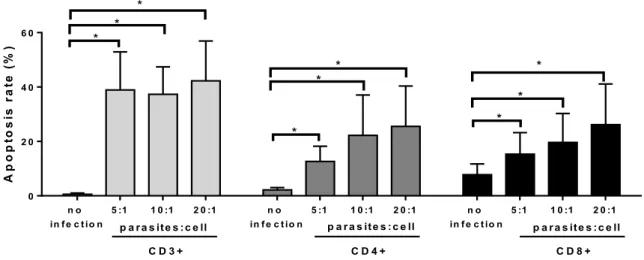
To confirm that L. infantum infection induces T cell apoptosis, PBMCs from healthy dogs were infected with increasing doses of L.infantum
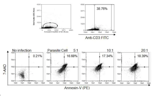
promastigotes and the levels of apoptosis were measured in CD3+, CD4+ and CD8+ cells. The infected cultures showed a gradual increase in apoptosis associated with parasite load in CD3+, CD4+ and CD8+ cells compared with non infected cells (p<0.05, paired Wilcoxon test; Figure 1A). A typical figure from flow cytometry analysis was shown (Figure 1B).
A p o p to s is r a te ( % ) n o in fe c tio n
5 :1 1 0 :1 2 0 :1 n o
in fe c tio n
5 :1 1 0 :1 2 0 :1 n o
in fe c tio n
5 :1 1 0 :1 2 0 :1
0 2 0 4 0 6 0
p a ra s ite s :c e ll
C D 3 + C D 4 + C D 8 +
p a ra s ite s :c e ll p a ra s ite s :c e ll
* * * * * * * * *
.
FIGURE 1B - Flow cytometry analysis: Gating scheme of forward vs side scatter to observe exclusion or inclusion of proper events, which is especially critical to live/dead staining. Fluorescence plot for gated PBMC from healthy dogs (Anti-CD3 monoclonal antibody FITC). Percentage inside upper right quadrants correspond to PBMC CD3+ cells; stained for Annexin V-PE and 7-AAD fluorescence (double stained), indicating late stage of apoptotic cells. Apoptosis in the presence of L. infantum: PBMC (5:1, 10:1, 20:1). Similar dot plot figures were observed using anti-CD4 monoclonal antibodies FITC or FITC- conjugated anti-CD8 monoclonal antibodies.
3.2. Levels of sFAS, sFASL and active Caspase-3 from the spleens of dogs infected with VL
42
and active Caspase-3 levels were higher (p<0.05) in spleen of dogs infected with VL compared with controls (Figures 2A, 2B, 2C).
H e a l t h y I n f e c t e d
0 2 0 4 0 6 0 a s F A S p g /m l *
H e a l t h y I n f e c t e d
0 5 0 1 0 0
1 5 0 b
s F A S L p g /m l *
H e a l t h y I n f e c t e d
0 . 0 0 . 2 0 . 4 0 . 6
c C a s p a s e 3 n g /m l *
3.3. Evaluaton of parasite load in spleen samples of dogs with VL and correlation with sFAS and sFASL
To assess whether the levels of sFAS and sFASL interfere in the control of parasitic load the correlation analysis were performed between sFAS and sFASL with parasitic load.
The values of the linear cycle (Threshold cycle-Ct), obtained from 10 to 100 ng of DNA from each sample, were analyzed with reference to the standard curve obtained for parasite load. The curve for quantifying parasite load determined r2=0.98 and the reaction showed a slope of -3.286. The parasite
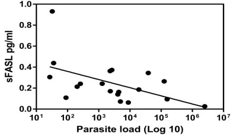
load showed variation in spleen samples from infected dogs. A weak negative correlation was observed between parasite load in the spleen and sFASL levels (p<0.05, r -0.52; Figure. 3). The correlation between sFAS levels and parasite load showed no significant results (data not shown).
FIGURE 3 - Negative correlation between sFASL and parasite load in the spleen of dogs with VL (p<0.05, r= -0.52, Spearman correlation).
101 102 103 104 105 106 107
0.0 0.2 0.4 0.6 0.8 1.0
Parasite load (Log 10)
sF
AS
L p
g/
m
44
4. Discussion
We observed increased rates of apoptosis in CD3+, CD4+ and CD8+ cells from peripheral blood in the presence of L. infantum infection and changes in the concentration of sFAS, sFASL and active Caspase-3 in extracts of spleen from infected compared to healthy dogs that suggest the involvement these molecules in the apoptotic mechanism previously observed in spleen tissue of infected dogs (Lima et al., 2012). A negative correlation between sFASL molecules and the parasitic load was also observed, which implies the involvement of this molecule in soluble form in the effector mechanisms responsible for the elimination of the parasite.
In vitro infection of mononuclear cells from healthy dogs with L. infantum
resulted in increased rates of apoptosis, confirming the preliminary results of an increase in apoptosis in CD4+ and CD8+ cells observed in the naturally infected dogs (SILVA et al., 2013), indeed, it has been shown in vitro that the parasite
Leishmania spp. and its membrane constituents can induce apoptosis in PBMC (WOLDAY et al.,1999).
To investigate the mechanisms of apoptosis induction in dogs, sFAS and sFASL were measured in spleen tissue. A decrease in sFAS in the infected group was observed. An in vitro study suggested that the soluble isoform inhibits apoptosis induced by agonist antibodies (CASCINO et al., 1996); in fact, higher rates of apoptosis were observed in the infected group compared with healthy controls (SILVA et al., 2013).
Silva et al. (2013) observed a decrease in mFASL in CD4+ and CD8+ cells in the spleen of dogs with LV. In contrast, an increase in sFASL was observed in extracts of spleen from infected dogs, which suggests the action of MMPs. In fact, in the serum of dogs with VL, increased levels of MMP-2 and MMP-9 were detected (MELO et al., 2012).Further research is required to determine which MMP are involved in the production of sFASL in CVL.
The high levels of sFASL determined in the spleen of infected dogs is similar to that observed in patients with acute VL, who show high levels of sFASL in the serum (EIDSMO et al., 2002). The infected group did show higher levels of sFASL compared with the uninfected group, but we observed a negative correlation between parasite burden and sFASL molecules in the spleen, suggesting that in canine VL the increase in sFASL is related to the reduction of parasite burden.
The weak negative correlation between spleen parasite load and the high level of sFASL suggests that sFASL stimulates inflammatory mechanisms in the spleen that reduce the parasitic load. In fact, an increase of TNF-α was observed in extracts of spleen from dogs with VL, which correlated with decreased parasite load in the spleen (MICHELIN et al., 2011).Recent investigation has suggested that sFASL activity is not restricted to programmed cell death. On the contrary, it may mainly account for inflammation in various diseases (BLANCO-COLIO et al., 2008; CARDINAL et al., 2010). sFASL has been used as a marker of inflammation (MUSIAL; ZWOLINSKA, 2012), and human sFASL exhibited chemotactic activity toward murine and human polymorphonuclear leukocytes (neutrophils) in vitro (OTTONELLO et al., 1999; SEINO et al., 1998). The inflammatory role of sFASL not yet been characterized in dogs with VL. Further studies need to be performed to clarify how sFASL induces a decrease in parasite load.
Besides the function of sFASL in apoptosis, this molecule has other known functions. Macrophages infected by the parasite Leishmania (L.) major
46
correlating with stimulation of NO production by these cells. Treatment with either substance alone failed to affect the parasite (CHAKOUR et al., 2003), suggesting that sFASL affects macrophage function, a fact that seems to be associated with the weak negative correlation observed between sFASL and splenic parasite load.
The high levels of active Caspase-3 and sFASL in the spleen of dogs with VL suggest that apoptosis occurs in spleen tissue, as observed in previous studies (LIMA et al., 2012; SILVA et al., 2013). sFASL can stimulate apoptosis and inflammatory mechanisms in the spleen that decrease parasitic load. Indeed, when the apoptotic bodies are not promptly phagocytosed, they can rapidly lose membrane integrity and release their intracellular constituents into the extracellular environment, behaving like necrotic cells, activating inflammatory mechanisms (SAVILL et al., 2002), leading to a reduction in parasite load. It also has been observed that apoptosis can stimulate a proinflammatory response in splenic macrophages by ingestion of intact apoptotic cells (LORIMORE et al., 2001).
Similar to our findings, in human HIV infection, high levels of sFASL were observed in the serum, associated with decreased numbers of CD4+ T cells. It has also been suggested that sFASL can be used as a factor in disease progression (HOSAKA et al., 2000). In this study, all the VL positive dogs evaluated presented clinical signs of disease. Future studies involving asymptomatic dogs may clarify whether this molecule can be used as a factor associated with the progression of canine VL.
Acknowledgments
The authors would like to thank the São Paulo Research Foundation (Fundação de Amparo a Pesquisa do Estado de São Paulo, FAPESP) for its financial support of this research (process no. 2011/06214-7).
References
ALEXANDER, C.E.; KAYE, P.M.; ENGWERDA, C.R. CD-95 is required for the early control of parasite burden in the liver of Leishmania donovani-infected mice. Eur. J. Immunol., v. 31, n.4, p. 1199-1210, 2001.
ASHFORD, R.W.; DESJEUX, P.; DE RAADT, P. Estimation of population at risk of infection and number of cases of Leishmaniasis. Parasitol. Today, v. 8, n.3, p. 104-105, 1992.
BADLEY, A.D.; DOCKRELL, D.; SIMPSON, M.; SCHUT, R.; LYNCH, D.H.; LEIBSON, P.; PAYA, C.V. Macrophage-dependent apoptosis of CD4+ T lymphocytes from HIV-infected individuals is mediated by FASL and tumor necrosis factor. J. Exp. Med., v. 185, n.1, p. 55-64, 1997.
BLANCO-COLIO, L.M.; MARTIN-VENTURA, J.L.; TUNON, J.; GARCIA-CAMARERO, T.; BERRAZUETA, J.R.; EGIDO, J. Soluble FAS ligand plasma levels are associated with forearm reactive hyperemia in subjects with coronary artery disease: a novel biomarker of endothelial function? Atherosclerosis, v. 201, n.2, p. 407-412, 2008.
BOURDOISEAU, G.; BONNEFONT, C.; MAGNOL, J.P.; SAINT-ANDRÉ, I.; CHABANNE, L. Lymphocyte subset abnormalities in canine leishmaniasis. Vet. Immunol.Immunopathol., v. 56, n.3/4, p. 345-351, 1997.
48
diagnostic accuracy and prognosis for acute coronary syndromes. Am. J. Cardiol., v. 105, n.6, p. 797-803 2010.
CASCINO, I.; PAPOFF, G.; ERAMO, A.; RUBERTI, G. Soluble FAS/Apo-1 splicing variants and apoptosis. Front. Biosci., v. 1, p.12-18, 1996.
CHAKOUR, R.; GULER, R.; BUGNON, M.; ALLENBACH, C.; GARCIA, I., MAUËL, J.; LOUIS, J.; TACCHINI-COTTIER, F. Both the FAS ligand and inducible nitric oxide synthase are needed for control of parasite replication within lesions in mice infected with Leishmania major whereas the contribution of tumor necrosis factor is minimal. Infect. Immun., v. 71, n.9, p. 5287-5295, 2003.
DAS, G.; VOHRA, H.; RAO, K.; SAHA, B.; MISHRA, G.C. Leishmania donovani
infection of a susceptible host results in CD4+ T-cell apoptosis and decreased Th1 cytokine production. Scand. J. Immunol., v. 49, n.3, p. 307-310, 1999.
DEANE, L.M.; DEANE, M.P. Observações preliminares sobre a importância comparativa do homem, do cão e da raposa (Lycalopexvetulus) como reservatórios de Leishmania donovani em área endêmica de calazar no Ceará. Hospital, v. 48, p. 61-76, 1955.
DESJEUX, P. Leishmaniasis: current situation and new perspectives. Comp. Immunol. Microbiol. Infect. Dis., v. 27, n.5, p. 305-318, 2004.
EIDSMO, L.; WOLDAY, D.; BERHE, N.; SABRI, F.; SATTI, I.; EL HASSAN, A.M.; SUNDAR, S.; CHIODI, F.; AKUFFO, H. Alteration of FAS and FAS ligand expression during human visceral leishmaniasis.Clin. Exp. Immunol., v.130, n.2, p. 307-313, 2002.
HOSAKA, N.; OYAIZU, N.; THAN, S.; PAHWA, S. Correlation of loss of CD4 T cells with plasma levels of both soluble form FAS (CD95) and FAS Ligand (FASL) in HIV-Infected Infants. Clin. Immunol., v. 95, n.1, p. 20-25, 2000.
LIMA, V.M.F.; FATTORI, K.R.; SOUZA, F.; EUGÊNIO, F.R.; SANTOS, P.S.P.; ROZZA, D.B.; MACHADO, G.F. Apoptosis in T lymphocytes from spleen tissue and peripheral blood of L. (L.) chagasi naturally infected dogs. Vet. Parasitol., v. 184, n.2/4, p. 147-153, 2012.
LIMA, V.M.F.; GONÇALVES, M.E.; IKEDA, F.A.; LUVIZOTTO, M.C.R.; FEITOSA, M.M.; Anti-Leishmania antibodies in cerebrospinal fluid from dogs with visceral leishmaniasis. Braz. J. Med. Biol. Res., v. 36, n.4, p. 485-489, 2003.
LORIMORE, S. A.; COATES, P. J.; SCOBIE, G. E.; MILNE, G.; WRIGHT, E. G. Inflammatory-type responses after exposure to ionizing radiation in vivo: a mechanism for radiation-induced bystander effects? Oncogene, v.20, n.48, p. 7085-7095, 2001.
MATUTE-BELLO, G.; LILES, W.C.; STEINBERG, K.P.; KIENER, P.A.; MONGOVIN, S.; CHI, E.Y.; JONAS, M.; MARTIN, T.R. Soluble FAS ligand induces epithelial cell apoptosis in humans with acute lung injury (ARDS). J. Immunol., v.163, n.4, p. 2217-2225, 1999.
MAURICIO, I.L., STOTHARD, J.R., MILES, M.A. The strange case of
Leishmania chagasi. Parasitol. Parasitol. Today, v.16, n.5, p.188-189, 2000.
MELO, G.D.; MARCONDES, M.; MACHADO, G.F. Canine cerebral leishmaniasis: potential role of matrix metalloproteínase-2 in the development of neurological disease. Vet. Immunol. Immunopathol., v. 148, n.3/4, p. 260-266, 2012.
50
BARATA, R.A.; ROMANHA, A.J.; FORTES-DIAS, C.L.; DIAS, E.S. Infectivity of seropositive dogs, showing different clinical forms of leishmaniasis, to
Lutzomyia longipalpis phlebotomine sandflies. Vet. Parasitol., v. 147, n.1/2, p. 67-76, 2007.
MICHELIN, F. A.; PERRI, S.H.V.; LIMA, V.M.F. Evaluation of TNF-alpha, IL-4, and IL-10 and parasite density in spleen and liver of L. (L.) chagasi naturally infected dogs. Ann. Trop. Med. Parasitol., v.105, n.5, p. 373-383, 2011.
MUSIAŁ, K.; ZWOLIŃSKA, D. The sFAS/sFASL ratio as a novel marker of inflammation in children with chronic kidney disease. Clin. Chim. Acta, v. 414, p. 7-11, 2012.
OTTONELLO, L.; TORTOLINA, G.; AMELOTTI, M.; DALLEGRI, F. Soluble FAS ligand is chemotactic for human neutrophilic polymorphonuclear leukocytes. J. Immunol., v.162, n.6, p. 3601-3606, 1999.
POTESTIO, M.; D'AGOSTINO, P.; ROMANO, G.C.; MILANO, S.; FERLAZZO, V.; AQUINO, A.; DIBELLA, G.; CARUSO, R.; GAMBINO, G.; VITALE, G.; MANSUETO, S.; CILLARI, E. CD4+ CCR5+ and CD4+ CCR3+ lymphocyte subset and monocyte apoptosis in patients with acute visceral leishmaniasis. Immunology, v.113, n.2, p.260-268, 2004.
RETHI, B.; EIDSMO, L. FASL and TRAIL signaling in the skin during cutaneous leishmaniasis - implications for tissue immunopathology and infectious control. Front. Immunol., v. 3, p.163, 2012.
SAVILL, J.; DRANSFIELD, I.; GREGORY, C.; HASLETT, C. A blast from the past: clearance of apoptotic cells regulates immune responses. Nat. Rev. Immunol., v. 2, n.12, p. 965-975, 2002.
activity of soluble FAS ligand against phagocytes. J. Immunol., v. 161, n.9, p. 4484-4488, 1998.
SHI, Y. Mechanisms of caspase activation and inhibition during apoptosis. Mol.Cell., v. 9, n.3, p. 459-470, 2002.
SILVA, K.L.; MELO, L.M.; PEROSSO, J.; OLIVEIRA, B.B.; SANTOS, P.S.; EUGÊNIO, F.R.; LIMA, V.M. CD95 (FAS) and CD178 (FASL) induce the apoptosis of CD4+ and CD8+ cells isolated from the peripheral blood and spleen of dogs naturally infected with Leishmania spp. Vet. Parasitol., v.197, n.3/4, p. 470-476, 2013.
SOLANO-GALLEGO, L.; KOUTINAS, A.; MIRÓ, G.; CARDOSO, L.; PENNISI, M.G.; FERRER, L.; BOURDEAU, P.; OLIVA, G.; BANETH, G. Directions for the diagnosis, clinical staging, treatment and prevention of canine leishmaniosis. Vet. Parasitol., v.165, n.1/2, p.1-18, 2009.
TANAKA, M.; SUDA, T.; TAKAHASHI, T.; NAGATA, S. Expression of the functional soluble form of human FAS ligand in activated lymphocytes. EMBO. J., v. 14, n.6, p. 1129-1135, 1995.
VERÇOSA, B.L.; MELO, M.N.; PUERTO, H.L.; MENDONÇA, I.L.; VASCONCELOS, A.C. Apoptosis, inflammatory response and parasite load in skin of Leishmania (Leishmania) chagasi naturally infected dogs: A histomorphometric analysis. Vet. Parasitol., v.189, n.2/4, p. 162-170, 2012.