Ancient Dispersal of the Human Fungal Pathogen
Cryptococcus gattii
from the Amazon Rainforest
Ferry Hagen1,2¤a, Paulo C. Ceresini3, Itzhack Polacheck4, Hansong Ma5¤b, Filip van Nieuwerburgh6, Toni Gabaldo´n7, Sarah Kagan4, E. Rhiannon Pursall5, Hans L. Hoogveld8, Leo J. J. van Iersel9, Gunnar W. Klau9, Steven M. Kelk9¤c, Leen Stougie9,10, Karen H. Bartlett11, Kerstin Voelz5, Leszek P. Pryszcz7, Elizabeth Castan˜eda12, Marcia Lazera13, Wieland Meyer14, Dieter Deforce6, Jacques F. Meis2,15, Robin C. May5, Corne´ H. W. Klaassen2, Teun Boekhout1,16,17*
1Department of Yeast and Basidiomycete Research, CBS-KNAW Fungal Biodiversity Centre, Utrecht, The Netherlands,2Department of Medical Microbiology and Infectious Diseases, Canisius-Wilhelmina Hospital, Nijmegen, The Netherlands,3Department of Phytopathology, UNESP - University of Sa˜o Paulo State, Ilha Solteira Campus, Ilha Solteira, Brazil,4Department of Clinical Microbiology and Infectious Diseases, Hadassah-Hebrew University Medical Center, Jerusalem, Israel,5School of Biosciences, University of Birmingham, Edgbaston, Birmingham, United Kingdom,6Laboratory of Pharmaceutical Biotechnology, Faculty of Pharmaceutical Sciences, Ghent University, Ghent, Belgium,7Centre for Genomic Regulation (CRG) and UPF Doctor Aiguader, Barcelona, Spain,8Netherlands Institute of Ecology (NIOO-KNAW), Centre for Limnology, Wageningen, The Netherlands,9Centrum Wiskunde & Informatica (CWI), Amsterdam, The Netherlands,10Department of Operations Research, VU University, Amsterdam, The Netherlands,11School of Environmental Health, University of British Columbia, Vancouver, British Columbia, Canada,12Instituto Nacional de Salud, Grupo de Microbiologı´a, Zona 6 CAN, Bogota´, Colombia,13Laboratorio de Micologia, Instituto de Pesquisa Clinica Evandro Chagas, Fundac¸ao Oswaldo Cruz – FIOCRUZ, Rio de Janeiro, Brazil,14Molecular Mycology Research Laboratory, Center for Infectious Diseases and Microbiology, Westmead Millennium Institute, Sydney Emerging Disease and Biosecurity Institute, Sydney Medical School – Westmead Hospital, University of Sydney, Westmead, NSW, Australia,15Department of Medical Microbiology, Radboud University Nijmegen Medical Centre, Nijmegen, The Netherlands,16Shanghai Key Laboratory of Molecular Medical Mycology, Changzheng Hospital, Second Military Medical University, Shanghai, China,17Department of Internal Medicine and Infectious Diseases, University Medical Center, Utrecht, The Netherlands
Abstract
Over the past two decades, several fungal outbreaks have occurred, including the high-profile ‘Vancouver Island’ and ‘Pacific Northwest’ outbreaks, caused by Cryptococcus gattii, which has affected hundreds of otherwise healthy humans and animals. Over the same time period,C. gattiiwas the cause of several additional case clusters at localities outside of the tropical and subtropical climate zones where the species normally occurs. In every case, the causative agent belongs to a previously rare genotype ofC. gattiicalled AFLP6/VGII, but the origin of the outbreak clades remains enigmatic. Here we used phylogenetic and recombination analyses, based on AFLP and multiple MLST datasets, and coalescence gene genealogy to demonstrate that these outbreaks have arisen from a highly-recombiningC. gattiipopulation in the native rainforest of Northern Brazil. Thus the modern virulentC. gattiiAFLP6/VGII outbreak lineages derived from mating events in South America and then dispersed to temperate regions where they cause serious infections in humans and animals.
Citation:Hagen F, Ceresini PC, Polacheck I, Ma H, Nieuwerburgh Fv, et al. (2013) Ancient Dispersal of the Human Fungal PathogenCryptococcus gattiifrom the Amazon Rainforest. PLoS ONE 8(8): e71148. doi:10.1371/journal.pone.0071148
Editor:Joseph Schacherer, University of Strasbourg, France
ReceivedMay 6, 2013;AcceptedJune 25, 2013;PublishedAugust 7, 2013
Copyright:ß2013 Hagen et al. This is an open-access article distributed under the terms of the Creative Commons Attribution License, which permits unrestricted use, distribution, and reproduction in any medium, provided the original author and source are credited.
Funding:This publication was made possible by NPRP grant 5-298-3-06 from the Qatar National Research Fund (a member of Qatar foundation) to TB, JFM, CHWK, and TG. Computational experiments were sponsored by the NCF (Netherlands Computer Facility Foundation) for the use of supercomputer facilities, with financial support from NWO (Nederlandse Organisatie voor Wetenschappelijk Onderzoek). Contributions of FH were supported by the Odo van Vloten Foundation. The funders had no role in study design, data collection and analysis, decision to publish, or preparation of the manuscript.
Competing Interests:The authors have declared that no competing interests exist.
* E-mail: [email protected]
¤a Current address: Department of Medical Microbiology and Infectious Diseases, Canisius-Wilhelmina Hospital, Nijmegen, The Netherlands
¤b Current address: Department of Biochemistry and Biophysics, University of California San Francisco, San Francisco, California, United States of America ¤c Current address: Department of Knowledge Engineering, Maastricht University, Maastricht, The Netherlands
Introduction
During the past two decades, fungal outbreaks caused by previously rare genotypes or even novel species have emerged that affect humans, mammals and amphibians. Among them are the white-nose-syndrome in bats due to Geomyces destructans [1], the killing of frogs byBatrachochytrium dendrobatidis[2], and the ongoing
Cryptococcus gattii outbreaks in Canada (the so-called ‘Vancouver Island’ outbreak) and the Pacific Northwest of the USA, as well as
several case clusters ofC. gattiiinfections in Western Australia, and Brazil [3–8].
Among the most prominent of these outbreaks is the Vancouver Island outbreak (British Columbia, Canada), caused by C. gattii, which expanded from Vancouver Island (Canada) to the mainland of British Columbia and the Pacific Northwest [3,6,7]. Until the Vancouver Island outbreak,C. gattiiwas almost exclusively known as the cause of rare cryptococcal infections in otherwise healthy humans and animals living in tropical and subtropical regions, mainly in Australasia and South America [5,7,9,10]. However,
over the past decade hundreds of apparently healthy people and animals in the affected Vancouver Island outbreak area have been infected by C. gattii strains belonging to the AFLP6/VGII genotypic lineage [4]. Since the onset of this outbreak, the incidence ofC. gattiiinfections on Vancouver Island was estimated to be approximately 27 times higher than in Northern Australia whereC. gattiiis endemic [5,7]. Over this period, 19 patients died due to aC. gattiiinfection, which is a case-fatality rate of 8.7% [5]. Concurrently with the Vancouver Island outbreak, there have been clusters of AFLP6/VGII C. gattii-related disease reported from captive psittacine birds in Sa˜o Paulo, Brazil (this study; [8]), a higher incidence of C. gattii AFLP6/VGII infections among immunocompetent children in Northern Brazil [11–13] and a case cluster among sheep in Western Australia [4,14].
All these outbreaks and case clusters are caused by genotype AFLP6/VGII strains, one of the five known genotypes within the
C. gattii complex [6,7,15–17]. This genotype AFLP6/VGII has been further differentiated into three major subgenotypes, the so-called ‘major’ genotype AFLP6A/VGIIa, the ‘minor’ genotype AFLP6B/VGIIb, and a novel genotype AFLP6C/VGIIc that emerged in the Pacific Northwest [3,6,7,17]. Comparative virulence studies suggested that C. gattii strains of the major genotype AFLP6A/VGIIa have a higher virulence potential than strains that belong to AFLP6B/VGIIb [6,18,19], whilst the recently discovered AFLP6C/VGIIc strains from the Pacific Northwest outbreak have a similar virulence as those of genotype AFLP6A/VGIIa strains from the Vancouver Island outbreak [3]. Trees are the natural habitat ofC. gattii[3,7,9,20–25], and soil, trees, water and air in the Vancouver Island outbreak region contain high quantities of C. gattiipropagules [3,25]. The recent range expansion of this pathogen to the Canadian mainland is likely caused by traffic-based transport of soil and dust-contam-inated individuals and motor vehicles [25].
The molecular epidemiology of theC. gattiicomplex has been extensively studied and led to several hypotheses to explain the origin of the Vancouver Island outbreak [3,6,7,15,17]. Two hypotheses emerged from these studies, firstly it has been proposed that same-sex mating between low virulent mating-typeastrains within the Australasian population or while in transit to North America gave rise to the highly virulent genotype that subse-quently caused the Canadian outbreak [6]. The same authors suggested also that the Vancouver Island outbreak may have been originated from other geographic localities. Ngamskulrungroj et al. [17] observed that the South AmericanC. gattiipopulation demonstrated the highest genetic diversity and these authors suggested that the origin of this species may be in South America. Thus, the origin of the highly virulent lineages within C. gattii
genotype AFLP6/VGII remains unclear.
Understanding the genetic and geographic origin of these outbreaks is critical in order to control the spread ofC. gattii. Here we provide an evolutionary model for the origin of the C. gattii
epidemics using extensive molecular population analyses that includes amplified fragment length polymorphisms (AFLP) anal-ysis, a novel multi-locus microsatellite panel, and coalescence gene genealogy analysis based on data obtained from a novel multi-locus sequence typing scheme based on sequence characterized AFLP regions (SCAR-MLST). In addition, we performed additional sequencing and re-analyzed recently published MLST data [3,8] by expanding the geographic range through the inclusion of more strains from South America. These analyses revealed that South American C. gattii AFLP6/VGII strains represent the ancestral lineages within the global population, which is in agreement with the observed high genetic diversity at that locality. Coalescence gene genealogy analysis showed that the
basal lineages within genotype AFLP6/VGII have a South American origin. Thus we provide strong evidence that South America is the cradle for the global population, including the expandingC. gattii AFLP6/VGII outbreaks in British Columbia (Canada) and in the Pacific Northwest of the USA [3,6,7].
Results
South America is the Origin of the GlobalC. gattiiAFLP6/
VGII Population
The historical population structure ofC. gattiiAFLP6/VGII was assessed by application of coalescence gene genealogy analysis using the SCAR-MLST dataset which was analyzed after removing 59 homoplasious sites in the 3,357 nucleotides alignment which resulted in 32 haplotypes (indicated as ‘H’ in Fig. 1; Table 1, 2 and S1) compared to 49 sequence types (STs) when all recombination breakpoints remained intact. The most basal lineage within the coalescence gene genealogy was found to be of South American descent and is represented by an environmen-tal strain isolated from a tree in the pristine Brazilian Amazon rainforest (LMM645; haplotype H17, mating-type a). The next diverging lineage is the South American lineage H24 (LMM265 and LMM266; Brazil, mating-typea) (Fig. 1; Table 1). Mutations deep in the coalescence gene genealogy divided the two basal lineages from three younger branches, which were differentiated from each other by more recent mutations. Two of these ancient lineages had an African and South American origin (H27 and H28). The four basal lineages diverged subsequently over time into 28 haplotypes associated with the occurrence of 22 mutations in the ‘Fragment 549locus (Fig. 1; Table 1). Compared to the other case cluster and North American outbreak lineages, the psittacine birdC. gattiicase cluster lineage H02 contained substantially more mutations due to a 20bp region in the non-coding locus ‘Fragment 159, which is highly polymorphic compared to the other four nuclear SCAR-MLST loci studied (Fig. 1; Table S1).
Generation time scaled coalescence analysis revealed that the major clade (H01–H16, H18–H23 and H25–H32) and the basal clade (H17 and H24) emerged approximately 176106generations before present (gbp) (Fig. 1; Table 1). The lineage representing the major genotype involved in the Vancouver Island outbreak (H01) emerged approximately 1.46106gbp. The estimated divergence time of the two closely related C. gattii strains A1M-R265 and CBS7750 (both lineage H01) was found to be approximately 51,449 years based on the number of synonymous nucleotide substitutions observed across 4,771 protein-coding orthologous genes.
The lineage representing the C. gattii case cluster among psittacine birds in Brazil (H02) emerged around 4.26106gbp. A major diversification event that gave rise to a global lineage (H03) occurred approximately 5.36106gbp and includes strains from theC. gattiioutbreak in the Pacific Northwest and strains belonging to the minor genotype from the Vancouver Island outbreak. The latter were found to be genetically indistinguishable from strains obtained from a case cluster among over 100 sheep in Western Australia based on the consensus multi-locus sequence typing scheme forC. neoformansandC. gattii[4] (Fig. 1).
Global Dispersal ofC. gattiiAFLP6/VGII
Ancient Dispersal ofCryptococcus gattii
dispersal event, which can be interpreted as a cascade of recombination and dispersal events rather than a single event. The most remarkable of these lineages (H03) was introduced into all inhabited continents and has resulted in recent outbreaks in North America and the case clusters in Australia and South America. Most of the lineages with a South American origin emerged during the past 3.4 – 4.06106 generations and were formed by a high rate of recombination events which seems to be the driving force behind the genetic diversification at this continent.
An Independent Origin for the various Outbreaks
The genetic diversity of C. gattii genotype AFLP6/VGII was assessed by SCAR-MLST, microsatellite analysis and AFLP fingerprint analysis (Fig. 2, S1 and S2; Table 1 and S3). The Simpsons diversity index demonstrated that microsatellite typing had the highest discriminatory power (D= 0.940), followed by AFLP (D= 0.928) and SCAR-MLST (D= 0.909). Using the individual AFLP, SCAR-MLST and STR datasets the South American population had the highest genetic diversity, which was corroborated when these datasets were combined resulting in 58 genotypes among 76 strains (D= 0.951) (Table 2). The South American population was also found to have the highest genotypic diversity (G0) for each of the datasets, resulting in an overall
significantly higherG0of 36.10 (P,0.05) compared to the other
four populations, supporting a South American origin of the global
C. gattiiAFLP6/VGII population (Table 2).
As independent datasets, AFLP fingerprinting, SCAR-MLST, microsatellite analysis and re-analysis of published MLST data [3,6] revealed that South America has the highest genetic diversity (Table 2). This is in agreement with previous investigations that showed the presence of all C. gattiigenotypes in South America [17,26,27]. Mean population sizes and the migration patterns between population pairs were determined by using the full SCAR-MLST dataset. This analysis showed that the actual mean population size of South America (h= 0.0224) is the largest, followed by Europe (h= 0.0098), Africa (h= 0.0094), North America (h= 0.0089) and Australia (h= 0.0062) (Fig. 3; Table
S2). However, the lower and upper boundaries of the population size analysis showed a large variation, with the African and European populations being most variable. The North American
C. gattiiAFLP6/VGII population has a higher influx than efflux of migrants from the other populations with South America being the main contributor, followed by Europe, Australasia and Africa (Fig. 3; Table S2). The South American C. gattii AFLP6/VGII population has a higher efflux than influx of migrants to and from the other populations, with the exception of the African population that has an extremely low efflux of 1.22610214Nmmcompared to no influx from South America (Fig. 3; Table S2). Efflux from the Australasian population is also higher to Africa, Europe and North America thanvice versa, with South America as the exception since it contributes more to the influx into Australasia (Fig. 3; Table S2). Maximum Likelihood phylogenetic analysis using the nuclear SCAR-MLST revealed that a highly supported basal lineage
contained strains from Africa and South America (Fig. 2). All basal African strains were mating-typea, while both mating-types were present among the South American lineages. Other well supported lineages arose from this basal cluster, including three major clades with mating-type a strains that originate from South America, a mixed clade of Australian and North American mating-type a
strains and a group of Vancouver Island outbreak mating-typea
strains with a diverse basal group of African, Australasian, European and South American strains comprising different mating-types. The major Vancouver Island outbreak cluster contained three strains that originated from localities in Thailand (MC-S-239), Argentina (LA295) and Brazil (IFM47258) which were, except for MC-S-239, genetically indistinguishable from each other.
One hundred and thirty five isolates ofC. gattiiwere used to re-analyze the updated ‘Fraser and Byrnes’ MLST dataset (original data taken from [3,6]; Fig. S3), which revealed that several clades emerged from a basal South American cluster. Using this dataset, the large cluster of the major genotypeC. gattiiVancouver Island outbreak strains (ST01) emerged from a diverse cluster containing Australasian (ST18, ST20), European (ST17) and South American (ST07, ST28, ST30) strains (Fig. S3).
High Virulence is not Restricted to Outbreak Strains
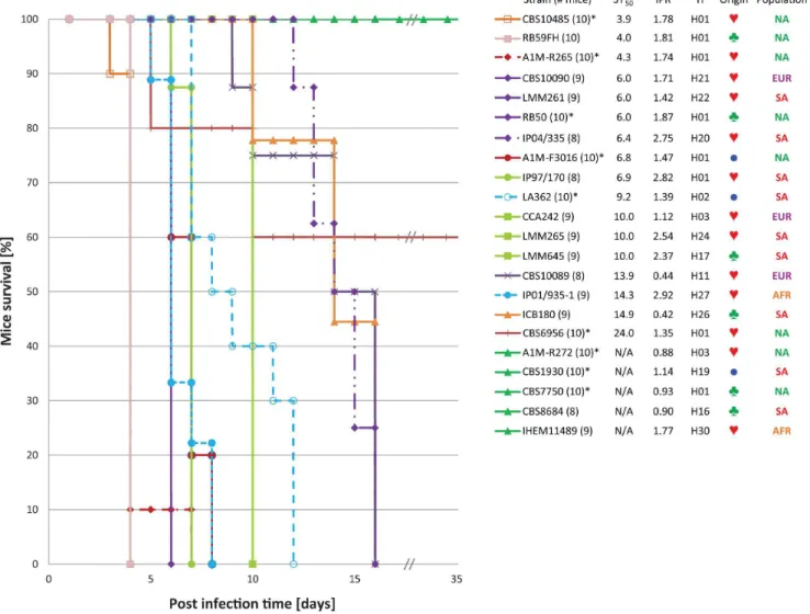
Previous studies indicated thatC. gattiiAFLP6A/VGIIa, the so-called major Vancouver Island outbreak genotype, and strains with genotype AFLP6C/VGIIc from the Pacific Northwest outbreak are highly virulent in different host models, while strains that belong to the minor Vancouver Island outbreak genotype AFLP6B/VGIIb were less virulent or non-virulent [3,6,18,19]. To further elucidate the virulence potential among strains within the basal lineages of the coalescence tree, we investigated the virulence of 22C. gattii strains using mice and macrophage pathogenicity assays ([3,19]; Figs. 1, 4 and 5; Table S3). This revealed that the ancestral lineages in the coalescence analysis represented by the South American strains LMM645 (H17; mating-type a), isolated from a tree in a pristine Amazon rainforest, and the clinical strain LMM265 (H24; mating-type a) were more virulent (ST50= 10.060.0 days each) when compared to other strains
(Figs. 4 and 5). Despite the fact these strains show a dramatic virulent potential to cause disease in the BALB/c mice model, these two South American strains were significantly (P,0.001) less virulent than strains with the major genotype AFLP6A/VGIIa from the Vancouver Island outbreak region, which had ST50
values ranging from 3.960.1 to 6.860.3 days (Figs. 4 and 5; Table S3). The macrophage intracellular proliferation rates (IPR) of these two South American strains were among the highest observed with values of 2.54 for LMM265 and 2.37 for LMM645 ([3,19]; Table S3). The highest IPR values were observed for the three mating-type a strains IP2001/935-1
(H27; Africa), IP1997/170 (H25; South America) and IP2004/ 335 (H20; South America) which had IPR values of 2.92, 2.82 and 2.75, respectively. These high IPR values were also reflected by high pathogenicity in BALB/c mice (Fig. 4; Table S3), although Figure 1. Coalescence gene genealogy based on SCAR-MLST data.The historical coalescence gene genealogy of the globalCryptococcus gattii AFLP6/VGII population structure was reconstructed by using five nuclear SCAR-MLST loci from which homoplasious sites were removed
resulting in a total number of 109 informative sites indicated on the branches as blue (‘Fragment 159locus), orange (‘Fragment 329locus), green
(‘Fragment 349locus), red (‘Fragment 549locus) and purple (IGS1 locus) dots. Numbers next to these dots represents the position of the site within
the sequence of informative sites (Table S1). Haplotypes that represent lineages currently involved in outbreaks or case cluster are indicated along the lines of these haplotypes. Numbers behind the populations indicate the number of strains within the given population/haplotypes. Mouse virulence and IPR results are shown as red or green mice and macrophages, respectively, to represent virulent and non-virulent strains as indicated in Fig. 4 and 5, and Table 1 and S3. The ‘generation time to the most common recent ancestor’ (GTMRCA) in ‘generations before present’ is provided (bold), including the lower and upper boundaries.
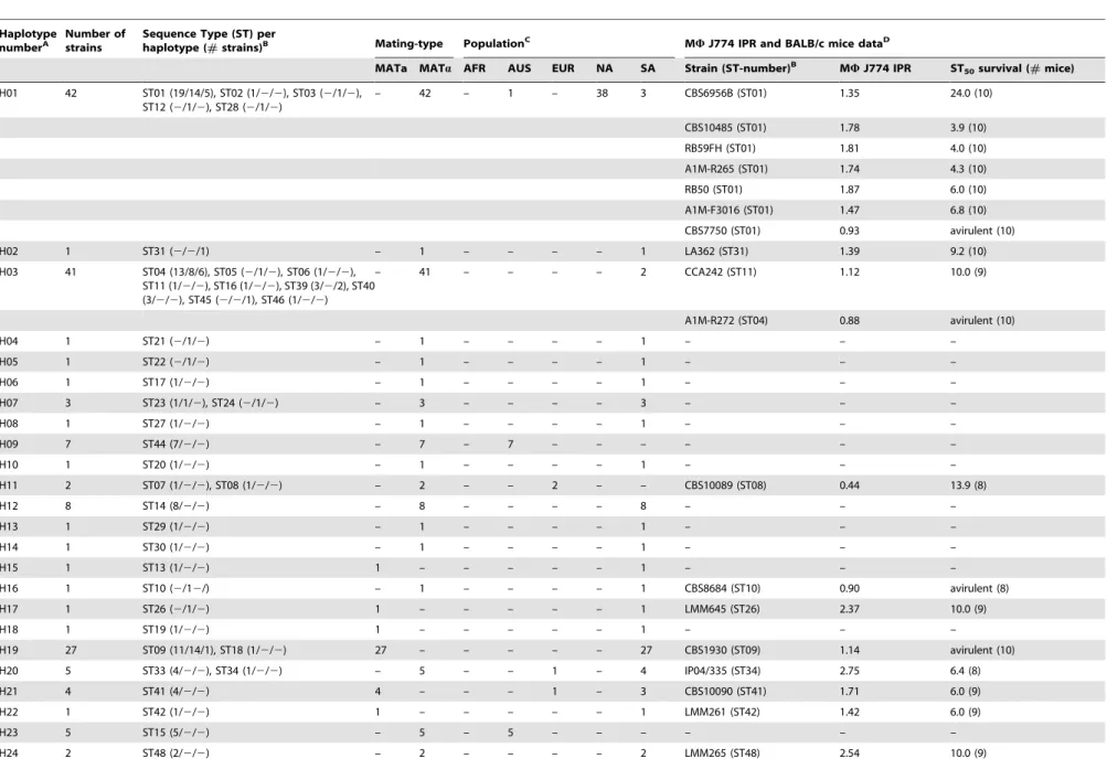
Table 1.Summarized overview of coalescence gene genealogy haplotypes, SCAR-MLST sequence types, mating-types and virulence related data.
Haplotype numberA
Number of strains
Sequence Type (ST) per
haplotype (#strains)B Mating-type PopulationC MWJ774 IPR and BALB/c mice dataD
MATa MATa AFR AUS EUR NA SA Strain (ST-number)B MWJ774 IPR ST
50survival (#mice)
H01 42 ST01 (19/14/5), ST02 (1/2/2), ST03 (2/1/2), ST12 (2/1/2), ST28 (2/1/2)
– 42 – 1 – 38 3 CBS6956B (ST01) 1.35 24.0 (10)
CBS10485 (ST01) 1.78 3.9 (10)
RB59FH (ST01) 1.81 4.0 (10)
A1M-R265 (ST01) 1.74 4.3 (10)
RB50 (ST01) 1.87 6.0 (10)
A1M-F3016 (ST01) 1.47 6.8 (10)
CBS7750 (ST01) 0.93 avirulent (10)
H02 1 ST31 (2/2/1) – 1 – – – – 1 LA362 (ST31) 1.39 9.2 (10)
H03 41 ST04 (13/8/6), ST05 (2/1/2), ST06 (1/2/2), ST11 (1/2/2), ST16 (1/2/2), ST39 (3/2/2), ST40 (3/2/2), ST45 (2/2/1), ST46 (1/2/2)
– 41 – – – – 2 CCA242 (ST11) 1.12 10.0 (9)
A1M-R272 (ST04) 0.88 avirulent (10)
H04 1 ST21 (2/1/2) – 1 – – – – 1 – – –
H05 1 ST22 (2/1/2) – 1 – – – – 1 – – –
H06 1 ST17 (1/2/2) – 1 – – – – 1 – – –
H07 3 ST23 (1/1/2), ST24 (2/1/2) – 3 – – – – 3 – – –
H08 1 ST27 (1/2/2) – 1 – – – – 1 – – –
H09 7 ST44 (7/2/2) – 7 – 7 – – – – – –
H10 1 ST20 (1/2/2) – 1 – – – – 1 – – –
H11 2 ST07 (1/2/2), ST08 (1/2/2) – 2 – – 2 – – CBS10089 (ST08) 0.44 13.9 (8)
H12 8 ST14 (8/2/2) – 8 – – – – 8 – – –
H13 1 ST29 (1/2/2) – 1 – – – – 1 – – –
H14 1 ST30 (1/2/2) – 1 – – – – 1 – – –
H15 1 ST13 (1/2/2) 1 – – – – – 1 – – –
H16 1 ST10 (2/12/) – 1 – – – – 1 CBS8684 (ST10) 0.90 avirulent (8)
H17 1 ST26 (2/1/2) 1 – – – – – 1 LMM645 (ST26) 2.37 10.0 (9)
H18 1 ST19 (1/2/2) 1 – – – – – 1 – – –
H19 27 ST09 (11/14/1), ST18 (1/2/2) 27 – – – – – 27 CBS1930 (ST09) 1.14 avirulent (10)
H20 5 ST33 (4/2/2), ST34 (1/2/2) – 5 – – 1 – 4 IP04/335 (ST34) 2.75 6.4 (8)
H21 4 ST41 (4/2/2) 4 – – – 1 – 3 CBS10090 (ST41) 1.71 6.0 (9)
H22 1 ST42 (1/2/2) 1 – – – – – 1 LMM261 (ST42) 1.42 6.0 (9)
H23 5 ST15 (5/2/2) – 5 – 5 – – – – – –
H24 2 ST48 (2/2/2) – 2 – – – – 2 LMM265 (ST48) 2.54 10.0 (9)
Ancient
Dispersal
of
Cryptococcus
gattii
PLOS
ONE
|
www.ploson
e.org
5
August
2013
|
Volume
8
|
Issue
8
|
we occasionally noted that a high IPR value does not necessarily predict a high virulent phenotype in the BALB/c mice model (Fig. 4; Table S3). Five strains had a non-virulent phenotype when tested in the BALB/c mice model, which could not always be predicted based on the macrophage intracellular proliferation assay as shown by strain IHEM11489 (IPR = 1.77; H30, Africa). The mice and macrophage model were in agreement for the non-virulent strains A1M-R272 (IPR = 0.88; H03, Vancouver Island outbreak), CBS7750 (IPR = 0.93; H01. Vancouver Island out-break) and CBS8684 (IPR = 0.90; H16, South America) (Table S3).
Recombination is High among South American Strains
The presence of both mating-types in a nearly equal rate (mating-type a : a= 0.8: 1) in the South American population suggests that recombination events are likely to occur at this locality. The majority of these mating-typeastrains sampled from various localities in Colombia were observed to be clonal.STE12
mating-type analysis of the C. gattii AFLP6/VGII strains confirmed that the population in Mediterranean Europe contained mating-typeaandastrains as well, while the African, Australasian and North American populations were mating-typeaonly (Table S3).
Ancestral recombination graph analysis of SCAR-MLST sequences showed the presence of extensive successive recombi-nation events in the history of the current global population ofC. gattiiAFLP6/VGII, as illustrated by the presence of 167 historical recombination events (Fig. S4). The level of recombination detected by the ancestral recombination graph analysis differed widely among populations. The number of historical recombina-tion events, based on ancestral recombinarecombina-tion graph analysis, was disproportionally high in South America (110 events), whereas this value was low in the Australasian, European and North American populations (8, 5 and 1 historical recombination events, respec-tively) and undetectable in the African population. Estimates of the minimum number of recombination events (RM) in the current
population of C. gattii AFLP6/VGII corroborated the ancestral recombination graph analysis results, indicating a high number of recombination events (RM= 22) in South America versus those in
the other populations (RM= 4 for Australasia;RM= 2 for Europe; RM= 0 for Africa and North America) (Table S4). TheWWtest
provided further evidence for actual signs of recombination in the SCAR-MLST dataset (P,0.001) but not within the extended ‘Fraser and Byrnes’ MLST dataset (P= 0.196). When the SCAR-MLST data was split into the five populations, evidence for recombination was found by the WW test in the Australian
(P= 0.033), European (P= 0.044), North American (P,0.001) and South American (P,0.001) populations. CASS network analysis also revealed highly supported signs of actual recombination in the SCAR-MLST dataset (Fig. S5). Thus, coalescence gene genealogy, ancestral recombination graph and phylogenetic approaches indicate that two large clades, including the Vancouver Island and Pacific Northwest outbreak lineages (H01 and H03, respec-tively) and a large cluster of strains from other geographical localities, were a result of ancient recombination events between ancestors of the current, but basally positioned, South American lineages and either the South American lineage H07 or one or more currently unknown ancestor(s) of the recombinant strain ENV133 (H01; Vancouver Island outbreak) (Fig. S5).
Discussion
Our study demonstrates that the current globalC. gattiiAFLP6/ VGII population has most likely a South American origin rather
than being introduced from another geographical locality such as Australia, Africa or Europe, as was proposed in previous studies [3,6]. The presence ofC. gattiiAFLP6/VGII in the natural and pristine environment of the Amazon rainforest [21] makes it highly unlikely that the fungus was introduced from Australia into South America via import of AustralianEucalyptusseeds containing
C. gattii, as suggested [3,6] and disputed [22] before. Therefore an ‘Out of the Amazon rainforest’ model is the most plausible scenario for the origin of the AFLP6/VGII outbreaks that emerged in British Columbia (Canada) and the Pacific Northwest (USA), as well as the case clusters among sheep in the Perth region (Australia) and psittacine birds in Brazil.
Migration patterns between theC. gattiiAFLP6/VGII popula-tions pairs showed that South America is the major donor of migrants for the other populations, with Africa being the exception due to a very small influx of genetic material into South America, while this populations seems not to contribute to the genetic influx into the African population (Fig. 3; Table S2). The mean South American population size was found to be the largest, distantly followed by the mean population sizes of Europe, Africa, North America and Australia, respectively. However, this needs to be interpreted cautiously because the variation in the lower and upper boundaries for the population size measurements were relatively large, especially for the populations of Africa and Europe that contained the smallest set of strains (Table S2). Taken together, the population size and migration analyses are pointing towards a South American origin of the global C. gattiiAFLP6/ VGII population.
Phylogenetic analyses demonstrated that the recently emerged Pacific Northwest outbreak lineage is only distantly related to the major Vancouver Island outbreak genotype AFLP6A/VGIIA, which is in agreement with previous studies [3,28]. The SCAR-MLST phylogenetic (Fig. 2), AFLP and microsatellite analyses (Fig. S1 and S2) showed that the Pacific Northwest outbreak strains are genetically not closely related to those from the Vancouver Island outbreak, but that they clustered together with strains from South America (Figs. 2, S1, S2, S3). This indicates that the genetic origin of the Pacific NorthwestC. gattiioutbreak strains is most likely in South America, and this seems also to be the case for the Pacific Northwest lineage that apparently emerged from recombination events with one or more South American historical lineages as depicted by our phylogenetic and recombi-nation network analyses (Fig. 2, S4 and S5). Notably, the
investigated population of South American strains were found to have a nearly equal rate of mating-type a and a (0.8: 1), as
opposite to the other populations were mating-typeastrains were absent or outnumbered by mating-typea strains, an indication
that regular mating events play a more important role in the South American population than so-called same-sex mating [6]. The implication of this observation is that the Pacific Northwest outbreak lineage was introduced into North America from South America independently from the Vancouver Island outbreak lineages. Similarly, theC. gattiiAFLP6/VGII case cluster among psittacine birds in Brazil is genetically not closely related to any of the other outbreaks (Fig. 2). In contrast, based on the data obtained using a consensus MLST scheme forC. neoformansandC. gattiithe isolates from theC. gattiiAFLP6/VGII case cluster among sheep in Western Australia were recently found to be genetically indistinguishable from strains with the minor genotype that also occur on Vancouver Island (Figs. 2, S1 and S2) [4].
Contrary to previous assumptions [3,6,18,19], the virulence trait is not restricted to Vancouver Island outbreak strains that belong to the major genotype AFLP6A/VGIIA, virulent and non-virulent phenotypes co-occur within recently emerged C. gattii
AFLP6/VGII lineages (Fig. 1).Cryptococcus gattii strains from the Pacific Northwest outbreak had a similar virulence potential in mice and macrophage pathogenicity assays as strains from British Columbia (this study; [3]).
The mechanism for the ancient global spread remains as yet unclear. Historical and actual recombination events shaped the genetic armamentarium ofC. gattiiAFLP6/VGII making it likely that the fungus became favorably adapted to ecological zones where the species has not been reported before [3]. Long distance dispersal may have contributed to the intercontinental movement of C. gattii. Unusual, extreme weather events, such as cyclonic winds, may have a great effect on the dispersal of microbial organisms [29,30]. Aerial movements of human pathogenic fungi have not been investigated at a large scale, but it has been shown that fungi, includingCryptococcus species, are present in samples from dust storms, which might be a possible mechanism of dispersal for global spread of C. gattii [30]. Moreover, sea- and freshwater movements may also have played a role in the dispersion.Cryptococcus gattii AFLP6/VGII yeast cells are able to survive in water with the same salinity as seawater, and the AFLP6/VGII genotype has been found to occur in seawater and marine mammals are regularly infected by this yeast [7,25,31]. Table 2.Genetic diversity ofC. gattiipopulations based on AFLP, SCAR-MLST and microsatellite typing.
Population Strains differential AFLP SCAR-MLST nuclear loci Microsatellite typing Combined datasets*
Allelic richness
nGENOTYPES D G0 nGENOTYPES D G0 nGENOTYPES D G0 nGENOTYPES D G0
Africa 10 5 0.822 5.56 3 0.689 2.63 6 0.844 4.17 9 0.978 7.14 (B) 2.95
Australasia 38 8 0.664 2.83 7 0.627 2.68 15 0.765 3.92 19 0.835 6.94 (BC) 3.42
Europe 6 5 0.933 4.46 6 1.000 5.44 5 0.933 4.46 6 1.000 7.00 (B) 3.73
North America 48 6 0.634 2.74 6 0.455 1.80 12 0.645 2.72 18 0.837 5.38 (C) 2.87
South America 76 33 0.877 7.44 31 0.870 7.06 37 0.929 12.03 48 0.951 36.10 (A) 5.85
All 178 52 0.928 13.14 49 0.907 10.23 72 0.940 15.55 97 0.969 37.74 N/A
Genetic diversity measured by Simpsons Diversity index (D) for each of the defined populations, as well as for allC. gattiiAFLP6/VGII strains. Differential AFLP refers to the matrix of arbitrarily scored AFLP markers. Diversity of the SCAR-MLST has been given for all six loci as well as for each of the five nuclear loci. Diversity values range from 0.000 to 1.000. The higher theDvalue, the higher the genetic diversity is. Next to this, the Stoddart and Taylor genotypic diversity (G0) was calculated. This revealed a significant (P,0.05) higher genetic diversity in the South AmericanC. gattiiAFLP6/VGII population, the asterisks in the column ‘combined datasets’ indicates that the genotypic diversity of the populations followed by the same letters (between brackets) are not significantly different (P.0.05) based on pairwise bootstrap tests with 1000 permutations.
doi:10.1371/journal.pone.0071148.t002
Ancient Dispersal ofCryptococcus gattii
During the past two decades several C. gattii AFLP6/VGII outbreaks have been observed in British Columbia (Canada), the Pacific Northwest (USA), case clusters among psittacine birds in Southern Brazil, an epidemic among sheep in Western Australia, and a higher prevalence of infections among immunocompetent native children in Para´ state, North East Brazil [3,4,6–8,12,13]. During the same period, another genotype ofC. gattii (AFLP4/ VGI) emerged in the European Mediterranean area where it caused several small outbreaks among goats [32]. Autochthonous
C. gattiiAFLP6/VGII infections were observed in Greece, and a
recent survey revealed that C. gattii AFLP4/VGI occurs wide-spread in indigenous tree species in the Spanish Mediterranean region [16,33–35]. These examples indicate thatC. gattiiis moving from its endemic tropical and subtropical regions towards more temperate climate zones, such as the European Mediterranean region [34,35]. As such the hypothesis of a sudden emergence of a single virulent lineage that may have given rise to the various North American outbreaks can be ruled out as all the strains involved in the various outbreaks and case clusters appear to have a different genetic composition.
Figure 2. Unrooted Maximum Likelihood phylogenetic analysis of the concatenated SCAR-MLST dataset.The STs corresponding to haplotypes H17, H24, H27 and H28, being the ancestral lineages in the coalescence gene genealogy analysis, were found to be the most basal
lineages when rooted withC. gattiiAFLP4. Populations are indicated as AFR (Africa), AUS (Australasia), EUR (Europe), NA (North America) and SA
(South America), followed by symbols indicating clinical (#), environmental (§) and/or veterinary (
N
) strains within the given ST. The most ancestrallineage H17 is indicated by a tree to highlight its environmental origin from a tree in pristine Amazon rainforest in Northern Brazil. STs that fell into
H01 as the major genotype AFLP6A/VGIIa Vancouver Island outbreak lineage, H02 as the case cluster ofC. gattiiinfections among psittacine birds in
Brazil, H03 the minor genotype Vancouver Island outbreak lineage (ST4 and ST5), and the recently emerged genotype AFLP6C/VGIIc outbreak in the Pacific Northwest (ST39) are indicated with symbols referring to their respective origin (e.g. human referring to human infections, cat representing veterinary cases, dolphin representing infections among sea animals) (Fig. 1; Table 1 and S3). Bootstrap values are given for branches that are highly
supported ($75).
Most Vancouver Island outbreakC. gattiistrains with the major genotype AFLP6A/VGIIa are highly virulent in two host models, irrespective of their clinical, veterinary or environmental source (this study; [17]). The clinical strain CBS6956 (H01), however, showed an attenuated virulence [6,19] and was isolated from sputum of a patient in Seattle in the 1970s. This strain was isolated from a patient approximately thirty years prior to the onset of the current Canadian and USA outbreaks [7]. This observation suggests that the genotype occurred in the outbreak region decades before the emergence of the outbreak. Moreover, a recent serological study revealed that approximately 65% of the residents of Vancouver Island, and 18% of the miners sampled between 1982–1984, had antibodies againstC. gattii[36]. Taken together, these observations suggest that the outbreak lineage was present in the region at least several decades before the emergence of the Canadian outbreak, but that it took until the end of the 20st century before the outbreaks emerged.
According to the recently updated Ko¨ppen-Geigen climate classification system [37] the outbreak and case cluster localities in Western Australia, Mediterranean Europe and North America are all situated in regions with a Mediterranean sea climate characterized by either dry summers or dry winters with an average temperature of 22uC during the warmest month. It is possible that recent changes in environmental conditions [38] favored the dispersal of the yeast into regions with more temperate climate zones and, thus, contributed to the development of the various outbreaks. The notion that severalC. gattiiinfections co-occurred in time in Mediterranean climate zones in Europe, Australia and North America, supports this latter supposition (this study; [3,4,7,14,33–35,37]. Estimates of the climate change velocity indicated that temperate and Mediterranean climate zones belong to the group with the highest velocity in temperature change [39]. Thus an increase in the average annual temperature
might influence the emergence and severity of pathogenic microorganisms in temperate climate zones [40]. The increase of global temperatures and factors that relate to this, such as precipitation, humidity, air and water temperature, has changed the distribution patterns of a variety of pathogenic microorganisms affecting plants and animals [40,41] and it seems plausible that this holds true for the recent range expansion ofC. gattiiAFLP6/VGII. However, the higher local prevalence among children in Northern South America and the case cluster of C. gattii AFLP6/VGII among psittacine birds in Brazil is a further indication that this yeast originated from this region thus reflecting its endemicity. Probably, these infections relate to yet unknown niche distur-bances of the pathogens that may be related to changed landscape use, which needs further assessment.
In conclusion, coalescence gene genealogy and phylogenetic analyses showed that the recent emergence of C. gattii AFLP6/ VGII into temperate Mediterranean climate regions was most likely due to ancient dispersal out of the tropical Amazon rainforest. The virulent trait is not restricted to the outbreak lineages from Canada (major genotype AFLP6A/VGIIa) and the Pacific Northwest (genotype AFLP6C/VGIIc), but is present among all globally sampled coalescence gene genealogy lineages, including the most basal lineages from South America. Non-virulent strains co-occurred with Non-virulent ones in the Vancouver Island outbreak lineage H01 (Fig. 1). Historical recombination events occurred in high rates within the South American population and are likely to be the driving force behind the diversification of the pathogen and the erratic phylogenetic presence of virulence among the lineages. We hypothesize that ecological change, such as alterations in global climate patterns, allowed C. gattii to emerge in temperate Mediterranean climate zones at various places on Earth, where apparently ideal, but yet Figure 3. Population sizes and global migration patterns ofC. gattii.TheC. gattiiAFLP6/VGII populations Africa (orange), Australasia (blue),
Europe (purple), North America (green) and South America (red) are indicated, together with their mean population size (h; Table S2), by similar
coloured ellipses. Bi-directional historical mean migration rates are provided for each of the population combinations (Table S2). The approximate geographical location of the most basal lineage of coalescence gene genealogy (H17) is indicated with a tree in the Amazon. The outbreaks in North
America are indicated with icons of human, cat and dolphin, and the parrot in the South American population indicates the case cluster ofC. gattii
infections among psittacine birds in Southern Brazil. Yellow coloured areas in the map represent localities with a Mediterranean climate according to the updated Ko¨ppen-Geigen classification (adapted from [37]).
doi:10.1371/journal.pone.0071148.g003
Ancient Dispersal ofCryptococcus gattii
unknown, conditions have led to population expansion and thus disease outbreaks in a diverse range of hosts.
Materials and Methods Ethics Statement
Procedures, care and treatment of the mice were carried out according to the principles of humane treatment outlined by the Guide for the Care and Use of Laboratory Animals of the Hebrew University, approved by the Committee for Ethical Conduct in the Care and Use of Laboratory Animals (approval number OPRR-A01–5011) and in full compliance with the Israel animal welfare act (law 5754–1994 and 5761–2001). The study was approved by the joint ethics committee for animal welfare (IACUC) of the Hebrew University and the Hadassah Medical Centre (Jerusalem). The facilities of the Hebrew University are accredited by the Association for Assessment and Accreditation of Laboratory Animal Care International (accreditation number 1285), experi-ments were performed in full compliance with national and international guidelines.
Strains and media
A global set of 178C. gattii strains was selected based on previous PCR fingerprinting and AFLP genotype analyses and included strains involved in the Vancouver Island outbreak (major and minor outbreak lineages AFLP6A/VGIIa and AFLP6B/ VGIIB, respectively), the Pacific Northwest outbreak AFLP6C/ VGIIc, the case cluster among psittacine birds in Brazil, strains
from Western Australian, and strains obtained from pediatric cryptococcosis cases (this study; [3,4,7,8,13,16,21,26,27]. The strains came from Africa (n= 10), Australasia (n= 38), Europe (n= 6), North America (n= 48) and South America (n= 76) (Table S3). Eleven strains were used as reference strains for molecular studies: WM148 ( = CBS10085;aA; AFLP1/VNI), WM626Brown
( = CBS10083; aA; AFLP1A/VNII), WM626White
( = CBS10084; aA; AFLP1A/VNII), WM629 (CBS10079; aD;
AFLP2/VNIV), WM628 (CBS10080; aAaD; AFLP3/VNIII),
WM179 (CBS10078; aB; AFLP4/VGI), WM161 (CBS10081; aB; AFLP5/VGIII), WM779 ( = CBS10101;aC; AFLP7/VGIV),
WM276 ( = CBS10510; aB; AFLP4/VGI), A1M-R265
( = CBS10514; aB; AFLP6A/VGIIa) and A1M-R272
( = CBS10865;aB; AFLP6B/VGIIb) [16]. Strains were cultured
and maintained on YPGA-medium (1% w/v yeast-extract, 1% w/ v peptone, 2%D-glucose and 2% w/v agar) at 25uC and stored at 4uC on slants or in glycerol stocks at2196uC in liquid nitrogen.
Intracellular Macrophage Proliferation Assay (IPR)
A subset ofC. gattiiAFLP6/VGII strains, indicated in Fig. 4 and Table S3, was tested for their ability to proliferate in J774 macrophages as a measurement of their virulence potential, as described previously by Ma et al. [19]. In brief, cryptococcal cells were opsonized with 18B7 antibody (a kind gift of Arturo Casadevall) and then exposed to J774 macrophage cells for two hours. Each well was then repeatedly washed with phosphate-buffered saline (PBS) to remove extracellular yeast cells before 1 ml of fresh serum-free DMEM was added. At time points of 0 Figure 4. Pathogenicity based on macrophage J774 Intracellular Proliferation Rate (IPR) and survival of BALB/c mice.The fifty percent
survival time (ST50) in days for BALB/c mice infected withC. gattiiis presented on the x-axis and plotted against the Intracellular Proliferation Rate
(IPR) of the sameC. gattiistrain inside murine J774 macrophage cells. Strains that were not virulent in the BALB/c mice experiment after 45 days are
excluded from this graph (A1M-R272, IPR = 0.88; CBS1930, IPR = 1.14; CBS7750, IPR = 0.93; CBS8684, IPR = 0.90; and IHEM11489, IPR = 1.77). The origins of the strains are indicated as AFR (Africa), AUS (Australasia), EUR (Europe), NA (North America) and SA (South America). For detailed IPR and ST50 survival values see Table S3.
hours (immediately after infection), 18 hours and 24 hours, media was removed and replaced with 200ml of sterile dH2O to lyse
macrophage cells. After 30 minutes, the intracellular yeast were released and collected and another 200ml dH2O was added to
collect the remaining yeast cells. Intracellular yeast were then mixed with Trypan Blue at a 1:1 ratio and the live yeast cells were counted. The IPR value was calculated by dividing the maximum intracellular yeast number (either at 18 or 24 hours) by the initial intracellular yeast number at T = 0. Each assay was repeated at least three times on different occasions.
Mice Virulence Study
Virulence of theC. gattiistrains was studied in a BALB/c mice model as described by Ma et al. [19]. Briefly, a single bolus injection of a 0.2 ml yeast suspension (2–56106yeast cells from a 48 h culture on Sabouraud Dextrose agar at 30uC) in PBS were injected into the tail veins of male albino BALB/c mice (body weight of 20–23 g). The tail vein injection method was preferred
above the use of the inhalation model since the former method provides a more controlled measurement of the number of yeast cells entering the blood system compared to the inhalation model. A hemocytometer was used to determine the concentration of yeast cells, while the viable count was measured as the number of CFU on Sabouraud Dextrose agar plates after 24–48 h of incubation at 30uC. For each of the testedC. gattiistrains, a set of 8–10 mice were included, based on IPR outcome of the inoculated yeast strain, that were maintained in separate cages. The genome sequenced reference strain A1M-R265 was used as a control for each experiment. The survival rate of the mice was recorded daily, for up to 45 days, in such a way that the animal handlers were unaware of the corresponding IPR values for the strains tested. Statistical analysis was performed using the Kaplan-Meier method in SPSS v21 (SPSS, Chicago, IL).
Figure 5. BALB/c mice survival curves.The Kaplan-Meier survival curves for each of the BALB/c mice virulence experiments carried out with 22
strains as listed from highest to lowest virulence potential in mice based on the ST50values (Fig. 4; Table 1 and S3). Strains that showed identical
progression of survival have similar curve profiles. The number of included mice per test are indicated between brackets, an asterisk after the strain number indicates that the data was from a previous mice virulence study [19]. Next to the strain number, the ST50, macrophage IPR and the
coalescence gene genealogy haplotype number (H) are provided (Fig. 1; Table 1 and S3), followed by a symbol indicating a clinical (#),
environmental (§) or veterinary (
N
) origin of the strain, as well as the population to which the strain belongs (AFR = Africa; AUS = Australasia;EUR = Europe; NA = North America; SA = South America). Note that strains A1M-R272, CBS1930, CBS7750, CBS8684 and IHEM11489 are non-virulent until day 45.
Ancient Dispersal ofCryptococcus gattii
Ploidy Analysis and Generation Time Determination
Strains were cultivated for 48 h on malt-extract agar at 25uC. The carbon sourcemyo-inositol, typically assimilated by tremellac-eous yeasts, was chosen for the generation experiment since this is an important factor for the sexual development and virulence potential ofCryptococcus, and it is abundantly present in the human central nervous system [42]. The complete procedure is described in Material and Methods S1 and includes Figure S6.
The ploidy of selectedC. gattiistrains (Table S3) was analyzed according to the method described by Bovers et al. [15]. The two haploid reference strains A1M-R265 and A1M-R272 and the homozygote diploid C. gattii strain RB59JF were included [6]. Further details are provided in Material and Methods S1.
DNA Extraction, Mating-type Determination, various Genotyping Approaches
Genomic DNA was extracted as described in Material and Methods S1. All C. gattii strains were subjected to mating-type determination by partial amplification of the STE12 gene, as described previously [16], and was performed to determine mating-type a anda specific regions of all strains using C. gattii
strains CBS1930 (aB) and CBS6956 (aB) as a reference. Strains
were genotyped using various approaches as described in Material and Methods S1, and include amplified fragment length polymorphism (AFLP) analysis, an approach called sequence characterized AFLP regions multi-locus sequence typing (SCAR-MLST) that was developed based on sequenced polymorphic markers obtained by differential AFLP fingerprinting analysis [43], a set of strains (Table S3) were used to supplement the MLST dataset of Byrnes et al. [3] and Fraser et al. [6] and, finally, a novel microsatellite typing scheme was established consisting of ten markers. A detailed workflow for each of the molecular techniques is described in Material and Methods S1 and includes Table S5.
DNA-based Analyses
SCAR-MLST and ‘Fraser & Byrnes’ MLST datasets were subjected to various sequence based analyses that include a search for the best fitting nucleotide substitution model, bootstrapped Maximum Likelihood phylogenetic analysis, genetic diversity analyses, recombination analyses, ancestral recombination graph analysis and coalescence gene genealogy analysis. The detailed workflow for these analyses is described in Material and Methods S1 and includes Table S5.
Whole Genome Sequencing
To estimate the divergence time of two closely relatedC. gattii
AFLP6/VGII strains the environmental and non-virulentC. gattii
strain CBS7750 and the virulent clinical reference strain A1M-R265 were subjected to whole genome sequencing with an approximately 350 fold coverage of the genomes. The workflow that consists of the MIRA mapping assembly, de novo assembly using SOAP, SNP and indel detection using Varscan and the genome-wide divergence time estimate is provided in Material and Methods S1.
Supporting Information
Figure S1 Microsatellite analysis of C. gattii AFLP6/
VGII strains. Minimum spanning tree analysis based on ten microsatellite loci showing the genetic diversity of the complete set of C. gattiiAFLP6/VGII strains and especially that of the South American population (red). Case cluster and outbreak related clusters are indicated. Colours represents the populations of Africa (yellow), Australasia (blue), Europe (purple), North America (green)
and South America (red). Circle sizes are relative to the number of strains that are represented by that microsatellite type. Connecting lines between circles indicate the number of different microsatellite between both groups. Thick black lines represent one different locus, thin black two loci, dashed black line three loci, dashed dark grey line four loci and five or more different loci are indicated with dashed light gray lines plus number of different loci.
(TIF)
Figure S2 Neighbour Joining analysis of arbitrarily
scored AFLP markers.The matrix of absent (0) and present (1) differential AFLP markers (Table S3) has been used to generate a Neighbour Joining tree including an outgroup ofC. neoformans
andC. gattiireference strains. Haplotype numbers are provided as shown in the coalescence gene genealogy and populations are indicated behind the strain numbers as AFR (Africa), AUS (Australasia), EUR (Europe), NA (North America) and SA (South America).
(TIF)
Figure S3 Phylogenetic analysis of the extended ‘Fraser
& Byrnes’ MLST dataset. Unrooted Maximum Likelihood phylogenetic analysis of the sequence types found within the concatenated dataset of seven nuclear MLST loci as provided by Byrnes et al. [S14] and Fraser et al. [S4] and extended with additional strains in the current study. The most ancestral lineage (here ST07) from the SCAR-MLST based coalescence gene genealogy analysis is indicated in this figure with a tree to highlight its origin from pristine Amazon rainforest. When rooted withC. gattiigenotype AFLP4/VGI, AFLP5/VGIII and AFLP7/VGIV, strains within ST18 are basal to the other lineages. The outbreaks in North America are indicated with icons of human, cat and dolphin, and the parrot in the South American population highlight the case cluster ofC. gattii infections among psittacine birds in Southern Brazil. Populations are indicated behind strain and haplotype numbers as AFR (Africa), AUS (Australasia), EUR (Europe), NA (North America) and SA (South America). (TIF)
Figure S4 Ancestral recombination graph analysis
based on SCAR-MLST. The ancestral recombination graph shows historical recombination events that gave rise to the current population structure represented by the clone corrected 49 sequence types (STs) observed within the concatenated nuclear SCAR-MLST dataset (Table S3). The ST01/ST02 lineage represents strains with the major Vancouver Island outbreak genotype AFLP6A/VGIIa (indicated with a human, cat and dolphin symbol to represent the clinical and veterinary strains), similarly indicated is ST04 representing the minor genotype AFLP6B/VGIIb. The recently emergedC. gattiiAFLP6C/VGIIc Pacific Northwest outbreak (ST39) has been indicated with a human and cat symbol. The tree icon indicates the most ancestral lineage that was isolated from a tree in the pristine Amazon rainforest (ST26). The 167 blue circles indicate historical recombination breakpoint positions within the concatenated nuclear SCAR-MLST dataset.
(TIF)
Figure S5 CASS network analysis based on
with icons of human, cat and dolphin, and the parrot-icon in the South American population highlight the case cluster ofC. gattii
infections among psittacine birds in Southern Brazil. (TIF)
Figure S6 Growth curves of C. gattii AFLP6/VGII
strains onmyo-inositol.Growth curves of the three replicated growth rate experiments for A1M-R265 (major genotype AFLP6A/VGIIa Vancouver Island outbreak lineage) and CBS1930 (Aruba). The formula for each of the trendlines is provided as this is part of the formula to calculate the generation time. For the coalescence gene genealogy generation time scale calculation, the average value is used from all six growth rate experiments.
(TIF)
Table S1 Map of informative sites used for the
coales-cence gene genealogy analysis.The clone corrected SCAR-MLST data has been collapsed into haplotypes after removal of homoplasious sites. Informative sites within the complete dataset, and among the 32 haplotypes identified, are provided per nuclear SCAR-MLST locus that has been provided as the Fragment-number. IGS1 refers to the Intergenic Spacer 1 region (see Table S5). Colours used for each of the loci correspond with those used to mark the mutation events along the coalescence gene genealogy in Fig. 1.
(PDF)
Table S2 Population sizes and historical migration rate
estimates among Cryptococcus gattii populations. The population sizes and migration rates were estimated using Migrate v2.3 (http://popgen.scs.fsu.edu) based on the five nuclear SCAR-MLST loci. The centre column shows the mean Nmmvalue, while the left flanking values represents the lower (left value) and upper (right value) 95% confidence interval values.
(PDF)
Table S3 List of strains and background information.
(XLSX)
Table S4 Overview of molecular variation, including
number of haplotypes, nucleotide diversity, estimates of theta based on the number of segregating sites and recombination parameters.The genetic diversity is provided per population and for all strains, as well as for each locus
independently and the mean value for the given values. Given values are for the number of sites within an alignment with or without alignment gaps, the nucleotide diversity per site (p) corresponding to the average number of nucleotide differences per site between two sequences, the number of segregating or polymorphic sites (S), the Watterson’s estimatedhper locus and per nucleotide, the ‘Hud4Nc per site’ value representing the recombination rate per generation between the most distant nucleotides. The last two values listed the number of recombina-tion events based on the ‘four gametic test’ and the minimum number of recombination events in the history of the sample (Note that provides an underestimation of the number of recombination events).
(PDF)
Table S5 Primers used for SCAR-MLST and
microsat-ellite typing. (PDF)
Materials and Methods S1 Supporting Information.
(DOCX)
Acknowledgments
We are grateful for the contributions and helpful comments of Saulo Aflitos, Maria do Amparo Salmito, Rolf Boesten, Joost van den Brink, Arjan Burggraaf, Eddie Byrnes III, William Chew, Maria Francisca Colom Valiente, Gijs Dingemans, Patricia Escando´n, Silvana T. Fortes, Dave Gerits, Ewald Groenewald, Joe Heitman, Alwin Kamermans, Richard van Leeuwen, Claudette Paula, Ran-Nir Paz, Bart Theelen, Aristea Velegraki and Timon Wyatt.
Sequence data
Sequences were deposited in Genbank with the accession numbers HM997344–HM998285 and HQ706505–HQ706637. Data obtained by whole genome sequencing can be accessed via http://www.cbs.knaw.nl/ index.php/online-protocols.
Author Contributions
Conceived and designed the experiments: FH PCC IP FvN TG HLH LJJvI GWK SMK LS DD RCM CHWK TB. Performed the experiments: FH PCC HM FvN TG SK HLH LJJvI GWK SMK KV LPP CHWK. Analyzed the data: FH PCC IP HM FvN TG SK ERP HLH LJJvI GWK SMK KV LPP JFM CHWK TB. Contributed reagents/materials/analysis tools: PCC IP HLH LJJvI GWK SMK FvN TG LS KB EC ML WM DD JFM RCM CHWK TB. Wrote the paper: FH PCC RCM CHWK TB.
References
1. Blehert DS, Hicks AC, Behr M, Meteyer CU, Berlowski-Zier BM, et al. (2009) Bat white-nose syndrome: an emerging fungal pathogen? Science 323: 227. 2. James TY, Litvintseva AP, Vilgalys R, Morgan JA, Taylor JW, et al. (2009)
Rapid global expansion of the fungal disease chytridiomycosis into declining and healthy amphibian populations. PLoS Pathog 5: e1000458.
3. Byrnes III EJ, Li W, Lewit Y, Ma H, Voelz K, et al. (2010) Emergence and pathogenicity of highly virulentCryptococcus gattiigenotypes in the northwest United States. PLoS Pathog 6: e1000850.
4. Carriconde F, Gilgado F, Arthur I, Ellis D, Malik R, et al. (2011) Clonality and
a-a recombination in the Australian Cryptococcus gattiiVGII population - an
emerging outbreak in Australia. PLoS One 6: e16936.
5. Datta K, Bartlett KH, Baer R, Byrnes E, Galanis E, et al. (2009) Spread of
Cryptococcus gattiiinto Pacific Northwest region of the United States. Emerg Infect Dis 15: 1185–1191.
6. Fraser JA, Giles SS, Wenink EC, Geunes-Boyer SG, Wright JR, et al. (2005) Same-sex mating and the origin of the Vancouver IslandCryptococcus gattii
outbreak. Nature 437: 1360–1364.
7. Kidd SE, Hagen F, Tscharke RL, Huynh M, Bartlett KH, et al. (2004) A rare genotype ofCryptococcus gattiicaused the cryptococcosis outbreak on Vancouver Island (British Columbia, Canada). Proc Natl Acad Sci USA 101: 17258–17263. 8. Raso TF, Werther K, Miranda ET, Mendes-Giannini MJ (2004) Cryptococcosis
outbreak in psittacine birds in Brazil. Med Mycol 42: 355–362.
9. Springer DJ, Chaturvedi V (2010) Projecting global occurrence ofCryptococcus gattii. Emerg Infect Dis 16: 14–20.
10. Kwon-Chung KJ, Bennett JE (1984) High prevalence ofCryptococcus neoformans
var. gattiiin tropical and subtropical regions. Zentralbl Bakteriol Mikrobiol Hyg A 257: 213–218.
11. Costa Sdo P, Laze´ra M, Santos WR, Morales BP, Bezerra CC, et al. (2009) First isolation ofCryptococcus gattiimolecular type VGII and Cryptococcus neoformans
molecular type VNI from environmental sources in the city of Bele´m, Para´, Brazil. Mem Inst Oswaldo Cruz 104: 662–664.
12. Santos WR, Meyer W, Wanke B, Costa SP, Trilles L, et al. (2008) Primary endemicCryptococcus gattiiby molecular type VGII in the state of Para´, Brazil. Mem Inst Oswaldo Cruz 103: 813–818.
13. Trilles L, Laze´ra M, Wanke B, Oliveira RV, Barbosa GG, et al. (2008) Regional pattern of the molecular types ofCryptococcus neoformansandCryptococcus gattiiin Brazil. Mem Inst Oswaldo Cruz 103: 455–462.
14. McGill S, Malik R, Saul N, Beetson S, Secombe C, et al. (2009) Cryptococcosis in domestic animals in Western Australia: a retrospective study from 1995–2006. Med Mycol 47: 625–639.
15. Bovers M, Hagen F, Kuramae EE, Boekhout T (2008) Six monophyletic lineages identified within Cryptococcus neoformans and Cryptococcus gattii by multi-locus sequence typing. Fungal Genet Biol 45: 400–421.
16. Hagen F, Illnait-Zaragozi MT, Bartlett KH, Swinne D, Geertsen E, et al. (2010)
In vitroantifungal susceptibilities and amplified fragment length polymorphism genotyping of a worldwide collection of 350 clinical, veterinary, and environmental Cryptococcus gattiiisolates. Antimicrob Agents Chemother 54: 5139–5145.
Ancient Dispersal ofCryptococcus gattii
17. Ngamskulrungroj P, Gilgado F, Faganello J, Litvintseva AP, Leal AL, et al. (2009) Genetic diversity of the Cryptococcus species complex suggests that
Cryptococcus gattiideserves to have varieties. PLoS One 4: e5862.
18. Ngamskulrungroj P, Serena C, Gilgado F, Malik R, Meyer W (2010) Global VGIIa isolates are of comparable virulence to the major fatalCryptococcus gattii
Vancouver Island outbreak genotype. Clin Microbiol Infect 17: 251–258. 19. Ma H, Hagen F, Stekel DJ, Johnston SA, Sionov E, et al. (2009) The fatal fungal
outbreak on Vancouver Island is characterized by enhanced intracellular parasitism driven by mitochondrial regulation. Proc Natl Acad Sci USA 106: 12980–12985.
20. Ellis DH, Pfeiffer TJ (1990) Natural habitat ofCryptococcus neoformansvar.gattii. J Clin Microbiol 28: 1642–1644.
21. Fortes ST, Laze´ra MS, Nishikawa MM, Macedo RC, Wanke B (2001) First isolation ofCryptococcus neoformans var.gattiifrom a native jungle tree in the Brazilian Amazon rainforest. Mycoses 44: 137–140.
22. Chowdhary A, Randhawa HS, Prakash A, Meis JF (2012) Environmental prevalence ofCryptococcus neoformansandCryptococcus gattiiin India: an update. Crit Rev Microbiol 38: 1–16.
23. Hagen F, Boekhout T (2010) The search for the natural habitat ofCryptococcus gattii. Mycopathologia 170: 209–211.
24. Kidd SE, Bach PJ, Hingston AO, Mak S, Chow Y, et al. (2007)Cryptococcus gattii
dispersal mechanisms, British Columbia, Canada. Emerg Infect Dis 13: 51–57. 25. Kidd SE, Chow Y, Mak S, Bach PJ, Chen H, et al. (2007) Characterization of environmental sources of the human and animal pathogenCryptococcus gattiiin British Columbia, Canada, and the Pacific Northwest of the United States. Appl Environ Microbiol 73: 1433–1443.
26. Meyer W, Castan˜eda A, Jackson S, Huynh M, Castan˜eda E (2003) Molecular typing of IberoAmericanCryptococcus neoformansisolates. Emerg Infect Dis 9: 189– 195.
27. Trilles L, Laze´ra M, Wanke B, Theelen B, Boekhout T (2003) Genetic characterization of environmental isolates of theCryptococcus neoformansspecies complex from Brazil. Med Mycol 41: 383–390.
28. Byrnes III EJ, Bildfell RJ, Frank SA, Mitchell TG, Marr KA, et al. (2009) Molecular evidence that the range of the Vancouver Island outbreak of
Cryptococcus gattiiinfection has expanded into the Pacific Northwest in the United States. J Infect Dis 199: 1081–1086.
29. Brown JKM, Hovmøller MS (2002) Aerial dispersal of pathogens on the global and continental scales and its impact on plant disease. Science 297: 537–541.
30. Griffin DW (2007) Atmospheric movement of microorganisms in clouds of desert dust and implications for human health. Clin Microbiol Rev 20: 459–477. 31. Duncan C, Schwantje H, Stephen C, Campbell J, Bartlett K (2006)Cryptococcus
gattiiin wildlife of Vancouver Island, British Columbia, Canada. J Wildl Dis 42: 175–178.
32. Baro´ T, Torres-Rodrı´guez JM, De Mendoza MH, Morera Y, Alı´a C (1998) First identification of autochthonousCryptococcus neoformans var.gattiiisolated from goats with predominantly severe pulmonary disease in Spain. J Clin Microbiol 36: 458–461.
33. Colom MF, Hagen F, Gonzalez A, Mellado A, Morera N, et al. (2012)Ceratonia siliqua(Carob) trees as natural habitat and source of infection byCryptococcus gattii
in the Mediterranean environment. Med Mycol 50: 67–73.
34. Hagen F, Colom MF, Swinne D, Tintelnot K, Iatta R, et al. (2012) Autochthonous and dormant Cryptococcus gattiiinfections in Europe. Emerg Infect Dis 18: 1618–1624.
35. Chowdhary A, Randhawa HS, Boekhout T, Hagen F, Klaassen CH, et al. (2012) Temperate climate niche forCryptococcus gattiiin Northern Europe. Emerg Infect Dis 18: 172–174.
36. Griffiths A (2006) M.Sc. thesis ‘Assessing exposure to Cryptococcus gattii’, University of British Columbia, Vancouver, BC, Canada. Available: https:// circle.ubc.ca/handle/2429/17839. Accessed 4 July 2013.
37. Peel MC, Finlayson BL, Mahon TA (2007) Updated world map of the Ko¨ppen-Geiger climate classification. Hydrol Earth Syst Sci 11: 1633–1644. 38. Levitus S, Antonov JI, Wang J, Delworth TL, Dixon KW, et al. (2001)
Anthropogenic warming of Earth’s climate system. Science 292: 267–270. 39. Loarie SR, Duffy PB, Hamilton H, Asner GP, Field CB, et al. (2009) The
velocity of climate change. Nature 462: 1052–1055.
40. Harvell CD, Mitchell CE, Ward JR, Altizer S, Dobson AP, et al. (2002) Climate warming and disease risks for terrestrial and marine biota. Science 296: 2158– 2162.
41. Jones KE, Patel NG, Levy MA, Storeygard A, Balk D, et al. (2008) Global trends in emerging infectious diseases. Nature 451: 990–993.
42. Xue C (2012)Cryptococcusand beyond - inositol utilization and its implications for the emergence of fungal virulence. PLoS Pathog 8: e1002869.