Serial Change in Cervical Length for the
Prediction of Emergency Cesarean Section in
Placenta Previa
Jae Eun Shin, Jong Chul Shin, Young Lee, Sa Jin Kim*
Department of Obstetrics and Gynecology, College of Medicine, The Catholic University of Korea, Seoul, Republic of Korea
Abstract
Purpose
To evaluate whether serial change in cervical length (CL) over time can be a predictor for emergency cesarean section (CS) in patients with placenta previa.
Methods
This was a retrospective cohort study of patients with placenta previa between January 2010 and November 2014. All women were offered serial measurement of CL by transvagi-nal ultrasound at 19 to 23 weeks (CL1), 24 to 28 weeks (CL2), 29 to 31 weeks (CL3), and 32 to 34 weeks (CL4). We compared clinical characteristics, serial change in CL, and out-comes between the emergency CS group (case group) and elective CS group (control group). The predictive value of change in CL for emergency CS was evaluated.
Results
A total of 93 women were evaluated; 31 had emergency CS due to massive vaginal bleed-ing. CL tended to decrease with advancing gestational age in each group. Until 29–31
weeks, CL showed no significant differences between the two groups, but after that, CL in the emergency CS group decreased abruptly, even though CL in the elective CS group con-tinued to gradually decrease. On multivariate analysis to determine risk factors, only admis-sions for bleeding (odds ratio, 34.710; 95% CI, 5.239–229.973) and change in CL (odds
ratio, 3.522; 95% CI, 1.210–10.253) were significantly associated with emergency CS.
Analysis of the receiver operating characteristic curve showed that change in CL could be the predictor of emergency CS (area under the curve 0.734, p<0.001), with optimal cutoff
for predicting emergency cesarean delivery of 6.0 mm.
Conclusions
Previous admission for vaginal bleeding and change in CL are independent predictors of emergency CS in placenta previa. Women with change in CL more than 6 mm between the
OPEN ACCESS
Citation:Shin JE, Shin JC, Lee Y, Kim SJ (2016) Serial Change in Cervical Length for the Prediction of Emergency Cesarean Section in Placenta Previa. PLoS ONE 11(2): e0149036. doi:10.1371/journal. pone.0149036
Editor:Rogelio Cruz-Martinez, Hospital de Especialidades del Niño y la Mujer de Queretaro, MEXICO
Received:July 5, 2015
Accepted:January 26, 2016
Published:February 10, 2016
Copyright:© 2016 Shin et al. This is an open access article distributed under the terms of theCreative Commons Attribution License, which permits unrestricted use, distribution, and reproduction in any medium, provided the original author and source are credited.
Data Availability Statement:Most of our data are within the paper. Some data cannot be made publicly available for ethical or legal reasons, because public availability would compromise patient confidentiality or participant privacy. An anonymized dataset will be available upon request.
Funding:The authors received no specific funding for this work.
second and third trimester are at high risk of emergency CS in placenta previa. Single mea-surements of short CL at the second or third trimester do not seem to predict emergency CS.
Introduction
Placenta previa is one of the most serious complications during pregnancy, because of possible emergency cesarean section (CS) if abrupt and massive hemorrhage occurs in the antepartum period. Massive bleeding can lead to serious maternal morbidity and even mortality, and emer-gency preterm delivery can contribute to neonatal morbidity or mortality. However, it is not easy to predict the risk of maternal bleeding and premature delivery in asymptomatic women. It is known that emergency CS due to uncontrollable bleeding is performed in 37% of patients
with placenta previa, before the date of the elective CS [1]. Thus, maternal and neonatal
mor-bidity would improve in patients with placenta previa, if the risk of bleeding during pregnancy could be predicted.
In theory, such complications (maternal hemorrhage, prematurity) could be expected more
frequently in patients whose risk of preterm labor is increased [2]. Ultrasound measurement of
cervical length (CL) has been shown to predict preterm delivery [3–5] and the date of delivery
in asymptomatic women [6,7]. An inverse relationship between CL measured by transvaginal
ultrasound and the risk of spontaneous preterm labor has been clearly demonstrated [8,9].
However, results regarding placenta previa are conflicting. Some studies reported an
associa-tion between short CL and the risk of emergency CS in patients with placenta previa [2,10–12],
but another study failed to demonstrate the correlation [1]. Furthermore, CL was measured
only once in the third trimester in women at a wide range of gestational ages in previous reports. In addition to a single measurement of CL, a recent study focused on the relationship between the change in CL over time and preterm CS following massive hemorrhage in women
with placenta previa [13]. However, no previous studies evaluated the clinical value of cervical
changes in predicting adverse perinatal outcomes.
Therefore, the aim of this study was to assess the value of serial transvaginal CL measurements as predictors of emergency CS in placenta previa. We also sought to explore whether change in CL leads to a better prediction than single CL measurements in the second and third trimester.
Methods
This was a retrospective cohort study in which pregnant women with placenta previa were
recruited at Bucheon St. Mary’s Hospital, Kyonggido, Korea, between January 2010 and
November 2014. This study was approved by the ethics committee of the Clinical Research Coordinating Center of the Catholic Medical Center (XC14RISI0095). The institutional review board waived the need for written informed consent from the participants for their informa-tion, because this study was retrospective study and data were analyzed anonymously.
Inclusion criteria were women who delivered neonates at our hospital and who were diag-nosed as having placenta previa at delivery. We excluded patients with any of the following: low-lying placenta, multifetal gestation, preterm delivery without vaginal bleeding, premature rupture of membrane, history of conization, presence of cerclage, maternal disease or hyper-tensive disorder during pregnancy, and clinical chorioamnionitis. For participants, the medial charts were then reviewed.
covered or reached the margin of the internal os [14]. The primary placental location was described as anterior or posterior.
In our practice, we routinely assess serial CL measurements in women with placenta previa, from early gestation to delivery. All women in the study were offered serial measurement of CL by transvaginal ultrasound at 19 to 23 weeks (CL1), 24 to 28 weeks (CL2), 29 to 31 weeks (CL3), and 32 to 34 weeks (CL4). CL was measured by a straight line from the internal os to the external os, on a clear view of the cervical canal. At least three measurements were obtained and the shortest measurement was recorded. Transvaginal ultrasound was performed with Accuvix XQ and V10 (Samsung Medison Co., Ltd., Seoul, Korea) equipped with a 5 MHz transvaginal transducer.
Elective CS was planned between 37 and 38 completed weeks of gestation. If a patient visited due to vaginal bleeding before the planned CS, she was admitted for observation. Emergency CS was performed according to the clinical status of the patient. In a case of massive bleeding, emergency CS was performed. If vaginal bleeding stopped after admission, the patient was dis-charged until elective CS.
Maternal characteristics were compared between elective and emergency CS groups. Serial changes in CL during the second and third trimester of the two groups were also analyzed. The change in CL (expressed as mm) was defined as the difference between the initial CL (CL1) and follow-up measurement at the third trimester (CL4). A short CL was defined as a single
measurement less than 25 mm. We used CL1<25 mm as a short CL at the second trimester
(19–23 weeks), and CL4<25 mm at the third trimester (32–34 weeks). The relationship
between change in CL and emergency CS for hemorrhage was examined.
Sample size estimates were based on the assumption that among women with placenta pre-via, the ratio of the emergent CS and elective CS groups would be 1:2. For the ratio of CL change, a sample size of 99 would be sufficient to identify an absolute increase of 26% between the groups, from 10% with elective CS group to 36% with emergent CS group, with 80% power.
All statistical analyses were performed using SPSS version 20.0 (SPSS Inc., Chicago, IL,
USA). Demographics of women were compared using chi-square analysis or Fisher’s exact test
for categorical variables, and the Studentttest or Mann-WhitneyUtest for continuous
vari-ables. The significance level was defined as a p value less than 0.05. The odds ratio (OR) for emergency CS was calculated, using logistic regression analysis to control for possible con-founders, which were maternal age, admission for bleeding, complete placenta previa, anterior
placenta, change in CL, CL1<25 mm, CL4<25 mm, nulligravida, nullipara, prior CS, and
prior preterm delivery. The adjusted OR and 95% confidence interval (CI) were analyzed. A receiver operating characteristic (ROC) curve was plotted to assess the value of change in CL for predicting women at high risk for emergency CS due to massive bleeding. The optimal cut-off point of CL was determined according to the highest sensitivity and specificity.
Results
During the study period, 1401 deliveries were recorded in the hospital; of these, 143 (10.2%) were in women with placenta previa. A total of 50 women were excluded due to low lying pla-centa (n = 23), multifetal gestation (n = 3), preterm delivery without vaginal bleeding (n = 4), premature rupture of membrane (n = 5), history of conization (n = 2), presence of cerclage (n = 1), maternal disease or hypertensive disorder during pregnancy (n = 3), clinical chorioam-nionitis (n = 1), and loss of follow-up (n = 8), which left 93 women for evaluation. Of these, emergency CS was performed in 31 (33.3%) women.
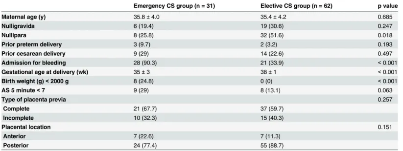
The clinical characteristics of patients are presented inTable 1, with a comparison between
groups with regard to maternal age, percentage of nulligravida, prior preterm or CS, low Apgar score, type of placenta previa, and placental location. The emergency CS group was associated with a lower percentage of nullipara (25.8% versus 51.6%, p = 0.018), and more admissions for
vaginal bleeding (90.3% versus 33.9%, p<0.001), earlier gestational age at delivery (35 ± 3
ver-sus 38 weeks ± 1, p<0.001), and higher neonatal birth weight<2000 g (24.8% versus 0%,
p<0.001) than the elective CS group.
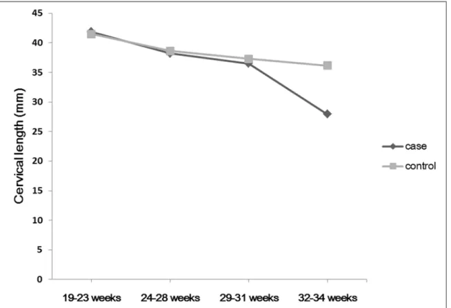
Fig 1shows the serial changes in cervical length from 19 to 34 weeks in the two groups. All 4 CL measurements were available for all participants. The mean CL of the emergency CS group
at 19–23, 24–28, 29–31, and 32–34 weeks was 41.83, 38.21, 36.50, and 27.93 mm, respectively.
The mean CL of the elective CS group was 41.46, 38.61, 37.27, and 36.15 mm, respectively. CL
tended to decrease with advancing gestational age in each group. Until 29–31 weeks, CL
showed no significant differences between the two groups, but after that, CL in the emergent CS group decreased abruptly, even though CL in the elective CS group continued to gradually decrease.
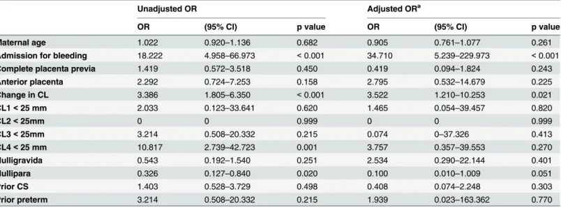
To determine whether the association between change in CL and risk of emergency CS is independent, or is the result of confounding effects, multivariable logistic regression analysis
was used.Table 2shows associations for risk factors for emergency CS in patients with placenta
previa. In the univariate analysis, women in the emergency CS group were more likely to have
admissions for vaginal bleeding (OR, 18.222; 95% CI, 4.958–66.973; p<0.001), change in CL
(OR, 3.386; 95% CI, 1.805–6.350, p<0.001), CL4<25 mm (OR, 10.817; 95% CI, 2.739–
42.723, p = 0.001), and less nullipara (OR, 0.326; 95% CI; 0.127–0.840; p = 0.020) than women
in the control group. In the multivariate analysis that controlled for maternal age, admission
for bleeding, complete placenta previa, anterior placenta, change in CL, CL1<25 mm,
CL4<25 mm, nulligravida, nullipara, prior CS, prior preterm delivery, admission for vaginal
bleeding and change in CL were still significantly associated with a higher risk of emergency CS with an odds ratio of 34.7 and 3.5, respectively. No significant difference was noted in the
rate of short CL at the third trimester (CL4<25 mm) or nullipara.
Table 1. Clinical characteristics of the patients.
Emergency CS group (n = 31) Elective CS group (n = 62) p value
Maternal age (y) 35.8±4.0 35.4±4.2 0.685
Nulligravida 6 (19.4) 19 (30.6) 0.247
Nullipara 8 (25.8) 32 (51.6) 0.018
Prior preterm delivery 3 (9.7) 2 (3.2) 0.193
Prior cesarean delivery 9 (29) 14 (22.6) 0.497
Admission for bleeding 28 (90.3) 21 (33.9) <0.001
Gestational age at delivery (wk) 35±3 38±1 <0.001
Birth weight (g)<2000 g 8 (24.8) 0 (0) <0.001
AS 5 minute<7 9 (29) 8 (13.1) 0.063
Type of placenta previa 0.257
Complete 21 (67.7) 37 (59.7)
Incomplete 10 (32.3) 15 (40.3)
Placental location 0.151
Anterior 7 (22.6) 7 (11.3)
Posterior 24 (77.4) 55 (88.7)
CS, cesarean section; AS 5, Apgar score at 5 minute
Values are expressed as mean±standard deviation or numbers (%).
The ROC curve for change in CL in the prediction of emergency CS for vaginal bleeding is
shown inFig 2. Based on the ROC curve at a cutoff point>6.0 mm for change in CL, the
sensi-tivity, specificity, positive predictive value (PPV), and negative predictive value (NPV) were 77.4, 67.74, 54.5, and 85.7%, respectively, for predicting cases at high risk for emergency CS,
with area under the curve (AUC) 0.734 (p<0.001).
Discussion
Our study shows that a previous admission for vaginal bleeding and change in CL are indepen-dent predictors of emergency CS in placenta previa. Women with a change in CL greater than 6 mm between the second and third trimester are at high risk of emergency CS in placenta pre-via. However, it may not be recommended to use it as the only diagnostic clinical parameter, because its predictive value is moderate. Single measurements of short CL at the second or third trimester do not seem to predict emergency CS. This study might help to identify asymp-tomatic patients with placenta previa at high-risk of emergency CS.
Previous studies showed that CL can be a tool to predict hemorrhage in placenta previa. Ghi et al. reported that CL in the third trimester was significantly shorter in patients with placenta previa who underwent CS before 34 weeks, compared with patients who underwent elective CS
[2]. Others also reported that a decrease in CL<30 or 35 mm in any gestation was associated
with increased risk of preterm CS due to massive hemorrhage [10,13]. However, these studies
had limitations, in that CL was checked at various points of gestational age; single measure-ments may not be representative of CL, because some women have a short CL initially, and
Fig 1. Serial changes in cervical length from 19 to 34 weeks comparing two groups.The black line represents the cervical length of women in the emergent cesarean delivery group, and the gray line represents that of the elective cesarean group.
Table 2. Unadjusted and adjusted odds ratio of risk factors for emergent cesarean delivery in placenta previa.
Unadjusted OR Adjusted ORa
OR (95% CI) p value OR (95% CI) p value
Maternal age 1.022 0.920–1.136 0.682 0.905 0.761–1.077 0.261
Admission for bleeding 18.222 4.958–66.973 <0.001 34.710 5.239–229.973 <0.001
Complete placenta previa 1.419 0.572–3.518 0.450 0.419 0.094–1.824 0.243
Anterior placenta 2.292 0.724–7.253 0.158 2.795 0.532–14.679 0.225
Change in CL 3.386 1.805–6.350 <0.001 3.522 1.210–10.253 0.021
CL1<25 mm 2.033 0.123–33.641 0.620 1.465 0.054–39.457 0.820
CL2<25mm 0 0 0.999 0 0 0.999
CL3<25mm 3.214 0.508–20.332 0.215 0.074 0–37.326 0.413
CL4<25 mm 10.817 2.739–42.723 0.001 3.757 0.357–39.553 0.270
Nulligravida 0.543 0.192–1.540 0.251 2.534 0.290–22.144 0.401
Nullipara 0.326 0.127–0.840 0.020 0.100 0.010–1.009 0.051
Prior CS 1.403 0.528–3.729 0.498 0.408 0.074–2.248 0.303
Prior preterm 3.214 0.508–20.332 0.215 1.939 0.023–163.362 0.770
OR, odds ratio; CI, confidence interval; CL, cervical length; CS, cesarean section Values are expressed as odds ratio (95% confidence interval)
aAdjusted for maternal age, admission for bleeding, complete placenta previa, anterior placenta, change in CL, CL1<25 mm, CL4<25 mm, nulligravida,
nullipara, prior CS, and prior preterm delivery
doi:10.1371/journal.pone.0149036.t002
Fig 2. Receiver operating curve for change in cervical length in the prediction of emergency cesarean section in women with placenta previa.
mean values reported in the literature at fixed points in pregnancy are quite variable [9,15,16]. For example, mean values for nulliparas vary from 34 to 41 mm at around 24 weeks, and from
23 to 30 mm at term [9,15,16]. Another study also argued that a single measurement of CL is
not sufficient to predict the outcome [13]. A recent study showed that CL in women with
pla-centa previa is a useful predictor of emergency CS due to bleeding or labor, and improved its
utility by using CLZ-scores adjusted for gestational age [17].
Instead of a single measurement of CL, we used the change in CL over 2 measurements between the second and third trimester as a significant predictor of emergency CS in asymp-tomatic pregnancies. Another study of twin pregnancies also showed that one CL demonstrat-ing a short cervix has predictive value for preterm birth, and serial CL measurements that demonstrate shortening can identify another high-risk group for spontaneous preterm birth,
even in the absence of a short CL [18]. In placenta previa, a previous study performed serial
measurements of CL every 2 weeks, but the change in CL was not taken into account as a
pre-dictor of emergency CS [13]. To our knowledge, this is the first study to evaluate change in CL
as a predictor of adverse outcome in women with placenta previa.
We also showed serial changes in CL during the second and third trimester in women with placenta previa. Serial change in CL is characterized by three patterns in normal patients: unchanged prior to delivery, progressive shortening within 2 weeks prior to delivery, and
steady shortening starting before 2 weeks prior to delivery [19]. In this study, change in CL in
the emergency CS group showed that CL shortened abruptly. With this result, we can assume that the rate of change from second to third trimester, rather than a single measurement during a gestation, can help to predict emergency CS. Other studies also showed that cervical
shorten-ing was a significant independent predictor of preterm delivery [20,21].
The mechanism of hemorrhage in placenta previa is not well known, but cervical effacement and labor are thought to be the cause of bleeding.The uterine cervix undergoes a series of changes during pregnancy, resulting in gradual reduction of mechanical strength, and eventual
effacement and dilatation [22,23], resulting in placental vessel tearing [16,22,24]. Inability of
the lower uterine segment myometrium to constrict the torn vessels [25] can cause massive
vaginal bleeding, necessitating emergency CS. This mechanism can justify use of CL shortening as a predictor of bleeding, because it represents cervical effacement and dilation, and is already known to be associated with preterm labor. For this reason, hemorrhage in placenta previa typ-ically occurs in the third trimester, as the lower uterine segment becomes more defined, and
the internal os dilates [11].
Our study also showed that a previous admission for vaginal bleeding was associated with increased risk of emergency CS, because of recurrent massive vaginal bleeding. A previous study showed that patients with episodes of antenatal bleeding had more emergency CS than
patients without bleeding [1]. For patients discharged after observation for bleeding, a warning
of the possibility of emergent CS should be given. Hospital admission for vaginal bleeding was a stronger predictor parameter than change in CL, which supports the hypothesis that the highest value of change in CL would be found in asymptomatic patients without vaginal bleeding.
There are some limitations to our study. First, this was a retrospective review, which pre-cludes control for additional risk factors for emergency CS. Second, our study only recruited a small number of women.
In cases of CL shortening without symptoms, we can educate patients earlier, to prevent massive bleeding. Prenatal assessment of factors associated with a high risk of emergency CS would improve the preparation and management of such patients, and can help decide whether to prepare for blood in advance, and when to schedule CS. For asymptomatic women with pla-centa previa, our study suggests that a change in CL greater than 6 mm from the second to third trimester gives useful clinical information on the likelihood of emergency CS.
Acknowledgments
The authors would like to thank the Clinical Research Coordinating Center of the Catholic Medical Center for assisting with the statistical analysis.
Author Contributions
Conceived and designed the experiments: SJK JES. Performed the experiments: JES. Analyzed the data: JES. Contributed reagents/materials/analysis tools: JCS. Wrote the paper: JES YL.
References
1. Hasegawa J, Higashi M, Takahashi S, Mimura T, Nakamura M, Matsuoka R, et al. Can ultrasonography of the placenta previa predict antenatal bleeding? J Clin Ultrasound. 2011; 39: 458–62. doi:10.1002/
jcu.20849PMID:21671240
2. Ghi T, Contro E, Martina T, Piva M, Morandi R, Orsini LF, et al. Cervical length and risk of antepartum bleeding in women with complete placenta previa. Ultrasound Obstet Gynecol. 2009; 33: 209–12. doi:
10.1002/uog.6301PMID:19173235
3. Palacio M, Sanin-Blair J, Sanchez M, Crispi F, Gomez O, Carreras E, et al. The use of a variable cut-off value of cervical length in women admitted for preterm labor before and after 32 weeks. Ultrasound Obstet Gynecol. 2007; 29: 421–6. PMID:17330832
4. Berghella V, Roman A, Daskalakis C, Ness A, Baxter JK. Gestational age at cervical length measure-ment and incidence of preterm birth. Obstet Gynecol. 2007; 110: 311–7. PMID:17666605
5. Sotiriadis A, Papatheodorou S, Kavvadias A, Makrydimas G. Transvaginal cervical length measure-ment for prediction of preterm birth in women with threatened preterm labor: a meta-analysis. Ultra-sound Obstet Gynecol. 2010; 35: 54–64. doi:10.1002/uog.7457PMID:20014326
6. Morales Rosello J, Hervas Marin D, Perales Marin A. Can we predict delivery date with obstetric ultra-sound? J Matern Fetal Neonatal Med. 2013; 26: 1807–11. doi:10.3109/14767058.2013.804049PMID:
23682599
7. Bayramoglu O, Arslan M, Yazici FG, Erdem A, Erdem M, Bayramoglu K, et al. Prediction of spontane-ous onset of labor at term: the role of cervical length measurement and funneling of internal cervical os detected by transvaginal ultrasonography. Am J Perinatol. 2005; 22: 35–9. PMID:15668842
8. Colombo DF, Iams JD. Cervical length and preterm labor. Clin Obstet Gynecol. 2000; 43: 735–45.
PMID:11100291
9. Iams JD, Goldenberg RL, Meis PJ, Mercer BM, Moawad A, Das A, et al. The length of the cervix and the risk of spontaneous premature delivery. National Institute of Child Health and Human Development Maternal Fetal Medicine Unit Network. N Engl J Med. 1996; 334: 567–72. PMID:8569824
10. Stafford IA, Dashe JS, Shivvers SA, Alexander JM, McIntire DD, Leveno KJ. Ultrasonographic cervical length and risk of hemorrhage in pregnancies with placenta previa. Obstet Gynecol. 2010; 116: 595–
600. doi:10.1097/AOG.0b013e3181ea2debPMID:20733440
11. Fukushima K, Fujiwara A, Anami A, Fujita Y, Yumoto Y, Sakai A, et al. Cervical length predicts placen-tal adherence and massive hemorrhage in placenta previa. J Obstet Gynaecol Res. 2012; 38: 192–7.
doi:10.1111/j.1447-0756.2011.01669.xPMID:21995621
12. Zaitoun MM, El Behery MM, Abd El Hameed AA, Soliman BS. Does cervical length and the lower pla-cental edge thickness measurement correlates with clinical outcome in cases of complete placenta pre-via? Arch Gynecol Obstet. 2011; 284: 867–73. doi:10.1007/s00404-010-1737-1PMID:21113721
13. Sekiguchi A, Nakai A, Okuda N, Inde Y, Takeshita T. Consecutive cervical length measurements as a predictor of preterm cesarean section in complete placenta previa. J Clin Ultrasound. 2015; 43: 17–22.
14. Dashe JS, McIntire DD, Ramus RM, Santos-Ramos R, Twickler DM. Persistence of placenta previa according to gestational age at ultrasound detection. Obstet Gynecol. 2002; 99: 692–7. PMID:
11978274
15. Cook CM, Ellwood DA. A longitudinal study of the cervix in pregnancy using transvaginal ultrasound. Br J Obstet Gynaecol. 1996; 103: 16–8.
16. Heath VC, Southall TR, Souka AP, Novakov A, Nicolaides KH. Cervical length at 23 weeks of gestation: relation to demographic characteristics and previous obstetric history. Ultrasound Obstet Gynecol. 1998; 12: 304–11. PMID:9819867
17. Garcia-Espinosa M, Cisneros-Bedoy E, Rosales-Ortiz S, Martinez-Rodriguez O, Lopez-Jimenez S, Moreno-Alvarez O. OC07.04: Adjusted cervical length for gestational age as predictor of urgent delivery in pregnancies complicated by placenta previa. Ultrasound in Obstetrics & Gynecology. 2014; 44: 16–
7.
18. Fox NS, Rebarber A, Klauser CK, Peress D, Gutierrez CV, Saltzman DH. Prediction of spontaneous preterm birth in asymptomatic twin pregnancies using the change in cervical length over time. Am J Obstet Gynecol. 2010; 202: 155.e1-4. doi:10.1016/j.ajog.2009.09.004PMID:19846054
19. Meijer-Hoogeveen M, Van Holsbeke C, Van Der Tweel I, Stoutenbeek P, Visser GH. Sonographic lon-gitudinal cervical length measurements in nulliparous women at term: prediction of spontaneous onset of labor. Ultrasound Obstet Gynecol. 2008; 32: 652–6. doi:10.1002/uog.5291PMID:18702094
20. Souka AP, Papastefanou I, Michalitsi V, Salambasis K, Chrelias C, Salamalekis G, et al. Cervical length changes from the first to second trimester of pregnancy, and prediction of preterm birth by first-trimester sonographic cervical measurement. J Ultrasound Med. 2011; 30: 997–1002. PMID:21705733
21. Naim A, Haberman S, Burgess T, Navizedeh N, Minkoff H. Changes in cervical length and the risk of preterm labor. Am J Obstet Gynecol. 2002; 186: 887–9. PMID:12015503
22. Word RA, Li XH, Hnat M, Carrick K. Dynamics of cervical remodeling during pregnancy and parturition: mechanisms and current concepts. Semin Reprod Med. 2007; 25: 69–79. PMID:17205425
23. Jenkins SM, Kurtzman JT, Osann K. Dynamic cervical change: is real-time sonographic cervical short-ening predictive of preterm delivery in patients with symptoms of preterm labor? Ultrasound Obstet Gynecol. 2006; 27: 373–6. PMID:16565995
24. Oppenheimer L. Diagnosis and management of placenta previa. J Obstet Gynaecol Can. 2007; 29: 261–73. PMID:17346497