Delayed Presentation of Catheter-Related Subclavian
Artery Pseudoaneurysm
Hwa Rim Kang, M.D.1
, Jin Yong Park, M.D.1
, Jee Hyun Kim, M.D.1
, Yook Kim, M.D.2
, Min Ho Kang, M.D.2 , YouJin Chang, M.D.1
, Kang Hyeon Choe, M.D., Ph.D.1
, Ki Man Lee, M.D., Ph.D.1
, and Jin Young An, M.D.1
1
Department of Internal Medicine, 2
Department of The Radiology, College of Medicine, Chungbuk National University, Cheongju, Korea
Central venous catheterization is a common diagnostic and therapeutic procedure in modern clinical practice. Pseudoaneurysms of the subclavian artery are rare and usually occur immediately after the causative event, whether the cause was trauma or a medical procedure. Here we report the rare case of a 71-year-old woman with delayed presentation of catheter-related subclavian pseudoan-eurysm. The patient was treated for aspiration pneumonia with respiratory failure in another hospital. The patient’s chest wall swelling began two weeks after the initial catheterization in the other hospital, probably because of slow leakage of blood from the injured subclavian artery caused by incomplete compression of the puncture site and uremic coagulopathy. She was successfully treated with ultrasound-guided thrombin and angiography-guided histoacryl injection without stent insertion or surgery. Her condition improved, and she was discharged to her home.
Key Words: aneurysm, false; enbucrilate; subclavian artery; thrombin.
Central venous catheters are commonly used for diagnostic and therapeutic purposes. However, they may sometimes be the cause of a number of catheter-related complications ranging from mild to severe.[1] Rare, potentially serious complications
include development of a pseudoaneurysm of the subclavian artery and its subsequent progression and eventual rupture. Most of the complications occur and present immediately after the causative event, but in rare cases, presentation could be delayed.
Here we report the rare case of a 71-year-old woman with a delayed presentation of catheter-related subclavian pseudoaneu-rysm who was successfully treated with ultrasound-guided thrombin and angiography-guided histoacryl injection.
Case Report
A 71-year-old woman was transferred to the emergency department for specialized intensive care including assisted
ventila-tion and renal replacement therapy. Her medical history included diabetes and a previous cerebrovascular incident. She had been admitted at another hospital one month earlier because
of aspiration pneumonia. Although broad-spectrum antibiot-ics were administered, her condition had gradually worsened;
ventilation was initiated in the intensive care unit because of acute respiratory failure. Her doctor had attempted to place a central line via the left subclavian vein. Vessel access was
performed using the standard needle and guidewire technique
cc This is an Open Access article distributed under the terms of the Creative Commons Attribution Non-Commercial License (http://creativecommons.org/ licenses/by-nc/3.0/) which permits unrestricted non-commercial use, distribution, and reproduction in any medium, provided the original work is properly cited.
Received on May 7, 2015 Revised on July 3, 2015 Accepted on July 4, 2015
Correspondence to: Jin Young An, Department of Internal Medicine, College of Medicine, Chungbuk National University, 776 1sunhwan-ro, Seowon-gu, Cheongju 28644, Korea
Tel: +82-43-269-6308, Fax: +82-43-269-6804 E-mail: [email protected]
*No potential conflict of interest relevant to this article was reported.
without ultrasonographic guidance, but catheter
place-ment via the left subclavian vein failed after several trials. Even with aggressive treatment, her symptoms gradually
worsened, and she developed acute kidney injury. She was transferred to our hospital for management of pneumonia
and continuous renal replacement therapy. In the emergency department, laboratory diagnostics revealed the following:
leukocyte count, 22,160/mm3; C-reactive protein level, 3.95
mg/dL; and pro-calcitonin quantitative level, 2.15 μg/L. Other blood chemistry values were within reference ranges
except increased blood urea nitrogen (70 mg/dL) and cre-atinine (4.7 mg/dL). In order to protect her renal function,
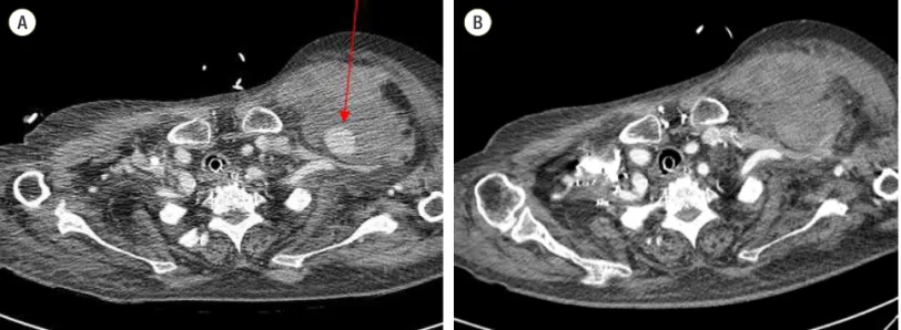
we changed her antibiotic to a less nephrotoxic one with a dose adjustment. Fortunately, her respiratory symptoms Fig. 1. Chest computed tomography (CT) scanning. It revealed a 2.6-cm enhancing nodular lesion, suggesting a pseudoaneurysm at the left subclavicular area with surrounding hematoma (arrow) (A). CT scanning after embolization revealed no bleeding and a smaller hematoma (B).
A B
Fig. 2. An ultrasound-guided thrombin injection was performed at the pseudoaneurysm of the left subclavian artery (A: pre-injection, B: post-injection).
improved along with her renal function. However, on the
ifth day after she was transferred to our hospital, we found
swelling in her left upper anterior chest area. We performed a computed tomography (CT) examination of the chest. CT
scanning revealed a 2.6-cm enhancing nodular lesion, sug-gesting a pseudoaneurysm at the left subclavicular area with
surrounding hematoma (Fig. 1A). Because the patient was on a ventilator and her vital signs were unstable, we chose
a bedside procedure to resolve this problem. We immedi-ately performed an ultrasound-guided thrombin injection at the pseudoaneurysm of the left subclavian artery (Fig. 2).
The swelling gradually decreased over the next four days; her medical condition, including respiratory failure and
renal failure, also continued to improve. However, on the eighth day after the procedure, swelling of the chest wall
had begun to increase, and we performed ultrasonography.
On ultrasonography, blood low could not be visualized at
the pseudoaneurysm because the vessel was blocked by a thrombus. Because the patient’s condition, including renal
function, was relatively stable, angiography was performed
using luoroscopy to ind the cause of the swelling. Angiog -raphy revealed active bleeding of a branch of the left
sub-clavian artery (Fig. 3A). Embolization was performed at the same branch of the subclavian artery using
n-butyl-2-cyano-acrylate (Histoacryl, B. Braun Medical Inc., Bethlehem, PA, USA). Follow-up angiography, performed after four days,
revealed no visible extravasation from the subclavian artery (Fig. 3B).
Both the patient’s medical condition and left chest swell-ing gradually improved. Follow-up CT scannswell-ing revealed no
evidence of active bleeding or pseudoaneurysm (Fig. 1B). The patient was continuously treated for aspiration
pneu-monia and respiratory failure, and her condition ultimately improved. One month after the procedure, she was
dis-charged to her home.
Discussion
A central venous line may be useful for monitoring the cardiovascular function of patients in critical condition and
for administering vasoactive drugs or solutions that would irritate peripheral veins, such as total parenteral nutrition.
[2] However, central line insertion may cause a number of complications such as skin infection, thrombophlebitis,
pneumothorax, thrombosis, central-line-associated sepsis, hemorrhage, and arrhythmia.[1] Pseudoaneurysm of the
subclavian artery can also develop after inadvertent punc-ture of the artery. Subclavian pseudoaneurysms are rare, and
their true incidence is unknown. Most cases are secondary to arterial catheterization, surgical procedures, and radiol-ogy interventions, but in a few cases described in the
lit-erature, injury to the subclavian artery was caused by blunt trauma.[3] Patients with subclavian pseudoaneurysm may
be asymptomatic or they may present with chest pain, Horn-er syndrome, paresthesia, hoarseness, uppHorn-er limb ischemia,
a pulsatile mass, or hemoptysis.[4]
Early recognition and optimal diagnostic tools are very
important in such cases. Various diagnostic methods may
be used. Sonography is highly sensitive and speciic for ac -Fig. 3. Angiography indings. It revealed active bleeding of a branch of the left subclavian artery (arrow) (A). After embolization, angiog -raphy showed no evidence of extravasations of contrast media (arrow) (B).
cessible arterial injuries, and it is often useful for rapid bed-side assessment in the intensive care unit. Spiral
contrast-enhanced computed tomography usually provides a good diagnostic yieldand was used to make the initial diagnosis
in this case. Angiography not only enables accurate diagno-sis but also provides endovascular treatment options without surgery.[5]These diagnostic tools are complementary; their
use depends on the condition of the patient.
In the case described here, signs of subclavian artery
pseu-doaneurysm were not seen until approximately two weeks after catheterization. We suggest the following possible
ex-planations of the delayed presentation. First, the patient was a quiet, overweight, geriatric woman with ample free space
for a hematoma reservoir. Second, she could not complain of chest discomfort because of intubation and deep sedation
during ventilator care. Third, uremic coagulopathy played a role in the continuous leakage of small amounts of blood from the injured vessel.[6]
The classical treatment of pseudoaneurysm is open sur-gery for resection and end-to-end anastomosis, venous graft,
suture, or bypass.[7] However, open surgery involves con-siderable morbidity and mortality, particularly with
high-risk patientsand urgent surgeries. Therefore, less invasive
procedures have been developed. Marin et al. published the
irst report of the use of covered stents to treat pseudoaneu -rysms.[8] Since the 1990s, ultrasound-guided compression repair and ultrasound-guided thrombin injections have been
used. Ultrasound-guided thrombin injection has a primary success rate of 97% and a low complication rate of 1.3%,
with an embolic rate of 0.5%.[9] Moreover, the use of endo-vascular stent treatment or embolization of endo-vascular injuries
via angiography has become an acceptable and less invasive alternative to surgical repair in subclavian artery injury.[10]
If the patient’s vital signs had been stable and anticoagula-tion for stent placement had been possible, we could have
considered stent insertion. However, her condition did not allow for this option; her vital signs were unstable and she had a cerebrovascular accident with severe disabilities. We
therefore chose to use treatment modalities that did not include stent placement. Fortunately, we have also
success-fully performed ultrasound-guided thrombin injection and histoacryl embolization by angiography without stent
inser-tion.
In conclusion, overweight and geriatric patients with
uremic coagulopathy could be predisposed to bleeding and delayed presentation of hematoma and pseudoaneurysm.
Therefore, such patients require accurate and relatively long compression times compared with other patients.
Ultra-sound-guided access, if available, should also be considered in order to avoid unnecessary damage to blood vessels.
ORCID
Hwa Rim Kang http://orcid.org/0000-0003-1472-8687
Jin Yong Park http://orcid.org/0000-0002-7487-3352
Jee Hyun Kim http://orcid.org/0000-0003-1336-3620
Min Ho Kang http://orcid.org/0000-0002-4441-7196
YouJin Chang http://orcid.org/0000-0002-4838-466X
Kang Hyeon Choe http://orcid.org/0000-0001-7197-6770
Jin Young An http://orcid.org/0000-0002-6553-8579
References
1) McGee DC, Gould MK: Preventing complications of
central venous catheterization. N Engl J Med 2003; 348: 1123-33.
2) Schwengel DA, McGready J, Berenholtz SM, Kozlows-ki LJ, Nichols DG, Yaster M: Peripherally inserted central catheters: a randomized, controlled, prospective
trial in pediatric surgical patients. Anesth Analg. 2004; 99: 1038-43.
3) Piffaretti G, Tozzi M, Lomazzi C, Rivolta N, Caronno R, Laganà D, et al: Endovascular treatment for
trau-matic injuries of the peripheral arteries following blunt trauma. Injury 2007; 38: 1091-7.
4) Brown HA, Aruny JE, Elefteriades JA, Sumpio BE: Subclavian aneurysm presenting with massive
hemop-tysis: a case report and review of the literature. Int J Angiol 2013; 22: 69-74.
5) Jeganathan R, Harkin DW, Lowry P, Lee B: Iatrogenic
subclavian artery pseudoaneurysm causing airway com-promise: treatment with percutaneous thrombin
injec-tion. J Vasc Surg. 2004; 40: 371-4.
6) Boccardo P, Remuzzi G, Galbusera M: Platelet
dys-function in renal failure. 2004; 30: 579-89.
endovas-cular grafts for the treatment of aneurysmal, occlusive and traumatic arterial lesions. Cardiovasc Surg. 1998; 6:
552-65.
8) Marin ML, Hollier LH, Avrahami R, Parsons R:
Vary-ing strategies for endovascular repair of abdominal and iliac artery aneurysms. Surg Clin North Am 1998; 78: 631-45.
9) La Perna L, Olin JW, Goines D, Childs MB, Ouriel K: Ultrasound-guided thrombin injection for the treatment
of postcatheterization pseudoaneurysms. Circulation. 2000; 102: 2391-5.