Detachment of chemotherapy catheters:
a rare complication?
Desconexão de cateter para quimioterapia: uma complicação rara?
Alexandre Faraco de Oliveira1, Horácio de Oliveira Filho2
Abstract
Use of totally implantable catheters for chemotherapy treatment is a necessity, but one which brings with it risks of multiple complications, some inherent to insertion of the device and others related to the catheter itself. We describe three cases in which the catheter became disconnected from its reservoir. In the irst case, the catheter became completely detached from its reservoir and in the second and third cases the catheter itself underwent fragmentation. In all three cases it was necessary to withdraw the endovascular catheter. his event is described as rare, but it tends to be present in the majority of reviews and it is associated with the risk of serious complications, even though it is often asymptomatic. It is advisable to follow-up patients who have these catheters implanted in order to detect these complications early and to enable understanding of the factors that cause these situations to occur.
Keywords: central venous catheters; equipment failure; vascular access devices.
Resumo
A utilização de cateteres totalmente implantáveis no tratamento quimioterápico constitui uma necessidade que acarreta o risco de múltiplas complicações, algumas inerentes à inserção do dispositivo e outras relacionadas ao próprio cateter. Relatamos três casos nos quais o cateter apresentou-se desacoplado de seu respectivo reservatório. No primeiro caso, ocorreu a desconexão do cateter de seu respectivo reservatório, e nos outros dois casos, veriicou-se a fragmentação do cateter. Em todos os casos, foi necessária a retirada endovascular do cateter. Tal desfecho é apontado como raro, mas costuma estar presente na maioria das revisões e traz consigo o risco de complicações graves, ainda que frequentemente seja assintomático. É desejável o acompanhamento de pacientes que possuem tais cateteres a im de que se possa detectar precocemente tais complicações e compreender os fatores que determinam o aparecimento dessas situações.
Palavras-chave: cateteres venosos centrais; falha de equipamento; dispositivo de acesso vascular.
1 Universidade do Planalto Catarinense – UNIPLAC, Lages, SC, Brazil. 2 Clínica Ana Carolina, Lages, SC, Brazil.
Financial support: None.
Conlicts of interest: No conlicts of interest declared concerning the publication of this article. Submitted: August 09, 2016. Accepted: November 07, 2016.
INTRODUCTION
Catheters for infusion of chemotherapy are very useful instruments and are sometimes essential to enable provision of oncological treatment. In many cases, lack of adequate access makes treatment impossible, whether because of irritation of the veins of the upper limbs by the drugs or because of the need for multiple sessions.1
Totally implantable catheters (TICs) of the port-a-cath type tend to be the most popular choice because once installed they allow permanent access to a deep vein, which is gained by puncturing the port rather than a vein. In addition to enabling infusion of medications, they can also be used to take blood samples for tests. As the range of chemotherapy treatments available expands and patient survival constantly increases as a result of these treatments, more and more of these catheters are used and for longer periods.1
These devices are associated with many complications, related both to placement and to use. The most serious complications, such as pneumothorax or hemothorax, are related to device implantation, tend to manifest immediately, and are the result of choice of puncture site. More common complications, such as hematoma or infection of the puncture site, tend to occur later, but are generally of lower risk to the patient and easily detected.2
In this article we describe three cases in which a catheter became detached from its reservoir, with no apparent cause for this event. As a result, the catheter became a foreign body in the deep vein system,
speciically the superior vena cava, generating a risk of potentially serious and dificult to diagnose
complications. After describing the cases we present a review of existing reports in order to place these complications in relation to them.
CASE REPORTS
Case 1: a 28-year-old female patient was diagnosed
with a left breast tumor during her irst gestation
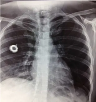
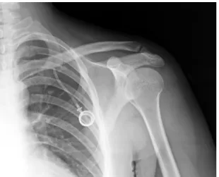
and underwent a left mastectomy at approximately 26 weeks. At 34 weeks, a cesarean delivery was performed, followed by placement of a chemotherapy catheter via puncture of the right subclavian vein and placement of the reservoir at the thorax (Figure 1). Around 10 days after implantation of the catheter, the patient complained of right-side cervical pain combined with tumefaction along the path of the right internal jugular vein and localized pain on palpation. A chest X-ray showed that the catheter had become detached from the reservoir (Figure 2). Cervical Doppler ultrasonography showed thrombosis of the internal
jugular vein. The patient was put on anticoagulation and exhibited complete regression of the symptoms related to thrombosis after 15 days. Anticoagulation was suspended for 72 hours. Endovascular removal of the catheter was then conducted by catheterization of the right common femoral vein and then anticoagulation was initiated once again.
Case 2: a 33-year-old female patient underwent a right mastectomy for right-side breast cancer.
Figure 1. Chest X-ray showing reservoir and catheter implanted
via puncture of the right subclavian vein.
Figure 2. Catheter detached from the reservoir implanted in
Approximately 90 days after surgery, a catheter was implanted for chemotherapy via puncture of the left subclavian vein, with placement of the reservoir in the left thorax. The access was used for
ive chemotherapy sessions over a 4-month period,
during which there were no reports of any problems utilizing the venous access. When she attended for the sixth treatment session, the catheter did not exhibit
relux when punctured, and the patient was referred
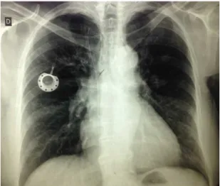
to the vascular service. A chest X-ray showed that the reservoir and a small segment of the catheter were still implanted in the left thorax, but a piece of the catheter measuring approximately 10 cm was visible on the cardiac projection (Figures 3-5). The patient was asymptomatic and reported that she felt no thoracic pain or discomfort, tachycardia, or tachypnea. Endovascular removal of the catheter was then performed via catheterization of the right common femoral vein.
Case 3: a 60-year-old female patient underwent a right mastectomy for right-side breast cancer. Approximately 60 days after surgery, she underwent implantation of a catheter for chemotherapy via
puncture of the right subclavian vein, with ixation of
the reservoir in the right thorax. The access was used for multiple chemotherapy sessions over a 19-month period, with no reports of problems utilizing the access. When she attended for the twentieth treatment
session, the catheter did not exhibit relux when
punctured, and the patient was referred to the vascular service. A chest X-ray showed that the catheter had
undergone fragmentation (Figure 6). The patient was asymptomatic and reported that she felt no thoracic pain or discomfort, tachycardia, or tachypnea and was sent for endovascular removal of the catheter.
DISCUSSION
Implantation of catheters for chemotherapy treatment is a procedure that is already well known and widely used, to the extent that the most frequent complications are also well known.1
Some complications are directly related to the puncture site and to implantation of the device. While
Figure 3. X-ray showing the reservoir and part of the catheter
implanted in the left thorax, with the greater part of the catheter embolized in the cardiac area.
Figure 4. X-ray showing the reservoir and part of the catheter
implanted in the left thorax, with the greater part of the catheter embolized in the cardiac area.
Figure 5. X-ray showing the reservoir and part of the catheter
there are many possibilities for deployment of these catheters, three accesses tend to be used more than
others: puncture of the subclavian vein with ixation
of the reservoir in the thorax, puncture or dissection
of the jugular vein with ixation of the reservoir in
the thorax, and dissection of an upper limb vein, the
cephalic or the basilic, with ixation of the reservoir
in the same upper limb. In all cases, the objective is to place the distal extremity of the catheter in the superior vena cava and to position the reservoir in a manner that facilitates its puncture locally.3
The most frequently reported complications, hematoma and infection of the surgical site, are common to all accesses. Insertion of the catheter by dissection, whether of a cervical vein or an upper limb vein, generally results in a longer operating time and dissection of a larger area and sometimes requires sedation combined with local anesthesia.2,4
Access via puncture of the subclavian vein
involves risks speciic to puncture of deep veins of
the thorax, i.e., pneumothorax and hemothorax, and also inadvertent puncture of arteries. However, it is a less traumatic access for the patient and it enables
ixation of the reservoir in the superior portion of the
thorax, in a site that facilitates puncture. Irrespective of which access is chosen, catheter-related thrombosis is always a possible complication, although it does appear to be more related to access performed by dissection of upper limbs.2,4,5
The risk inherent to puncture related to access via the subclavian vein tends to be minimized by the surgeon’s experience and by using ultrasonography to guide the procedure.6 Furthermore, problems related
to the puncture itself tend to manifest immediately
and are easily identiied.4
However, access via puncture of the subclavian vein
will always involve risk of a speciic complication,
known as pinch-off syndrome, which is caused by the catheter being subjected to a pincer movement
between the clavicle and the irst rib, with consequent
partial or total fracture of the catheter.7-9 A systematic
review of catheters implanted via the subclavian vein demonstrated that damage to the catheter with microruptures may be more common than is realized and that it may be related to the type of material utilized.10
Although all TICs have similar shapes, there
are speciic differences in the choice of materials.
The reservoirs may be made of plastic or metal and the catheters themselves are manufactured in silicone or polyurethane. There is evidence that polyurethane catheters are more prone to thrombotic and infectious complications, whereas those made from silicone are more susceptible to mechanical events such as detachment and rupture.10
The cases described here involved two different situations that have in common the same result: a foreign body loose in the deep vein system, near
to or within the cardiac chambers. In the irst case,
the entire catheter became disconnected from its reservoir, and in the second and third cases the catheter fractured. The catheters involved were from different
manufacturers, but were similarly made, while the irst
two were polypropylene and the third was silicone. Published reviews that cover complications related to chemotherapy catheters tend to agree on the frequency and severity of complications in general and show that catheter rupture occurs in around 1-4% of cases.1,2,11
This can progress to local thrombosis, as in the
irst case described, or may remain silent, as in the
second and third cases. Although all three cases had satisfactory outcomes, these events caused the patients morbidity and, in general, such events bring with them the risk of causing serious arrhythmia, precordial pain, and embolization into the pulmonary artery.12-14
In the three cases described, both disconnections and fractures occurred spontaneously. However, there are also reports of detachment and fragmentation of catheters related to removal procedures.15 Balsorano
conducted a study speciically designed to verify
the integrity of catheters after removal, whether because of malfunction or because treatment had been terminated, and found that the type of catheter and use of “heterodox” accesses were related to microruptures.16
Figure 6. Chest X-ray showing the fragmented catheter and the
The hospital in which the TICs were implanted in the cases described here is a referral center for cancer treatment that has implanted around 100 TICs per year since 2011. Over this period, complications such as local infection, hematoma, thrombosis at the catheter site and exteriorization of the catheter reservoir occurred in an occasional and rare manner, without causing serious risk or morbidity to patients, although we do not have precise data for all patients.
Until around 90 days ago, we were unaware of any cases of fragmentation or embolization of catheters
like those described here. A signiicant proportion of
the hospital’s population live in remote rural areas, which has created obstacles to our efforts to actively locate these patients and identify possible unreported complications. This factor is compounded by the fact that a proportion of these patients were given treatment with palliative objectives and had limited life expectancies.
In the cases described here, although it is not possible
to deinitively state the causes, pinch-off syndrome
is a probable candidate for the fractures, but there is no obvious explanation for the total detachment.7-9
Constant follow-up of all patients over the long term could lead to development of protocols that indicate which accesses and types of catheter are most appropriate, in order to minimize complications
or provide speciic prophylaxis for each type of
complication.
Although the complications related to use of catheters for chemotherapy can be minimized by conducting individualized studies of the most appropriate access for each patient and by continued development of the materials, it is logical to conclude that, as with other invasive procedures, risk will always be present and complications will manifest from time to time.
REFERENCES
1. Schiffer CA, Mangu PB, Wade JC, et al. Central venous catheter care for the patient with cancer: American Society of Clinical Oncology clinical practice guideline. J Clin Oncol. 2013;31(10):1357-70. PMid:23460705. http://dx.doi.org/10.1200/JCO.2012.45.5733.
2. Bassi K, Pattanayak M, Pandey K, Giri A, Abraham S. Totally implantable venous access ports: Retrospective review of long-term complications in 81 patients. Indian J Cancer. 2012;49(1):114-8. PMid:22842178. http://dx.doi.org/10.4103/0019-509X.98934.
3. Debourdeau P, Farge D, Beckers M, et al. International clinical practice guidelines for the treatment and prophylaxis of thrombosis associated with central venous catheters in patients with cancer. J Thromb Haemost. 2013;11(1):71-80. PMid:23217208. http:// dx.doi.org/10.1111/jth.12071.
4. El Hammoumi M, El Ouazni M, Arsalane A, El Oueriachi F, Mansouri H, Kabiri EH. Incidents and complications of permanent venous central access systems: a series of 1,460 cases. Korean J Thorac Cardiovasc Surg. 2014;47(2):117-23. PMid:24782960. http://dx.doi. org/10.5090/kjtcs.2014.47.2.117.
5. Reddy MA, Natarajan R. Epigenetic mechanisms in diabetic vascular complications. Cardiovasc Res. 2011;90(3):421-9. PMid:21266525. http://dx.doi.org/10.1093/cvr/cvr024.
6. Zerati A, Figueiredo T, Moraes R, et al. Risk factors for infectious and noninfectious complications of totally implantable venous catheters in cancer patients. J Vasc Surg Venous Lymphat Disord. 2016;4(2):200-5. PMid:26993868. http://dx.doi.org/10.1016/j. jvsv.2015.10.008.
7. Morales-Victorino N, Damas de los Santos F, Kuri-Ayache M, López-Aguilar C. Pinch-off syndrome: case report and review of the literature. Gac Med Mex. 2015;151(4):529-32. PMid:26290031. 8. Sugimoto T, Nagata H, Hayashi K, Kano N. Pinch-off syndrome:
transection of implantable central venous access device. BMJ Case Rep. 2012;20:7-9. PMid:23203173.
9. Cho JB, Park IY, Sung KY, Baek JM, Lee JH, Lee DS. Pinch-off syndrome. J Korean Surg Soc. 2013;85(3):139-44. PMid:24020024. http://dx.doi.org/10.4174/jkss.2013.85.3.139.
10. Wildgruber M, Lueg C, Borgmeyer S, et al. Polyurethane versus silicone catheters for central venous port devices implanted at the forearm. Eur J Cancer. 2016;59:113-24. PMid:27023050. http:// dx.doi.org/10.1016/j.ejca.2016.02.011.
11. Ko SY, Park SC, Hwang JK, Kim SD. Spontaneous fracture and migration of catheter of a totally implantable venous access port via internal jugular vein: a case report. J Cardiothorac Surg. 2016;11(1):50. PMid:27067705. http://dx.doi.org/10.1186/ s13019-016-0450-y.
12. Faraj W, Zaghal A, El-Beyrouthy O, Kutoubi A. Complete catheter disconnection and migration of an implantable venous access device: the disconnected cap sign. Ann Vasc Surg. 2010;24(5):692-7. PMid:20413259. http://dx.doi.org/10.1016/j.avsg.2010.02.011.
13. Wassef AW, Kass M, Parmar G, Ravandi A. An unusual cause of ventricular tachycardia: Port-A-Cath fracture and embolization into the pulmonary artery. Heart Int. 2014;9(1):30-2. PMid:27004095.
14. Samad AM, Ibrahim YA. Complications of Port A Cath implantation: a single institution experience. Egypt J Radiol Nucl Med. 2015;907:11.
15. Ribeiro RC, Monteiro AC, Menezes QC, Schettini ST, Vianna SMR. Totally implantable catheter embolism: two related cases. Sao Paulo Med J. 2008;126(6):347-9. PMid:19274324. http://dx.doi. org/10.1590/S1516-31802008000600011.
*
Correspondence
Alexandre Faraco de Oliveira Clínica Ana Carolina Rua Marechal Deodoro, 856, sala 03 - Centro CEP 88501-001 - Lages (SC), Brazil Tel.: +55 (49) 3224-3872 E-mail: [email protected]
Author information
AFO - Board certiied in Vascular Surgery by Sociedade Brasileira de Angiologia e de Cirurgia Vascular (SBACV); Board certiied in Vascular Doppler Ultrasound by SBACV; MSc in Human Aging; Professor of the School of Medicine at Universidade do Planalto Catarinense (UNIPLAC). HOF - Board certiied in Angiology and Vascular Surgery by Sociedade Brasileira de Angiologia e de Cirurgia Vascular (SBACV), Clínica Ana Carolina.
Author contributions
Conception and design: AFO, HOF Analysis and interpretation: AFO, HOF Data collection: AFO Writing the article: AFO Critical revision of the article: AFO, HOF Final approval of the article*: AFO, HOF Statistical analysis: N/A. Overall responsibility: AFO