Effect of Epicatechin against Radiation-Induced Oral
Mucositis:
In Vitro
and
In Vivo
Study
Yoo Seob Shin1., Hyang Ae Shin2., Sung Un Kang1
, Jang Hee Kim3, Young-Taek Oh4, Keun Hyung Park1, Chul-Ho Kim1*
1Department of Otolaryngology, School of Medicine, Ajou University, Suwon, Republic of Korea,2Department of Otorhinolaryngology-Head and Neck Surgery, National Health Insurance Corporation Ilsan Hospital, Goyang, Republic of Korea,3Department of Patholog School of Medicine, Ajou University, Suwon, Republic of Korea, 4Department of Radiation Oncology, School of Medicine, Ajou University, Suwon, Republic of Korea
Abstract
Purpose:Radiation-induced oral mucositis limits the delivery of high-dose radiation to head and neck cancer. This study investigated the effectiveness of epicatechin (EC), a component of green tea extracts, on radiation-induced oral mucositis in vitroandin vivo.
Experimental Design: The effect of EC on radiation-induced cytotoxicity was analyzed in the human keratinocyte line HaCaT. Radiation-induced apoptosis, change in mitochondrial membrane potential (MMP), reactive oxygen species (ROS) generation and changes in the signaling pathway were investigated.In vivotherapeutic effects of EC for oral mucositis were explored in a rat model. Rats were monitored by daily inspections of the oral cavity, amount of oral intake, weight change and survival rate. For histopathologic evaluation, hematoxylin-eosin staining and TUNEL staining were performed.
Results:EC significantly inhibited radiation-induced apoptosis, change of MMP, and intracellular ROS generation in HaCaT cells. EC treatment markedly attenuated the expression of p-JNK, p-38, and cleaved caspase-3 after irradiation in the HaCaT cells. Rats with radiation-induced oral mucositis showed decreased oral intake, weight and survival rate, but oral administration of EC significantly restored all three parameters. Histopathologic changes were significantly decreased in the EC-treated irradiated rats. TUNEL staining of rat oral mucosa revealed that EC treatment significantly decreased radiation-induced apoptotic cells.
Conclusions:This study suggests that EC significantly inhibited radiation-induced apoptosis in keratinocytes and rat oral mucosa and may be a safe and effective candidate treatment for the prevention of radiation-induced mucositis.
Citation:Shin YS, Shin HA, Kang SU, Kim JH, Oh Y-T, et al. (2013) Effect of Epicatechin against Radiation-Induced Oral Mucositis:In VitroandIn VivoStudy. PLoS ONE 8(7): e69151. doi:10.1371/journal.pone.0069151
Editor:A. R. M. Ruhul Amin, Winship Cancer Institute of Emory University, United States of America ReceivedApril 23, 2013;AcceptedJune 4, 2013;PublishedJuly 18, 2013
Copyright:ß2013 Shin et al. This is an open-access article distributed under the terms of the Creative Commons Attribution License, which permits unrestricted use, distribution, and reproduction in any medium, provided the original author and source are credited.
Funding:This work was supported by the National Research Foundation of Korea (NRF) grant funded by the Korea government (MEST) (2010-0012821) and CCRB through the ‘‘GRRC’’ Project of Gyeonggi Provincial Government, Korea (GRRC Ajou-2012-A03). The funders had no role in study design, data collection and analysis, decision to publish, or preparation of the manuscript. The funders had no role in study design, data collection and analysis, decision to publish, or preparation of the manuscript.
Competing Interests:The authors have declared that no competing interests exist. * E-mail: [email protected]
.These authors contributed equally to this work.
Introduction
Radiotherapy has become increasingly important for the treatment of head and neck squamous cell carcinoma [1,2]. Oral mucositis, associated with both radiation and chemotherapy, is a very common, painful, and dose-limiting toxicity, with an incidence that can be as high as 90% [3]. Radiation-induced oral mucositis may be accompanied by altered taste, pain, dry mouth, decreased appetite, and even ulceration, and thus can lead to decreased nutritional uptake [3,4]. Oral mucositis, especially in its ulcerative form, increases morbidity, mortality, and costs of cancer therapy [5]. The presence of ulcerative oral mucositis in granulocytopenic patients often leads to systemic infections, and even to fatal consequences. Oral mucositis may significantly detract from the quality of life of cancer patients undergoing radiotherapy or chemotherapy. Furthermore, oral mucositis can
cause clinicians to interrupt the therapy of patients, which is quite undesirable as it can limit the success of therapy. Therefore, there have been a lot of efforts to mitigate radiation-induced oral mucositis. [6] [7] [8] [9] [10] [11] [12] [13,14] [3,15] Despite this, no intervention has yet been completely successful in preventing radiation-induced oral mucositis [16].
Radiation-induced oral mucositis develops because radiation therapy can interfere with the maturity and cellular growth of epithelial cells, causing changes to normal turnover and cell death. Irradiated epithelial, endothelial, and connective tissue cells in the buccal mucosa release free radicals, modified proteins, and proinflammatory cytokines, including interleukin-1B, prostaglan-dins, and tumor necrosis factor [17]. Biochemical changes such as an increase in the generation of reactive oxygen species (ROS) and depletion of glutathione can occur in irradiated cells prior to the loss of surface membrane integrity [18]. Studies using ROS
scavengers such as vitamin E, amifostine, and N-acetylcysteine have provided evidence of the importance of ROS as an early trigger leading to oral mucositis [19,20]. Another promising way to find a radioprotector is to find natural products with antioxidant actions. Among the various phytochemicals, green tea polyphenols showed strong anti-oxidative property and are considered to be powerful ROS scavenging agents [21]. The goal of the present study was to investigate the in vitro radio-protective effects of epicatechin (EC), a component of green tea extracts, on radiation-induced cell death and associative mechanisms in the HaCaT human keratinocyte line and thein vivoefficacy of EC in rats.
Materials and Methods
Cell Lines and Irradiation Conditions
Human keratinocytes (HaCaT cell line) were obtained from the American Type Culture Collection (ATCC, Manassas, VA, USA). HaCaT cells were maintained in high glucose Dulbecco’s modified Eagle’s medium (DMEM; Gibco, Grand Island, NY, USA) containing 10% fetal bovine serum (FBS; Gibco). The cells were cultured in a humidified incubator at 37uC in an atmosphere containing 5% CO2. Cells were prepared in 6-well or 24-well
plates and irradiated with single doses of 20 Gy. A single 20 Gy dose was delivered by opposed photon beams at a rate of 2 Gy/ min using the LINAC, 6MV (21EX; Varian, Palo Alto, CA, USA).
Cell Viability Assay
HaCaT cells were pre-treated with various concentrations of EC (0–200mM) for 1 h, and then washed twice with cold PBS and
prepared in 24-well plates. A single dose of 20 Gy was applied. To determine cell viability, the HaCaT cells were seeded in 96-well plates at densities of 56103cells/well in 1 ml complete medium
after being exposed to radiation. At 5 days interval after irradiation, 3-(4,5-dimethyl-thiazol-2-yl)-2. 5-diphenyl-2H-tetrazo-lium bromide (MTT; Sigma-Aldrich, St. Louis, MO, USA) was
added to 40ml of the cell suspension for 4 h. After three washes with phosphate buffered saline (PBS, pH 7.4), the insoluble formazan product was dissolved in dimethyl sulfoxide (DMSO). The optical density (OD) of each culture well was measured using a microplate reader (Bio-Tek, Winooski, VT, USA) at 540 nm.
Terminal Deoxynucleotidyl Transferase (TdT)-mediated dUTP-biotin Nick End Labeling (TUNEL) Assay
HaCaT apoptosis was determined by the TUNEL method using anin situcell detection kit (Roche Molecular Biochemicals, Mannheim, Germany). The cells were exposed to medium with EC (100mM) only, radiation only (20 Gy), or radiation (20 Gy) plus EC (100mM). Cells were processed as described previously [22]. The stained cells were analyzed using a fluorescence microscope (Carl Zeiss, Oberkochen, Germany). TUNEL-positive cells were manually counted under a 200X magnification field in triplicate.
Annexin V-fluorescein Isothiocynate (FITC)/Propidium Iodide (PI) Double Staining
The quantitative analysis of apoptotic cell death caused by irradiation was performed using the FITC Annexin V Apoptosis Detection Kit II (Becton Dickinson, Franklin Lakes, NJ, USA). The cells were stained with following the manufacturer’s protocol. HaCaT cells were plated at 16106 cells/well in a 6-well plate,
incubated for 16 h, and then treated with EC (100mM) only,
radiation only (20 Gy), or radiation (20 Gy) plus EC (100mM). The stained cells were analyzed by fluorescence-activated cell sorting (FACS) using an ARIA3 apparatus (BD Biosciences, San Jose, CA, USA) equipped with WinMDI 2.9 Software.
Mitochondrial Membrane Potential Assay
The mitochondrial membrane potential (MMP) of intact cells was measured by flow cytometry with the lipophilic cationic probe 5,5,V,6,6 V-tetrachloro-1,1 V 3,3 V-tetra
bocyanine iodide (JC-1; Molecular Probes). The cells were exposed to medium with EC (100mM) only, radiation only (20 Gy), or radiation (20 Gy) plus EC (100mM). Cells were processed as described previously [22]. The change in MMP was measured by flow cytometry and fluorescence microscopy at 72 h after irradiation.
Measurement of Intracellular ROS Production
Intracellular generation of ROS was quantified using 5-(and 6)-carboxyl-29,79-dichlorodihydro fluorescein diacetate (DCFDA; Molecular Probes, Eugene, OR, USA). For the assay, the HaCaT cells were cultured overnight in 6-well plates and then treated with 20 Gy of radiation in the presence or absence of EC (100mM) for
24 h. The cells were incubated in the dark with 10mM DCFDA in
serum-free medium for 10 min at 33uC. An oxidative burst (hydrogen peroxide, H2O2) was detected using a FACScan flow
cytometer (BD Biosciences) with excitation and emission settings at 488 and 530 nm, respectively.
Western Blot Assay
The HaCaT cells were pre-treated with EC (100mM) for 1 h,
and then the cells were washed twice with cold PBS. Subsequently, the cells were irradiated with 20 Gy or not.Total proteins were extracted using the ProteoExtractHSubcellular Proteome Extrac-tion Kit (Calbiochem, La Jolla, CA, USA) following the manufacturer’s instructions. Proteins were processed as described previously [22]. All primary antibodies were purchased from Cell Signaling Technology (Danvers, MA, USA) and antibody dilution concentrations were 1: 1000.
Animal Study
Thirty two female, 6-week-old, 145–160 g (mean 15263.5 g) Sprague-Dawley rats (Samtaco, Osan, Korea) were maintained in the central animal laboratory for at least 1 week. The animals were housed in small groups of three animals per cage and allowed free access to water and food. The temperature was maintained at 2161uC and lights were turned on from 8:00 AM to 8:00 PM. This study was approved by the Committee for Ethics in Animal Experiments, Ajou University School of Medicine (IACUC
Figure 2. Protective effect of epicatechin against radiation toxicity in HaCaT cells.(A) TUNEL assay of treated HaCaT cells. EC reduced radiation-induced apoptosis of HaCaT cells. Scale bar = 50mm. (B) Quantification of percentage of apoptosis seen in flow cytometry of Annexin V-FITC and PI double stained HaCaT cells. The data represent the mean6SD of three independent experiments. **p,0.01, by one-way ANOVA. doi:10.1371/journal.pone.0069151.g002
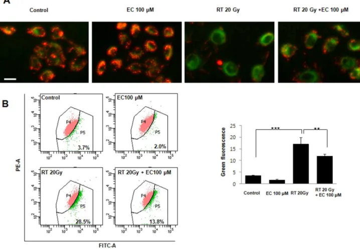
Figure 3. Effect of epicatechin on radiation-induced mitochondrial membrane potential in the HaCaT cells.(A) Cells were irradiated or not (20 Gy) in the absence or presence of EC (100mM), and stained with JC-1 (red fluorescence). Increase of green fluorescence in irradiated cells suggest loss of MMP. (B) The MMP change was objectively measured using the flow cytometry FACScan. The data represent the mean6SD of three independent experiments. **p,0.01, ***p,0.001. Scale bar = 50mm.
number: AMC115). The rats were randomly divided into four groups (n = 8 per group). The control group did not receive any radiation or EC. The EC alone group (EC group) was treated with EC (Sigma-Aldrich). The radiation+epicatechin group (RT+EC group) was treated with EC after irradiation. The radiation alone group (RT group) received radiation and no further treatment. The rats in the RT+EC and RT groups were sedated using an
intraperitoneal (IP) injection of 3.125 mg/kg tiletamine,
3.125 mg/kg zolazepam, and 11.5 mg/kg xylazine hydrochloride. The rats were placed and restrained in the prone position on an acryl plate. Radiation was restricted to the oral cavity to spare the rest of the body (superior border of radiation: transverse line between lower eyelid and lower margin earlobe; posterior border of radiation: imaginary vertical line of earlobe). A single 30 Gy dose was delivered by opposed photon beams at a rate of 2 Gy/ min bilaterally at a distance of 100 cm from the source to the axis
using the LINAC, 6MV (21EX; Varian). Tissue-equivalent material was placed on the irradiated regions to ensure a uniform distribution of the prescribed dose. Rats in the RT+EC group received oral treatment with EC using a feeding device each day at
9:00 AM, 1:00 PM, and 6:00 PM (2 mM, 100mL/dose, total
300mL/day) for 23 days after irradiation.
Euthanasia of rats was expected if rats demonstrated the conditions listed below, whether the animal had been manipulated or not. (1) Weight loss: loss of 20–25%. (2) Inappetance: complete anorexia for 24 hours and partial anorexia (less than 50% of caloric requirement) for 3 days. (3) Weakness/inability to obtain feed or water: Inability or extreme reluctance to stand which persists for 24 hours, assuming that the rat has recovered from anesthesia. (4) Moribund state: measured by a lack of sustained purposeful response to gentle stimuli (example of purposeful
Figure 4. Effect of epicatechin on radiation-induced intracellular ROS generation in HaCaT cells and radiation-induced NOX3 expression in rat oral mucosa.(A) Intracellular ROS generation was measured in HaCaT cells treated with 20 Gy of radiation in the presence or absence of EC (200mM). The level of intracellular ROS was measured by FACScan flow cytometry using the peroxide sensitive fluorescent probe, DCFDA. (B) The results (mean6SD) were calculated as a percent of the control group (not exposed to radiation). The data represent the mean6SD of five independent experiments. **p,0.01. (C) Immunohistochemistry of NOX-3 in control, radiation alone, EC alone and radiation plus EC treated rat oral mucosa sections from each experimental group were stained with anti-NOX-3 antibody. Scale bar: 100mm.
doi:10.1371/journal.pone.0069151.g004
response- weak attempt to get up; if animal is on its side, attempts should be asymmetrical in nature).
Assessment of Radiation Damage in Rats
All animals were monitored daily to examine status of the oral cavity, amount of oral intake, weight, and survival. To evaluate the histopathologic changes in the tongue and buccal mucosa, one rat in each group was sacrificed on the 4th and 9th day after irradiation, respectively. Mucosa of the tongue and buccal mucosa was harvested from rats that died during the experiment and sacrificed at end of the experiment. The tongue and both buccal mucosa were exposed and photographed. Tissue samples of tongue and buccal mucosa were fixed in 10% buffered formalin and embedded in paraffin blocks. Each paraffin-embedded specimen was sectioned at a thickness of 5mm. The paraffin was
removed by immersion in graded series of ethanol (from 100 to 70%), and each section was then stained with a hematoxylin-eosin solution and observed with an Olympus BX-51 microscope (Olympus, Tokyo, Japan). We also analyzed TUNEL staining as described below.
TUNEL Staining in Rats
Apoptosis of oral mucosa was determined by the TUNEL method using an in situ detection kit (TACS 2 TdT DAB kit; Trevigen, Gaithersburg, MD, USA) according to the manufac-turer’s instructions. The tissue slides were then immersed in quenching solution and 16terminal deoxynucleotidyl transferase
(TdT) labeling buffer and incubated in brominated nucleotide (BrdU)-labeling reaction mix. Visualization of apoptotic cells was achieved by incubating the sections in diaminobenzidine/hydro-gen peroxide solution. Finally, the slides were counterstained with
methyl blue solution, and the stained slides were observed and photographed using light microscopy.
Immunohistochemical Analysis in Rats
For immunohistochemistry study, DAKO immunohistochemis-try kit (DAKO LSAB Universal K680, Carpinteria, CA, USA) was used according to the manufacturer’s instructions. Specimen sections of 5mm were deparaffinized in xylene and rehydrated
through a gradual concentrations of ethanol. The endogenous peroxidase was blocked with 3% hydrogen peroxide for 5 min at room temperature following PBS washes. Nonspecific binding was blocked with 1% bovine serum albumin for 1 h. Anti-NOX-3 antibody (Sigma Aldrich) was added to the slides, and incubation proceeded for 1 h. After repeated washes with PBS, the sections were incubated with biotinylated secondary rabbit antibody for 1 h containing horseradish peroxidase. Finally, the sections were stained in a freshly prepared substrate solution (3 mg of 3-amino-9-ethylcarbazole in 10 ml of sodium acetate buffer pH 4.9, 500mL of dimethylformamide, 0.03% hydrogen peroxide) for 10 min.
Statistical Analyses
The Student’s t test and one-way ANOVA were used for the statistical analyses of thein vitrodata.In vivostudy, all values were expressed as mean6 standard deviation and statistical analysis was performed by the Kruskall-Wallis Test and the Mann-Whitney U Test. The survival rates were evaluated using the Kaplan Meier method (SPSS, version 17, Chicago, IL, USA). AP -value,0.05 was regarded as statistically significant.
Results
Pretreatment with EC Increased Viability of Irradiated HaCaT Cells
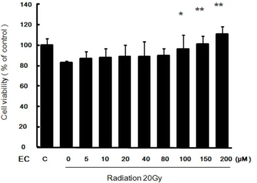
As shown in Figure 1, radiation decreased the viability of the HaCaT cells, but EC significantly protected the HaCaT cells from radiation induced cytotoxicity in a dose-dependent manner.
EC Protected HaCaT Cells Against Radiation-induced Apoptosis
Radiation induces HaCaT cell death by apoptosis, so the TUNEL assay and Hoechst 33258 staining were performed to determine if that could be prevented by treatment with 100mM EC. As shown in Figure 2, EC treatment decreased TUNEL-positive cells. Annexin V-FITC and PI double staining was used to analyze the percentage of apoptotic cells irradiated in the absence
or presence of EC (Figs. 2B). Radiation plus EC (100mM)
significantly decreased the number of total apoptotic cells compared to radiation (20 Gy) only.
EC Inhibited Radiation-induced Changes in the Mitochondrial Membrane Potential
As shown in Figure 3, in the control cells, a high MMP was maintained, as indicated by the predominantly red fluorescence of the JC-1 dye. However, radiation treatment decreased the green cell fluorescence indicating a loss of MMP and mitochondrial damage. Treatment with EC inhibited radiation-induced changes in the MMP and cell morphology (Fig. 3).
EC Inhibited Intracellular ROS Generated by Radiation
We examined the effects of EC on radiation-induced ROS generation. Irradiation significantly increased the intracellular ROS generation. EC significantly reduced radiation-induced the
Figure 5. Inhibition of radiation-induced phosphorylation of MAPKs and caspase-3 dependent pathway by epicatechin treatment in HaCaT cells.The cells were pretreated with 200mM of EC followed by the addition of 20 Gy radiation. Immunoblot of treated HEI-OC1 cells stained with antibodies against p38, JNK, ERK, and cleaved caspase-3.
generation of intracellular ROS generation (Fig. 4A, B), suggesting that increased ROS generation may be involved in radiation-induced HaCaT cell apoptosis. To demonstrate the effects of EC
on radiation-induced ROS generation in vivo rat model, we
performed an immunohistochemistry study for NOX. In immu-nohistochemistry study, radiation markedly induced the expression
Figure 6. Effect of epicatechin on oral intake and body weight of irradiated rats.(A) Effect of EC on rat oral intake. On the 1stday after irradiation, the oral intake of irradiated rats began to decrease. The oral intake in the rats in the RT+EC group started to increase on the 9thday after irradiation and was markedly increased on the 11thday. The mean oral intake of the RT
+EC group was similar to that of the control group between the 11thand 23rdday (P =0.09, Mann-Whitney U test). (B) Effect of EC on rat body weight after irradiation. On 2ndday after irradiation, the body weight of irradiated rats began to decrease. The RT+EC group displayed a statistically significant lower weight loss than the RT group on the 5th, 6th and 7thday (*p
,0.05). The rats in the RT+EC group started to gain weight on the 10thday and reached their original weight on the 17thday. Weight gain velocity was not significantly different as compared with the control group (p =0.75).
doi:10.1371/journal.pone.0069151.g006
of NOX-3 protein in the oral mucosa of rat. However, NOX-3 protein was significantly decreased in the oral tongue and buccal mucosa in rat treated with both EC plus radiation compared to rat treated with radiation alone. (Fig. 4C).
Inhibition of MAPK Activity by Epicatechin Rescued the HaCaT Cells from Radiation Induced Cytotoxicity
To clarify the associated mechanism regarding the activity of radiation and EC, we evaluated the radiation and EC-induced changes on protein expression of the MAPK pathway and the apoptotic signal pathway. The effects of EC treatment on the expression of p38, JNK, and cleaved caspase-3 were investigated by Western blot analysis. A representative Western blot is shown in Figure 5. The results obtained confirmed increased expression of p-JNK, p38, and cleaved caspase-3 after radiation treatment. These results suggest that EC blocked radiation-induced apoptosis via down-regulation of JNK and p-38 in the HaCaT cells.
EC Restored the Oral Intake, Weight Loss, and Survival Rate of Irradiated Rats
On the 1stday after irradiation, the oral intake of the irradiated
rats began to decrease and declined until day 8. The mean oral intake between irradiated groups was not significantly different until day 10. The oral intake of the rats in the RT+EC group started to increase on day 9 and was markedly increased on day 11. The mean oral intake of the RT+EC group was not statistically significantly different to that of the control group between 11thand 23rdday. The mean oral intake of the control group was similar to that of the EC group during the experimental period (Fig. 6A).
The body weight of irradiated rats started to decrease on post-irradiation 2ndday and significantly decreased between 2ndand 9th day as compared with the control and EC groups. The rats in the RT group did not gain weight before death. The RT+EC group showed a statistically significant less weight loss than the RT group on 5th, 6thand 7thdays (*p,0.05). The rats in the RT+EC group began to gain weight on 10thday, reached their original weight on 17th day, and continuously gained thereafter. The weight gain velocity of the RT+EC group was not significantly different as compared with the control and EC groups (Fig. 6B).
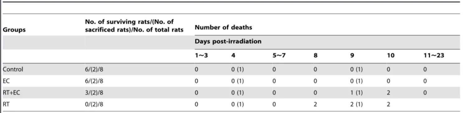
In the RT group, two rats died on 8thday, two rats on 9thday, and the other on 10thday. In the RT+EC group, one rat died on 9thday, and two rats died on 10thday. The survival rates of the control group, EC group, RT+EC group, and RT group on day 23 were 100% (6/6), 100% (6/6), 50% (3/6), and 0% (0/6), respectively (Table 1).
EC Inhibited Histopathologic Changes of Oral Mucosa by Irradiation in Rats
One rat in each group was sacrificed for histopathologic evaluation of the oral mucosa on post-irradiation 4thand 9thday. All remaining rats were examined after death during the experiment or, if still alive on 23rd day, were sacrificed and analyzed. In the control and EC groups, histopathologic findings of oral mucosa on 4th, 9th, and 23rddays were similar. On 4thday after irradiation, oral mucositis was not grossly evident, except for ulceration of the tongue tip in the RT group. However, histopathologically, the buccal mucosa of the RT group showed mild submucosal edema with inflammatory cell infiltration and tongue tip ulceration. High magnification views showed partial nucleopleomorphism and extracellular edema in the buccal mucosa of the RT group (Fig. 7A). However, in other groups, we could not find typical abnormal findings. On post-irradiation 9thday, grossly intact anterior buccal mucosa and mild ulceration in the posterior buccal mucosa and tongue were detected in the rats of the RT+EC group as compared to the RT group, which showed severe ulceration and necrosis in the whole tongue and oral mucosa. Histopathologically, the buccal mucosa of the
RT+EC group showed ulceration with epithelial change in
adjacent mucosa, and the buccal mucosa of the RT group was severely ulcerated with necrotic inflammatory exudates (Fig. 7B). On 23rdday after irradiation, the tongue and buccal mucosa of the RT+EC group were similar to those of the control group. Healed mucosa was observed in hematoxylin-eosin stained tissue and little difference was seen between the RT+EC group and the non-irradiated control and EC groups (Fig. 7C).
EC Reduced Radiation-induced Apoptosis in Rat Oral Mucosa
To determine the nature of cell death, TUNEL staining was carried out on all four groups. TUNEL-positive cells were not observed in any of the oral mucosa from rats in the non-irradiated control and EC groups. On post-irradiation 4th day in the RT group, weak TUNEL-positive cells were evident around the ulcerated region of the tongue tip (Fig. 8A). In contrast, the oral mucosa from radiation-treated rats showed marked TUNEL-positive labeling of a variety of cell types within the irradiated mucosa on post-irradiation 9th day (Fig. 8B). However, TUNEL analysis revealed the reduction of TUNEL-positive cells by EC treatment. On post-irradiation 23rdday, TUNEL findings of the RT+EC group were similar to those of the control and EC groups (Fig. 8C).
Table 1.Survival rate and time of death of each group of rats.
Groups
No. of surviving rats/(No. of
sacrificed rats)/No. of total rats Number of deaths
Days post-irradiation
1,3 4 5,7 8 9 10 11,23
Control 6/(2)/8 0 0 (1) 0 0 0 (1) 0 0
EC 6/(2)/8 0 0 (1) 0 0 0 (1) 0 0
RT+EC 3/(2)/8 0 0 (1) 0 0 1 (1) 2 0
RT 0/(2)/8 0 0 (1) 0 2 2 (1) 2
Abbreviations: EC group = epicatechin alone group, RT+EC group = radiation+epicatechin group, RT group = radiation alone group, (): One rat each in all groups was sacrificed for histopathologic evaluation on day 4 and 9, respectively.
Figure 7. Effect of epicatechin on histopathologic findings of irradiated rats.(A) Gross photographic images and histopathologic images of oral mucosa on the 4thday after irradiation. Each paraffin-embedded specimen was sectioned at a thickness of 5mm. (H&E staining: x100 and x400). Tongue tip ulceration (arrow). (B) The gross photographic images and histopathologic images (H&E staining: x100 and x400) of oral mucosa on the 9thday after irradiation. (C) The gross photographic images and histopathologic images (H&E staining: x100 and x400) of oral mucosa on the 23rdday after irradiation.
doi:10.1371/journal.pone.0069151.g007
Discussion
The exact pathophysiology of radiation-induced oral mucositis is not been fully known. The onset of oral mucositis is a biologically complex process that has only been partially elucidated at the molecular level [23]. Radiotherapy and chemotherapy are known to lead to the generation of ROS that activate several signaling pathways in the submucosa and epithelium [23,24]. These activated pathways lead, in turn, to abrogate epithelial cell renewal, spur apoptosis, increase atrophy, and trigger ulcer formation. Based on this ROS-dependent mechanism of radiation-induced oral mucositis, various antioxi-dant strategies can be considered. [25] We speculated that EC, which is well-known anti-oxidative phytochemical, could be a possible solution for ameliorating radiation induced oral mucositis. We previously reported about the otoprotective effect of EC against radiation and cisplatin through the reduction of ROS generation [26–28]. In the present study, we intended to investigate the protective effect of EC against radiation-induced cellular damage in vitro in the HaCaT cells and in vivo in a rat model.
In this investigation, we revealed that radiation-induced apoptosis of the HaCaT cells was dose-dependently prevented by EC treatment. EC reduced the intracellular ROS generation which induced by irradiation. These results indicate that radiation-induced cellular toxicity is closely connected with intracellular ROS generation and EC may ameliorate this toxicity by diminishing oxidative cellular damage. Regarding the associated mechanism, JNK and p38 in MAPKs were activated in irradiated cells. In addition, we demonstrated the possibility of EC to inhibit radiation-induced oral mucositis in a rat model. Radiation-induced oral mucositis decreased oral intake, weight, and survival rate, but these were significantly restored by oral administration of EC. Histopathologic changes and TUNEL positive cells induced by irradiation were significantly decreased in the oral mucosa of the EC-treated rats.
Green tea is a worldwide popular beverage and its main ingredients, green tea polyphenols have been reported to exert anticancer, antiinflammatory, antibacterial and antioxidative properties [29,30]. Tea polyphenols may exhibit antioxidant effects through direct radical scavenging, down regulation of radical production, elimination of radical precursors, metal chelation, or elevation of endogenous antioxidants [31]. Further-more, a growing body of evidence suggests that polyphenols regulate cell signaling and affect disease progress [31]. One of the important molecular mechanisms associated with the cellular protective activities of polyphenols are MAPK kinases. Epigallo-catechin gallate and EC were reported to reduce the phosphor-ylation of MEK1/2, ERK1/2, and c-Jun [32,33]. Schroeteret al.
reported that EC and its metabolite 39-O-methyl-(2)-EC activated ERK in neuronal cells, as well as the ERK downstream target CREB [34]. In our previous investigation, EC significantly reduced the radiation-induced expression of p-JNK, p-ERK, in cochlear organs of Corti-derived cell lines, HEI-OC1 [28]. EC also protected the auditory organ by attenuating cisplatin-induced ototoxicity through inhibition of ERK [27]. In this investigation, EC treatment significantly reduced the ROS generation and expression of p-JNK and p38 increased by irradiation. The protective effect of EC on radiation-induced cytotoxicity might be related to the inhibition of MAPK expression.
Figure 8. Protective effect of epicatechin against radiation in rat oral mucosa, as indicated by TUNEL staining for apoptosis. (A) The 4thday after irradiation, (B) The 9thday after irradiation, (C) The 23rd day after irradiation. TUNEL staining of rat mucosa revealed EC
treatment significantly decreased radiation-induced TUNEL-positive cells (arrow). Scale bar = 50mm.
In the present study, staining with the fluorescence dye JC-1 revealed that radiation induced the loss of MMP, which is a marker of mitochondrial membrane integrity. Mitochondria are known to be a producer of ROS as well as the primary target of ROS induced cytotoxicity. Therefore, there is an inextricable connection between mitochondrial damage and intracellular ROS generation, and consequent oxidative stress [35]. Regarding the mitochondrial protection efficacy of EC, Princeet alreported that pretreatment with EC could prevent cardiac mitochondrial damage in myocardial infarction [36]. Our results suggested the possible connection between the radio-protective effect of EC and mitochondrial protection.
In the present in vivo study using a rat model, EC treatment significantly improved the clinical features and histologic findings caused by irradiation. TUNEL staining of rat oral mucosa revealed that EC treatment significantly decreased radiation-induced TUNEL-positive apoptotic cells. The mean oral intake, body weight, and histopathologic findings of the EC group were similar to that of the control group, and none of the EC group died during the experiment. EC showed a considerable radio-protective effect on oral mucositis. In irradiated non–EC-treated rats on the 9th Day, the epithelial layer was severely disrupted, and thick pseudomembrane was formed with dense inflammatory cell infiltration. EC treatment ameliorated the distortion of the epithelial cell layer and reduced the associated inflammation. These histologic findings correlated with the increase of oral intake and body weight of EC-treated rats started on the 9th day. Improvement of oral mucositis may have contributed to the restoration of the oral intake and body weight, eventually leading to the survival of irradiated rats. All non-EC treated rats were not able to survive over 10 days from irradiation; in contrast, 50% of EC treated group survived through study period.
However, the presentin vivostudy was designed as a preliminary animal experiment for evaluation of EC in radiation-induced oral mucositis and an intermediate step toward large-scale animal studies using multi-fraction schemes of irradiation. Even though this study followed previous studies which used large single radiation dose scheme, [15,37] the small number of rats and the use of a large single dose could be potential limitations. A large
single dose using this study may induce tissue changes qualitatively different from that during fractionated radiotherapy in the clinical setting. Therefore, a well-designed animal study using multi-fraction irradiation animal study is warranted.
The advantage of green tea application as a radiation mitigator for oral mucositis is its chemopreventive properties and its well-proven safety. For clinical application of any agent as a radiation mitigator, absolute certainty about the protection factors for tumor and normal tissues is compulsory to avoid unpredictable disease progression or complications. Because EC is a dietary component of green tea which has been ingested by humans for a long time, the ingredients should be safe and non-toxic. The application of tea, as a cancer chemopreventive agent has been thoroughly investigated in the last two centuries. Not only epidemiological, but also laboratory studies have proven the protective effect of tea consumption in the development of certain cancer types [38–40]. The molecular mechanisms related to the chemopreventive properties of green tea component are known to be appreciated to the modulating cell signaling pathways such as NF-kB, or MAPK kinases [31]. By regulating signaling pathways, polyphe-nols could promote cell death and induce apoptosis in premalig-nant or maligpremalig-nant cells, consequently leading to the inhibition of cancer development or progression.
In conclusion, we demonstrated that EC protected the HaCaT cells in vitro from radiation by inhibiting ROS generation, maintaining the mitochondrial integrity, and blocking the MAPKs activation. Moreover, EC is an effective means of reducing oral mucosal side effects and facilitating wound healing after radiation exposure in the oral cavity of rats. Although larger numbers of animals and clinically relevant fractionation schemes are necessary to confirm the effect of EC, these results suggest the possibility of EC to inhibit radiation-induced oral mucositis, a common complication in radiotherapy of head and neck cancers.
Author Contributions
Conceived and designed the experiments: CHK. Performed the experi-ments: HAS SUK. Analyzed the data: JHK YTO. Contributed reagents/ materials/analysis tools: KHP. Wrote the paper: YSS.
References
1. Sankaranarayanan R, Masuyer E, Swaminathan R, Ferlay J, Whelan S (1998) Head and neck cancer: a global perspective on epidemiology and prognosis. Anticancer Res 18: 4779–4786.
2. Pignon JP, Bourhis J, Domenge C, Designe L (2000) Chemotherapy added to locoregional treatment for head and neck squamous-cell carcinoma: three meta-analyses of updated individual data. MACH-NC Collaborative Group. Meta-Analysis of Chemotherapy on Head and Neck Cancer. Lancet 355: 949–955. 3. Epstein JB, Gorsky M, Guglietta A, Le N, Sonis ST (2000) The correlation
between epidermal growth factor levels in saliva and the severity of oral mucositis during oropharyngeal radiation therapy. Cancer 89: 2258–2265. 4. Epstein JB, Robertson M, Emerton S, Phillips N, Stevenson-Moore P (2001)
Quality of life and oral function in patients treated with radiation therapy for head and neck cancer. Head Neck 23: 389–398.
5. Plevova P (1999) Prevention and treatment of chemotherapy- and radiotherapy-induced oral mucositis: a review. Oral Oncol 35: 453–470.
6. Antunes HS, de Azevedo AM, da Silva Bouzas LF, Adao CA, Pinheiro CT, et al. (2007) Low-power laser in the prevention of induced oral mucositis in bone marrow transplantation patients: a randomized trial. Blood 109: 2250–2255. 7. Leborgne JH, Leborgne F, Zubizarreta E, Ortega B, Mezzera J (1998)
Corticosteroids and radiation mucositis in head and neck cancer. A double-blind placebo-controlled randomized trial. Radiother Oncol 47: 145–148. 8. Epstein JB, Silverman S, Jr., Paggiarino DA, Crockett S, Schubert MM, et al.
(2001) Benzydamine HCl for prophylaxis of radiation-induced oral mucositis: results from a multicenter, randomized, double-blind, placebo-controlled clinical trial. Cancer 92: 875–885.
9. Trotti A, Garden A, Warde P, Symonds P, Langer C, et al. (2004) A multinational, randomized phase III trial of iseganan HCl oral solution for reducing the severity of oral mucositis in patients receiving radiotherapy for head-and-neck malignancy. Int J Radiat Oncol Biol Phys 58: 674–681.
10. Brizel DM, Wasserman TH, Henke M, Strnad V, Rudat V, et al. (2000) Phase III randomized trial of amifostine as a radioprotector in head and neck cancer. J Clin Oncol 18: 3339–3345.
11. Rosenthal DI, Trotti A (2009) Strategies for managing radiation-induced mucositis in head and neck cancer. Semin Radiat Oncol 19: 29–34. 12. Rovirosa A, Ferre J, Biete A (1998) Granulocyte macrophage-colony-stimulating
factor mouthwashes heal oral ulcers during head and neck radiotherapy. Int J Radiat Oncol Biol Phys 41: 747–754.
13. Hille A, Rave-Frank M, Pradier O, Damm C, Dorr W, et al. (2003) Effect of keratinocyte growth factor on the proliferation, clonogenic capacity and colony size of human epithelial tumour cells in vitro. Int J Radiat Biol 79: 119–128. 14. Spielberger R, Stiff P, Bensinger W, Gentile T, Weisdorf D, et al. (2004)
Palifermin for oral mucositis after intensive therapy for hematologic cancers. N Engl J Med 351: 2590–2598.
15. Lee SW, Jung KI, Kim YW, Jung HD, Kim HS, et al. (2007) Effect of epidermal growth factor against radiotherapy-induced oral mucositis in rats. Int J Radiat Oncol Biol Phys 67: 1172–1178.
16. Wilkes JD (1998) Prevention and treatment of oral mucositis following cancer chemotherapy. Semin Oncol 25: 538–551.
17. Sonis ST, Lindquist L, Van Vugt A, Stewart AA, Stam K, et al. (1994) Prevention of chemotherapy-induced ulcerative mucositis by transforming growth factor beta 3. Cancer Res 54: 1135–1138.
18. Sheng-Tanner X, Bump EA, Hedley DW (1998) An oxidative stress-mediated death pathway in irradiated human leukemia cells mapped using multilaser flow cytometry. Radiat Res 150: 636–647.
19. Lalla RV, Peterson DE (2006) Treatment of mucositis, including new medications. Cancer J 12: 348–354.
20. Suntharalingam M, Jaboin J, Taylor R, Wolf J, Banglore M, et al. (2004) The evaluation of amifostine for mucosal protection in patients with advanced
regional squamous cell carcinomas of the head and neck (SCCHN) treated with concurrent weekly carboplatin, paclitaxel, and daily radiotherapy (RT). Semin Oncol 31: 2–7.
21. Schroeder P, Klotz LO, Sies H (2003) Amphiphilic properties of (-)-epicatechin and their significance for protection of cells against peroxynitrite. Biochem Biophys Res Commun 307: 69–73.
22. Shin YS, Song SJ, Kang SU, Hwang HS, Choi JW, et al. (2012) A novel synthetic compound, 3-amino-3-(4-fluoro-phenyl)-1H-quinoline-2,4-dione, in-hibits cisplatin-induced hearing loss by the suppression of reactive oxygen species: In vitro and in vivo study. Neuroscience 232C: 1–12.
23. Sonis ST (2002) The biologic role for nuclear factor-kappaB in disease and its potential involvement in mucosal injury associated with anti-neoplastic therapy. Crit Rev Oral Biol Med 13: 380–389.
24. Siomek A, Tujakowski J, Gackowski D, Rozalski R, Foksinski M, et al. (2006) Severe oxidatively damaged DNA after cisplatin treatment of cancer patients. Int J Cancer 119: 2228–2230.
25. Cho SA, Park JH, Seok SH, Juhn JH, Kim SJ, et al. (2006) Effect of granulocyte macrophage-colony stimulating factor (GM-CSF) on 5-FU-induced ulcerative mucositis in hamster buccal pouches. Exp Toxicol Pathol 57: 321–328. 26. Kim CH, Kang SU, Pyun J, Lee MH, Hwang HS, et al. (2008) Epicatechin
protects auditory cells against cisplatin-induced death. Apoptosis 13: 1184–1194. 27. Lee JS, Kang SU, Hwang HS, Pyun JH, Choung YH, et al. (2010) Epicatechin protects the auditory organ by attenuating cisplatin-induced ototoxicity through inhibition of ERK. Toxicol Lett 199: 308–316.
28. Pyun JH, Kang SU, Hwang HS, Oh YT, Kang SH, et al. (2011) Epicatechin inhibits radiation-induced auditory cell death by suppression of reactive oxygen species generation. Neuroscience 199: 410–420.
29. Guo Q, Zhao B, Li M, Shen S, Xin W (1996) Studies on protective mechanisms of four components of green tea polyphenols against lipid peroxidation in synaptosomes. Biochim Biophys Acta 1304: 210–222.
30. Higdon JV, Frei B (2003) Tea catechins and polyphenols: health effects, metabolism, and antioxidant functions. Crit Rev Food Sci Nutr 43: 89–143.
31. de Mejia EG, Ramirez-Mares MV, Puangpraphant S (2009) Bioactive components of tea: cancer, inflammation and behavior. Brain Behav Immun 23: 721–731.
32. Chung JY, Huang C, Meng X, Dong Z, Yang CS (1999) Inhibition of activator protein 1 activity and cell growth by purified green tea and black tea polyphenols in H-ras-transformed cells: structure-activity relationship and mechanisms involved. Cancer Res 59: 4610–4617.
33. Chung JY, Park JO, Phyu H, Dong Z, Yang CS (2001) Mechanisms of inhibition of the Ras-MAP kinase signaling pathway in 30.7b Ras 12 cells by tea polyphenols (-)-epigallocatechin-3-gallate and theaflavin-3,39-digallate. FASEB J 15: 2022–2024.
34. Schroeter H, Bahia P, Spencer JP, Sheppard O, Rattray M, et al. (2007) (-)Epicatechin stimulates ERK-dependent cyclic AMP response element activity and up-regulates GluR2 in cortical neurons. J Neurochem 101: 1596–1606. 35. Cai J, Yang J, Jones DP (1998) Mitochondrial control of apoptosis: the role of
cytochrome c. Biochim Biophys Acta 1366: 139–149.
36. Stanely Mainzen Prince P (2013) (2) Epicatechin attenuates mitochondrial damage by enhancing mitochondrial multi-marker enzymes, adenosine triphosphate and lowering calcium in isoproterenol induced myocardial infarcted rats. Food Chem Toxicol 53: 409–416.
37. Murphy CK, Fey EG, Watkins BA, Wong V, Rothstein D, et al. (2008) Efficacy of superoxide dismutase mimetic M40403 in attenuating radiation-induced oral mucositis in hamsters. Clin Cancer Res 14: 4292–4297.
38. Artali R, Beretta G, Morazzoni P, Bombardelli E, Meneghetti F (2009) Green tea catechins in chemoprevention of cancer: a molecular docking investigation into their interaction with glutathione S-transferase (GST P1–1). J Enzyme Inhib Med Chem 24: 287–295.
39. Mann CD, Neal CP, Garcea G, Manson MM, Dennison AR, et al. (2009) Phytochemicals as potential chemopreventive and chemotherapeutic agents in hepatocarcinogenesis. Eur J Cancer Prev 18: 13–25.