S
ILVIAC
RISTINAB
ATEZATIA
LVESRessonância magnética funcional para avaliação do incômodo
do zumbido em pacientes com audiometria normal
Tese apresentada ao Departamento de Oftalmologia e Otorrinolaringologia da Faculdade de Medicina da Universidade de São Paulo para obtenção do título de Doutor em Ciências
Área de concentração: Otorrinolaringologia Orientadora: Prof. Dra. Tanit Ganz Sanchez
São Paulo
Dados Internacionais de Catalogação na Publicação (CIP)
Preparada pela Biblioteca da
Faculdade de Medicina da Universidade de São Paulo
©reprodução autorizada pelo autor
Alves, Silvia Cristina Batezati
Ressonância magnética funcional para a avaliação do incômodo do zumbido em pacientes com audiometria normal / Silvia Cristina Batezati Alves. -- São Paulo, 2008.
Tese(doutorado)--Faculdade de Medicina da Universidade de São Paulo. Departamento de Oftalmologia e Otorrinolaringologia.
Área de concentração: Otorrinolaringologia. Orientadora: Tanit Ganz Sanchez.
Descritores: 1.Zumbido 2.Imagem por ressonância magnética 3.Neuroanatomia 4.Vias auditivas 5.Emoções 6.Cognição
Ao meu noivo Ricardo, por seus conselhos sensatos quando este trabalho parecia inviável; e, principalmente, por seu amor e apoio incondicionais, que transformaram minha vida em um objetivo e meus sentimentos em um amor infinito.
Aos meus pais, Maria Célia e Sebastião, pelo exemplo de honestidade, amor e apoio em todas as fases da minha vida.
Às minhas irmãs, Ana Lívia e Maria Luíza, pelas nossas semelhanças que vão além dos traços físicos.
À minha amiga e segunda mãe Alda, por me integrar como uma verdadeira filha em sua família e pelo seu amor livre de julgamentos.
Agradecimentos
A Deus, por me colocar diante de obstáculos proporcionais à minha capacidade de superá-los.
À minha terapeuta e amiga Deborah Malvásio, por me mostrar o caminho da paz espiritual.
Aos meus tios adotivos, Edison e Jarci Chiaramelli, pelas suas orações para meu sucesso pessoal e profissional.
À equipe que trabalhou ativamente neste projeto, Antônio Cesário Monteiro Cruz Júnior, Vivian de Souza Sacomano, Mariana Penteado Nucci da Silva e Marcos Akiba, sem os quais este trabalho teria sido impossível. Particularmente à Vivian, agradeço seu companheirismo nos momentos de dificuldades técnicas que enfrentamos e por ter se revelado uma verdadeira amiga; à Mariana, por seu esforço em viajar de Santos à São Paulo para auxiliar-me na coleta dos dados; ao Marcos, pelo seu trabalho de alto padrão na análise dos dados; e ao meu amigo Cesário, "Prof. Pardal", por sua ajuda em absolutamente todos os momentos da coleta de dados deste projeto. Este trabalho não existiria sem o auxílio e a genialidade de Cesário.
À Prof. Dra. Tanit Ganz Sanchez, pela idéia original deste projeto, pela oportunidade de trabalhar no seu grupo de pesquisa e por sua orientação.
está listado como co-orientador oficial deste projeto. Entretanto, deixo gravado aqui meu reconhecimento da sua participação fundamental como co-orientador e amigo. Ao Prof. Dr. Ricardo Ferreira Bento, chefe do Departamento de Otorrinolaringologia, por sua mente visionária e apoio à pesquisa.
À Prof. Dra. Claudia da Costa Leite, diretora do Setor de Ressonância Magnética, por seu estímulo à parceria entre os Departamentos de Radiologia e Otorrinolaringologia.
À Dra. Margaret Bradley, coordenadora do Centro para Estudos em Emoção e Atenção da Universidade da Flórida (EUA), pela sua valorosa contribuição e comentários sobre os estímulos sonoros IADS no momento do delineamento da metodologia deste trabalho.
Ao Dr. Alexandre Felippu Neto, pelo incentivo no início desta pesquisa.
Ao Dr. Geraldo Busatto Filho, por gentilmente permitir o uso das dependências e dos equipamentos do simulador de ressonância magnética no Departamento de Psiquiatria.
Aos integrantes do Grupo de Pesquisa em Zumbido, Dra. Adriana da Silva Gürtler, Dra. Renata de Almeida Marcondes, Dr. Márcio Ricardo de Barros Pio, Rosa Maria Rodrigues dos Santos, Maria Elisabete Bovino Pedalini, Dra. Márcia Akemi Kii, Carina Andréa Bezerra Rocha, Dra. Jeanne de Rosa Oiticica Ramalho, Dr. Ítalo Roberto Torres de Medeiros e Dra. Savya Cybelle Milhomen Rocha, pelo apoio na seleção dos pacientes, pelo câmbio de experiências em zumbido e pela amizade.
Aos membros integrantes da banca de qualificação, Dra. Cristiana Borges Pereira, Dra. Roseli Saraiva Moreira Bittar e Dr. Rubens Vuono Brito de Neto, pelos seus valorosos conselhos a este projeto.
À minha amiga Dra. Adriana da Silva Gürtler, pela sua energia irradiante e por sua ajuda imprescindível nos detalhes de encerramento desta tese.
Ao meu amigo Dr. Márcio Ricardo de Barros Pio, pelo seu carinho e apoio em todas as horas e por haver se tornado um verdadeiro irmão.
Às minhas colegas de trabalho, Dra. Cristiana Borges Pereira e Dra. Mariana Fávero, pelo seu carinho, interesse e incentivo à pesquisa no campo da ressonância magnética funcional.
À equipe do Núcleo de Neuroimagem Funcional do HCFMUSP, João Ricardo Sato, Dr. Ellison Fernando Cardoso, Dra. Paula Ricci Arantes, Maria Ângela M. Barreiros, pelos conselhos e apoio a este projeto. Particularmente, aos colegas de pós-graduação, Dr. Jorge Renner Cardoso de Almeida, Dra. Maria da Graça Moraes Martin e Dr. Carlos Toledo Cerqueira, por compartilharem a viabilidade do aparelho de ressonância magnética durante a coleta dos dados.
Aos colegas do laboratório “Functional Neuroimaging in Emotional Disorders” da Universidade de Pittsburgh (EUA), Dr. Jorge Renner Cardoso de Almeida, Dra. Amélia Versace e Dra. Dalila Akkal pela ajuda e indicação de bibliografia pertinente à discussão deste trabalho.
Magnética do HCFMUSP, pelo auxílio com os pacientes durante a coleta de dados. Aos enfermeiros e secretários do ambulatório de Otorrinolaringologia do HCFMUSP pela assistência no atendimento aos pacientes.
Às bibliotecárias da Faculdade de Medicina da USP, Valéria de Vilhena Lombardi, Sueli Campos Cardoso, Marinalva Souza Aragão, Fabíola Rizzo Sanchez e Gildete Oliveira Batista, pela significante ajuda e aconselhamento sobre os detalhes de encerramento desta tese.
Ao altruísmo de todos os voluntários sem zumbido que participaram deste projeto, bem como aos pacientes, sempre disponíveis e interessados em participar da pesquisa.
À Fundação de Amparo e Ensino à Pesquisa do Estado de São Paulo – FAPESP – pelo apoio financeiro a este projeto.
À Coordenação de Aperfeiçoamento de Pessoal de Nível Superior – CAPES – pela concessão de bolsa nível doutorado (Programa Demanda Social).
"Live as if you were died tomorrow,
Mahatma Gandhi
Esta tese está de acordo com as seguintes normas, em vigor no momento desta publicação:
Referências: adaptado de International Committee of Medical Journals Editors (Vancouver)
Universidade de São Paulo, Faculdade de Medicina, Serviço de Biblioteca e Documentação. Guia de apresentação de dissertações, teses e monografias. Elaborado por Annelise Carneiro da Cunha, Maria Julia de A. L. Freddi, Maria F. Crestana, Marinalva de Souza Aragão, Suely Campos Cardoso, Valéria Vilhena. 2a ed. São Paulo: Serviço de Biblioteca e Documentação; 2005.
Sumário
Lista de figuras Lista de tabelas Lista de abreviaturas Lista de símbolos Lista de siglas Resumo Summary ARTIGO
Normas de publicação na “Hearing Research” Comprovante de submissão do artigo
Artigo submetido à “Hearing Research”
1 INTRODUÇÃO ...01
2 OBJETIVOS ...06
3 REVISÃO DA LITERATURA ...07
3.1 Princípios da RMf ...07
3.2 RMf em zumbido ...09
4 MÉTODOS ...14
4.1 Casuística ...14
4.2 Paradigma ...17
4.3 Procedimento ...19
4.4 Parâmetros de aquisição da RM ... 20
4.5 Seqüência de pulso com ruído minimizado ...20
4.6 Processamento dos dados e estatística ... 21
5 RESULTADOS ...23
6 DISCUSSÃO ... 30
6.1 Aspectos metodológicos ...30
6.1.1 Seqüência de pulso com ruído acústico minimizado ...30
6.1.2 Inclusão de pacientes com audiometria normal ...33
6.1.3Avaliação do grau de incômodo do zumbido ...34
6.1.4 Exclusão de indivíduos com histórico de depressão ...35
6.2 Neuroimagem funcional em zumbido ...36
6.3 Modelos que explicam o incômodo do zumbido ...38
6.4 Cerebelo e cognição ...42
6.5 BA 22 e BA 44 ...44
6.6 Percepção emocional de um estímulo ...45
7 CONCLUSÕES ...49
9 GLOSSÁRIO ...65 10 ANEXOS ...66
Lista de figuras
Lista de tabelas
Lista de abreviaturas
AI alça inferior de ativação neural do modelo neurofisiológico
AS alça superior de ativação neural do modelo neurofisiológico
BA “Brodmann area”, área de Brodmann
GC grupo controle – participantes sem zumbido
GZ grupo zumbido – pacientes com zumbido
CRH curva de resposta hemodinâmica
CoA córtex auditivo
CoF córtex frontal
CoPF córtex pré-frontal
CoPFDL córtex pré-frontal dorsolateral
CoPFDM córtex pré-frontal dorsomedial
CoPFVL córtex pré-frontal ventrolateral
D à direita
DP desvio padrão
Dr. doutor (a)
E à esquerda
et al. e outros
ENIZ escala numérica de incômodo do zumbido
FA “flip angle”, ângulo de inclinação
fig. figura
GCA giro do cíngulo anterior
GFI giro frontal inferior
GTS giro temporal superior
Inc. “Incorporate”
ME média etária
NEX “numbers of excitations”, número de excitações
OD orelha direita
OE orelha esquerda
p. página
PET “positron emission tomography”, tomografia por emissão de pósitrons
Prof. professor (a)
RM ressonância magnética
RMf ressonância magnética funcional
SPECT “single photon emission computed tomography”, tomografia computadorizada por emissão de fóton único
SPRAM seqüência de pulso com ruído acústico minimizado
SNA sistema nervoso autônomo
SNC sistema nervoso central
tab. tabela
TE “time to echo”, tempo ao eco
THI “Tinnitus Handicap Inventory”, inventário de severidade do zumbido
TR “time to repeat”, tempo de repetição
TRA “tinnitus-related neural activity”, atividade neural relacionada ao
v. versão
Lista de símbolos
cm centímetro
dB NA decibel nível de audição
dB NPS decibel nível de pressão sonora
° grau
Hz Hertz
> maior que
± mais ou menos
< menor que
≤ menor ou igual que
min minuto
mm milímetro
ms milisegundo
mT/m militesla por metro
p nível de significância
® registrado
s segundo
t valor calculado no teste de “Student”
Lista de siglas
CAPES Coordenação de Aperfeiçoamento de Pessoal de Nível Superior
CAPPesq Comissão de Ética para Análise de Projetos de Pesquisa
EUA Estados Unidos da América
FMUSP Faculdade de Medicina da Universidade de São Paulo
Resumo
Alves SCB. Ressonância magnética funcional para avaliação do incômodo do zumbido em pacientes com audiometria normal [tese]. São Paulo: Faculdade de Medicina, Universidade de São Paulo; 2008. 69p.
Descritores: Zumbido, Imagem por ressonância magnética, Neuroanatomia, Vias auditivas, Emoções, Cognição.
Summary
Alves SCB. Analysis of tinnitus-related annoyance in patients with normal audiometry using functional magnetic resonance imaging [thesis]. 2008. São Paulo: “Faculdade de Medicina, Universidade de São Paulo”; 2008. 69p.
the unpleasant sounds (via insula), or a lack of regulation of individual affective reaction (via hippocampus).
ARTIGO
HEARING RESEARCH
Guide for Authors
1. Aims and scope
The aim of the journal is to provide a forum for papers concerned with basic auditory mechanisms. Emphasis is on experimental studies, but theoretical papers will also be considered. The editor of the journal is prepared to accept original research papers in the forum of the full-length papers, methodological papers, letters to Editor, and reviews. Papers submitted should deal with neurophysiology, ultra-structure, psychoacoustics and behavioral studies of hearing in animals, and models of auditory functions. Papers on comparative aspects of hearing in animals and man, and on effects of drugs and environment contaminants on hearing function will also be considered. Clinical papers will not be accepted unless they contribute to the understanding of normal hearing functions. Authors may suggest one or two reviewers from the Editorial Board for consideration by the Editor. The act of submitting a manuscript to the Journal carries with it the right to publish that paper and implies the transfer of the copyright from the author to the Publisher.
2. Types of Papers
# Research papers should deal with original research not previously published or being considered for publication elsewhere. These papers should provide a survey, evaluation and critical interpretation of recent research results and concepts in the fields covered by the Journal.
# Methodological papers should describe the methods for the recording, collection, and/or analysis of data relevant to understanding how the auditory system works. Manuscripts must describe the method in sufficient detail to enable others to implement or replicate the method or procedure. Manuscripts must demonstrate that the method actually works; and should be applied to real data.
# Letters to the Editor should be comments on or clarifications of articles published in the Journal.
# Announcements that the Editor considers to be of interest of readers of the Journal will also be considered for publication.
3. Submission procedure
You are strongly urged to submit your manuscript to Hearing Research electronically via ees.elsevier.com/heares
All correspondence, including notification of the Editor’s decision and requests, take place by e-mail.
If you be unable to submit via the web, please contact the Editorial Office at [email protected] for advice.
Formats. We accept most word processing formats, but Word, WordPerfect or LaTeX is preferred. The file extension should provide information about the digital format used. The text should be in single-column format. Keep the layout as simple as possible. Do not embed graphically designed equations or tables. Put them on a separate page, and note in the manuscript where the equations should appear.
General. Please write your text in good English (American and British usage is accepted but not a mixture of these). Use double spacing and wide (3cm) margins. Check spelling carefully.
The title should be concise and informative. Titles are often used in information-retrieval systems.
The Author’s affiliation address (where the actual work was done) should be listed below the Author’s names. Indicate all affiliations with a lower-case superscript letter immediately after each Author’s name. Provide the full postal address of each affiliation, including the country name and the e-mail address of each Author.
Corresponding author. Clearly indicate who is willing to handle correspondence at all stages of referring and publication and post-publication. Provide telephone and fax numbers (with country and area code), e-mail address and complete postal address.
Abstract. For full-length research and review papers a concise and factual abstract is required, not exceeding 200 words (on page 2 of the manuscript). Avoid references in the abstract.
Keywords. The abstract should be followed by 3 to 6 key words which will be used for indexing purposes.
Sections. Research papers should be divided into sections: Introduction, Material and methods, Results, Discussion, References.
If the work that is reported involves experimentation on animals has been approved by a specific university’s Animal Care and Use Committee, and that studies involving humans have been approved by the Institutional Review Board of the university where the study is performed. If these options do not apply, please contact the Editor.
If drugs or other substances that are not commercially available are used in the studies that are reported, information on how to obtain these substances must be included so that other researchers can replicate the studies.
Tables. Tables should contain only horizontal lines and each should have a descriptive heading (legend) above the table. Footnotes and explanations if applicable should be placed underneath each table.
Figures (see below).
Acknowledgments. Please acknowledgments, including information on grants received in a separate section before the reference list.
the publisher and place of publication are also needed. Periodicals (i), books (ii) and edited volumes (iii) should appear in the reference list as follows:
(i) Zhong, S.-X., Liu, Z.-H., 2004. Immuno-histochemical localization of the epithelial sodium channel in the rat inner ear. Hear. Res., 193(1-2) 1-8.
(ii) Moller, A.R., 2000. Hearing: Its Physiology and Pathophysiology, Academic Press, San Diego, CA.
(iii) Mills, J.H., Boettcher, F.A., Dubno, J.R., Schmiedt, R.A., 1996. Psychophysical and evoked response studies of aged subjects: masking by low noise. In: Axelson, A., Borchgrevink, H., Hamernick, R., Hellstrom et al. (Eds), Scientific Basis of Noise-Induced Hearing Loss. Thieme Medical Press, New York, pp. 181-192.
Abbreviations of journal titles should conform to the List of Serial Word Abbreviations, International Serials Data System, 20, rue Bachaumont, 75002, Paris, France. ISBN: 2-904938-02-8.
For futher information, please go to www.elsevier.com/locate/guidepublication.
5. Figures
A detailed guide to electronic artwork is available on our website (http://www.elsevier.com/artworkinstructions). Submitting your artwork in an electronic format helps us to produce your work to the vest possible standards, ensuring accuracy, clarity and a high level of detail. Number the illustrations according to their sequence in the text. Use a logical naming convention for your artwork files, and supply a separate list of titles and the software used.
Use one of the following fonts in your illustrations: Arial, Courier, Helvetica, Times, Symbol. Make sure you use uniform lettering and sizing of your original artwork. Save text in illustrations as graphics, or enclose the font. Please do not embed graphics in your word processing file. Regardless of the application used, when your electronic artwork is finalized, please save as or convert the images to one of the following formats: .eps, .tiff, .jpg.
Size. For Hearing Research the figures should be prepared for either single (84mm) or double column (178mm) width. Ensure that the resolution of the figures at their correct size in the journal will be at least 1000 dpi for line drawings and 500 dpi for half-tone and colour illustrations.
Free web colour illustrations. If, together with your accepted article, you submit usable colour figures, then Elsevier will ensure, at no additional charge, which these figures will appear in colour on the Web (e.g., Science Direct and other sites) regardless of whether or not these illustrations are reproduced in colour in the printed version. For colour reproduction in print, you will receive information regarding the costs from Elsevier after receipt of your accepted article. Please indicate your preference for colour in print, or on the Web only. Because of technical complications which can arrive by converting colour figures to gray-scale, for the printed version you should not opt for colour in print. In addition, please submit usable black and white versions of all the colour illustrations. Please make sure that artwork files are in an acceptable format (.tiff; .eps or MS office files) and with the correct resolution (500 dpi).
Figure captions. Ensure that each illustration has a caption. A caption should comprise a brief title and a description. Keep text in the illustrations themselves to a minimum, but explain all symbols and abbreviations used. Supply all captions together on a separate page.
6. Preparation of supplementary data
Supplementary files offer the author additional possibilities to publish supporting applications, movies, animation sequences, high-resolution images, background datasets, sound clips and more. Supplementary files supplied will be published online alongside the electronic version of your article in Elsevier web products, including Science Direct: http://www.sciencedirect.com. To ensure that your submitted material is directly usable, please provide data in one of our recommended file formats. Authors should submit the material in electronic format together with the article and supply a concise and descriptive caption for each file. For more detailed instructions please visit www.elsevier.com/authors. When supplementary files are applied, an additional ‘supplementary’ figure list should also be submitted. Any supplementary material that is not directly referred to from within the text of your manuscript should be referred to via use of a footnote to the article title. In addition, it is also recommended that a short description is provided for each supplementary file supplied. When published online, the descriptive texts will appear as captions alongside links to the relevant supplementary files, an example layout of online supplementary material can be viewed at http://authors.elsevier.com/ArtworkInstructions.html?dc=A149. Please note that any supplementary material supplied is subject to the normal peer review process.
7. Proofs
When your manuscript is received by the Publisher, it is considered to be in its final form. Proofs are not regarded as ‘drafts’. One set of page proofs in PDF format will be sent by e-mail to the corresponding Author to be checked for typesetting/editing errors. No changes in, or additions to, the accepted (and subsequently edited) manuscript will be allowed at this stage. Proofreading is solely your responsibility. A form with queries from the copy editor accompanies your proofs. Please answer all queries and make any corrections required.
The proofs should be checked carefully and returned by e-mail or (air) mail within 48 hours of receipt (also in case of corrections).
As only one set of corrections will be accepted; please ensure that you send as all your corrections to us together in one communication. If changes in meaning are made, the manuscript may have to be re-reviewed.
8. Offprints
A PDF file or offprint may be ordered by filling in and returning to the Publisher the order form that is sent to the corresponding author. Per contribution 25 free offprints will be made available should the PDF file not be required.
Submission of an article implies that the work described has not been published previously (except in the forma of an abstract or as part of a published lecture or academic thesis), which is not under consideration for publication elsewhere, which its publication is approved by all Authors and tacitly or explicitly by the responsible authorities where the work was carried out, and that, it will not be published elsewhere in the same form, in English or in any other language, without the written consent of the Publisher. Upon acceptance of an article, Authors will be asked to transfer copyright (for more information, please go to http://www.elsevier.com/authorsrights). This transfer will ensure the widest possible dissemination of information. A 2 paper letter will be e-mailed to the corresponding Author, confirming receipt of the manuscript. A form facilitating transfer of copy right will be provided. The corresponding Author must sign it on behalf of all authors and return both pages to the Publisher.
10. Information on accepted papers
From: Hearing Research [[email protected]] Sent: Monday, October 06, 2008 12:53 PM To: Batezati, Silvia
Subject: A manuscript number has been assigned: HEARES-D-08-00176
Ms. Ref. No.: HEARES-D-08-00176
Title: Analysis of the Psychophysiological Models of Tinnitus using Functional Magnetic Resonance Imaging
Hearing Research
Dear Dr. Batezati-Alves,
Your submission entitled "Analysis of the Psychophysiological Models of Tinnitus using Functional Magnetic Resonance Imaging" has been assigned the following manuscript number: HEARES-D-08-00176.
You may check the progress of your paper by logging on to the Elsevier Editorial System as an Author. The URL is http://ees.elsevier.com/heares/.
Your username is: batezatis Your password is: batezati-a7487
Thank you for submitting your work to this journal.
With kind regards,
Analysis of the Psychophysiological Models of Tinnitus using
Functional Magnetic Resonance Imaging
Silvia C. Batezati-Alves, M.D.1, 2, [email protected]
Tanit Ganz Sanchez, M.D., Ph.D.1, [email protected]
Maria Elisabete Bovino Pedalini, Ph.D.1, [email protected]
Antonio Cesário Cruz Jr., B.S.2, [email protected]
Vivian de Souza Sacomano, M.S.2, [email protected]
Mariana Penteado Nucci da Silva, B.S.2, [email protected]
Marcos Akiba, M.D.2, [email protected]
Edson Amaro Jr., M.D., Ph.D. 2, [email protected]
1
Department of Otolaryngology, University of São Paulo School of Medicine, Brazil (255 Dr. Enéas de Carvalho Aguiar Ave, 6th floor, São Paulo, SP, Zip code: 05403-010, Phone: 55-11-30852278)
2
Corresponding author:
Silvia C. Batezati-Alves, Address: 125 Meyran Ave, Room 216, Loeffler Building, Pittsburgh, PA, USA, 15232, Phone: 1-412-973-9669, Fax: 1-412-383-8336, E-mail: [email protected]
ABSTRACT
Key words: Tinnitus; Functional MRI; Auditory pathways; Emotions; Cognition
INTRODUCTION
Tinnitus is a conscious perception of sound in the ears without the presence of a corresponding external sound (McFadden, 1982; Jastreboff, 1990); thus, it may be described as a phantom auditory perception (Colles, 1998). According to the National Institute of Health, 44 million Americans suffer from tinnitus (Sanchez, 1997a; Hoffman and Reed, 2004). In 15 to 25% of the cases the tinnitus may significantly interfere with the quality of the patients’ life, affecting their sleeping pattern, mental concentration, emotional equilibrium, and attendance to social activities (Sanchez T et al, 1997a; Coelho et al., 2004; Lima, 2005; Lima et al., 2007).
For many years, both clinicians and researchers neglected the study of tinnitus. Great effort has been spent of late, to correlate the different psychoacoustic characteristics of tinnitus to specific treatments or to the level of its annoyance (Sanchez, 1998, 2003; Henry and Meikle, 2000), without reaching valid conclusions. Likewise, in the medical approach, we lack an effective treatment, mostly because we do not have sufficient information about the symptom.
Although they are largely accepted in clinical practice, they still lack experimental support and validation.
Hallam et al. (1984) were the first to describe the psychological model, suggesting that the pathological basis of tinnitus is “some neurophysiological disturbance in the auditory system at any point between periphery and cortex” (Mckenna, 2004). In turn, the neurophysiological model states that tinnitus is the result of a dynamic interaction of some centers of the central nervous system, including auditory and non-auditory pathways (Jastreboff, 1990). Both models suggest that tinnitus becomes problematic because it may be associated with something negative or unpleasant in the patient’s life (Jastreboff et al., 1994; Mckenna, 2004).
In turn, our approach involves using fMRI to study the emotion perception of acoustic stimuli in patients suffering from tinnitus. Emotional perception is of interest, as others have correlated tinnitus to a multidirectional psychosomatic interaction (Hallam et al., 1984; Mckenna, 2004). In addition, our study focuses on the processing of unpleasant acoustic stimuli, which addresses the Jastreboff’s model that suggests a correlation between the degree of annoyance of tinnitus and its negative relevance in the patients’ life (Jastreboff, 1990; Mckenna, 2004).
Due to the arguments presented, we aimed to analyze those neural systems involved in emotion perception of acoustic stimuli, especially unpleasant emotional valence sounds, using fMRI in subjects with and without tinnitus. Analyzing the auditory and non-auditory areas, and correlating these findings to the theories asserted by both current models of developing tinnitus, we seek to correlate potential findings to a higher level of annoyance in tinnitus patients.
MATERIAL AND METHODS
The present study was developed through collaboration of the Tinnitus Research Group of the Department of Otolaryngology, and the Functional Neuroimaging Laboratory (NIF) of the Department of Radiology from the University of São Paulo School of Medicine (Brazil).
Participants
non-pulsatile chronic tinnitus from diverse etiologies for at least 3 months (tinnitus group, TG). Volunteers for the control group (CG) were included in the study if they reported good health and no history of neurological, psychiatric, and/or otological diseases. Additional inclusion criteria for both groups comprised the following: normal pure-tone audiogram (thresholds equal to or below 25dB HL from 250 to 8000 Hz), confirmed by the GSI 61 Clinical Audiometer ® (Grason-Stadler, Inc., Madison, USA); right-handedness as assessed by the Edinburgh inventory (Olfield, 1971; Knecht et al., 2000; table 1); Beck depression inventory score below 20 points (Beck et al., 1961, 1998; Gorestein and Andrade., 1998; table 1); and formal education level equal to or above 11 years. Subjects were excluded from the study if they had claustrophobia; suspected or proven pregnancy; metallic prosthesis or pacemaker devices; or current use of psychotropic medication.
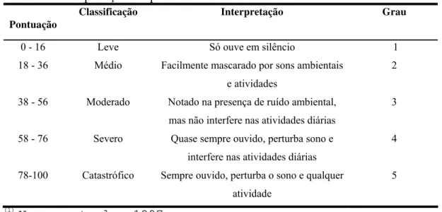
The TG comprised fifteen patients (3 men, 12 women), with ages ranging from 18 to 62 years (mean age 40.05±11.65 years; table 1), and the CG consisted of 20 volunteers (six men, 14 women), ranging in age from 27 to 59 years (mean age 40.20±7.85; table 1). Patients with tinnitus answered a Brazilian Portuguese validated questionnaire called the tinnitus handicap inventory (THI), which identifies the degree of annoyance that tinnitus is causing in the patient’s life (Newman et al., 1996; Ferreira et al., 2005; Schmidt et al., 2006). The THI score ranged from 14 to 54 points, classifying the patients as slightly handicapped (0-16 points, n=3), mildly handicapped (18-36 points, n=9), and moderately handicapped (38-56 points, n= 3; table 2).
The stimuli applied in the event-related paradigm consisted of sounds with validated emotional valences and arousal, as well as a visual analog scale that was modified for the fMRI environment. First, we selected 75 acoustic stimuli from the International Affective Digitized Sounds (IADS), which comprises an array of 116 naturalistic and daily sounds standardized to experimental investigations of emotion and attention (Bradley and Lang, 1999). The mean ratings for the pleasant, neutral, and unpleasant sounds were 7.02, 4.94, and 2.7, respectively, and mean arousal ratings were 5.16, 4.59, and 6.94 for pleasant, neutral, and unpleasant sounds, respectively, based on the IADS norms 1 (Bradley and Lang, 2000). Originally each IADS clip lasted six seconds. Due to the silent-event related (SER) design that required a stimulus’ duration of three seconds, we converted all selected IADS from six to three seconds using Cool Edit Software ® (Syntrillium Software, now Adobe Audition, Adobe Systems Inc., San Jose, California, USA). This adaptation of the original stimuli was not statistically different from the original ratings for valence, arousal and control dimensions of the 6-second sound in eleven normal volunteers inside a mock MRI scanner (unpublished data).
In addition, the Self Assessment Manikin (SAM) scale, developed by Peter Lang in 1980, was modified and applied in the present study. The original scale is comprised of three parts, each consisting of nine options, to classify an emotional stimulus, according to emotional valence, arousal and control dimensions of emotion
1
(Lang, 1980). Only the initial, middle and final figures of each part were chosen in this experiment because of time constraints of the SER fMRI paradigm design and button-type responses inside the MRI scanner. The modified SAM, according to the emotional valence dimension, classified the sound as pleasant (happy manikin), neutral (center’s manikin) and unpleasant (unhappy manikin) (figure 1A). The second part of the modified scale classified the arousal dimension of emotion as excited (left manikin), neutral (center’s manikin) and calm (right manikin) (figure 1B). The third part of the original scale that describes the control dimension was not used in the experiment (Bradley and Lang, 1994, 1999, 2000). The present work will analyze and describe specifically the brain activation during unpleasant sounds.
Study Design
In order to be familiar with the environment of the magnetic resonance machine, subjects heard 45 sounds selected from the IADS and practiced the tasks inside a mock scanner. This consisted of an original MRI scanner, without magnetic field, and equipped with exactly the same devices, including a recorded sound of the ambient noise from the MRI scanner used in the fMRI experiment. Next, all subjects listened to a new set of sounds selected from the IADS (30 acoustic stimuli) in the real fMRI experiment. A new set of sounds was applied to avoid a possible involvement of auditory memory, which would introduce a bias that may interfere with the classification of sounds.
MRI Scanning Parameters
The study was performed in a 1.5 T General Electric MR system ® (33mmT/m). Prior to the functional scanning, anatomical images were acquired to assist localization of activation using a T1-weighted images (FSGR/ TR: 15 ms/ TE: 5 ms/ FOV: 20 x 15 cm/ matrix: 256 x 192 voxels/ NEX: 1/ width: 1.5mm/ spacing: 0 mm/ flip angle: 25°), with the acquisition time of 5 min 26 s. The acquisition comprised twenty-four slices oriented according to the bi-commissural plane, providing full brain coverage. In addition, the functional image acquisition was performed by echo gradient sequence, Cartesian echoplanar BOLD (TR: 9 s/ TE: 40 ms/ FOV: 20 cm/ matrix: 64 x 64 voxels/ width: 5 mm/ spacing: 0.5 mm/ flip angle: 90°/silent time: 7 s) to produce a spatial resolution of 3.125 x 3.125 x 5 mm, and a temporal resolution of 2.25 s.
fMRI data were acquired using a specific technique to minimize the acoustic interference originated by the scanner noise, according to the silent event-related (SER) presentation scheme. The sounds were presented intermixed with the MRI scanner noise, minimizing significantly the scanner acoustic noise effect during the stimulus presentation and the brain activation detection. In addition, we used a variable jittered compressed acquisition (GRE-EPI, TR 2 s, silent gap 7 s; Amaro et al., 2001, 2006), in which the stimulus onset asynchronicities were varied according to a Poisson distribution, and the conditions were altered randomly. Each segment of the experiment took twelve minutes, and each individual was tested in three segments. The presentation order was balanced for all individuals. For each individual, sequences of three seconds for each condition of the same selected emotional valence (pleasant, neutral and unpleasant) were presented subsequently, thus providing two points in the hemodynamic response function (HRF) curve for each condition (Amaro et al., 2002; figure 2).
Data Processing
Pre-processing was performed for movement and spin history correction. The BOLD effect was modeled using Poisson functions, and statistical inference was based on the non-parametric approach with Talairach maps (Talairach and Tornoux, 1988) threshold at p<0.05 (XBAM_v3.4, Brain Activation Mapping, London, UK; Brammer and Bellmore, 1996). The authors considered the most activated area according to the smallest probability level, calculated individually.
RESULTS
Tinnitus Group Map Analysis for Unpleasant Sounds
The exposure to unpleasant sounds in the tinnitus group resulted in clusters found in the superior temporal gyrus (STG) bilaterally, right inferior-posterior temporal lobe, right middle temporal gyrus, insula bilaterally, primary visual cortex bilaterally, cerebellum bilaterally and left thalamus (table 3). The insula was more activated bilaterally, but predominantly on the left side, followed by the STG, also predominantly on the left (table 3). In addition, the thalamus and the cerebellum demonstrated high levels of BOLD activation bilaterally (table 3).
Control Group Map Analysis for Unpleasant Sounds
Presentation of unpleasant sounds was associated with positive BOLD effect in right STG, left inferior temporal gyrus, left infero-posterior temporal lobe, hippocampus bilaterally, right insula, cerebellum bilaterally, right primary visual cortex, right inferior frontal gyrus (ITG), right thalamus and right putamen (table 4). The left hippocampus was found in the center of the largest cluster detected during listening to unpleasant sounds, followed by the right primary visual (parastriate) cortex, and the right STG (table 4).
Analysis of Activation of the Limbic Structures
in this analysis (table 4). Also, the right insula was found activated for unpleasant sounds in the CG (table 4).
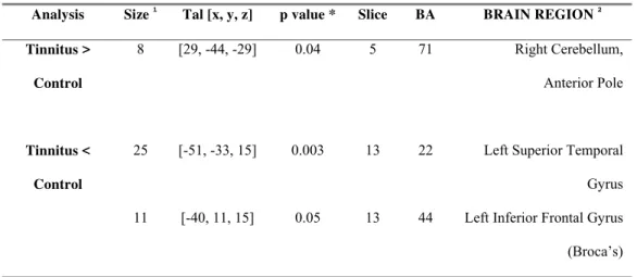
Comparative Analysis between Tinnitus and Healthy Patients
When both groups were compared, we found significantly higher activation of the cerebellum in the TG (table 5; figure 3). In addition, significantly low BOLD effect was found in the left STG, and in the left ITG for the patients with tinnitus (table 5; figure 3).
DISCUSSION
Methodological Aspects
Several methodological concerns had to be solved during the design phase of our study, especially related to the inclusion of patients with normal audiometry, and the survey of subjects using methods that measure the tinnitus-related annoyance.
et al., 2005). This functional finding in normal hearing patients with tinnitus may be correlated with the clinic, and the lower activation of the left STG may represent the first symptom of some alteration within auditory pathway.
In addition, the TG had a THI score ranging from 14 to 54 points, including patients who were slightly to moderately handicapped (table 2). We did not identify any patients with severe and catastrophic handicaps (58-76 and 78-100, respectively) during patient recruitment. Our findings suggested that the patients with tinnitus and normal hearing may have lower level of annoyance than those whom experience tinnitus associated with hearing loss. Others, using diverse tools to measure tinnitus-related annoyance, had already suggested that tinnitus in patients with normal hearing may be less annoyed by the symptom (Sanchez et al., 2005; Savastano, 2008). This may be responsible for the absence of expected brain activation and for a lower level of significance of findings at the limbic system. Even though the normal audiometry was an important inclusion criterion, that may have created a bias, and we selected more patients who suffer lower tinnitus annoyance.
Application of Functional Imaging in Tinnitus
The novel idea that tinnitus could be imaged was initially proposed by Sasaki et al. (1980), based on an animal model using autoradiography and a glucose tracer (14 C) 2-deoxyglucose. After his seminal work, other techniques for spatially mapping brain function, such as single photon computed tomography (SPECT), positron emission tomography (PET) and fMRI, have also been applied to individuals with tinnitus (Arnold et al., 1996; Cacace, 1999).
lateral areas of auditory cortex, associated with individuals presenting gaze-evoked tinnitus. Since then, fMRI has been applied to tinnitus in a few studies (Cacace et al. 1995, 1999; Cacace 1997, 1999; Levine et al. 1997, 1998; Guimarães et al., 1998; Melcher et al., 2000, 2002, 2005; Folmer et al. 2002; Sigalovsky et al., 2002; Domènech et al., 2005; Kovacs et al., 2005; Smits et al. 2005, 2007) searching for tinnitus-related neural activity (TRA) and abnormalities, using various paradigms.
Our approach differs slightly as our method was not designed to identify and isolate the TRA. We focused on studying the auditory processing of sounds that differs in emotional valence, based on the hypothesis that patients suffering from tinnitus present abnormalities in the emotion perception of unpleasant sounds. This hypothesis was developed based on observations by Hallam et al. (1984) who correlated tinnitus to a multidirectional psychosomatic interaction (psychological method of tinnitus), and Jastreboff (1990), who explained that the annoyance of tinnitus may be related to something negatively relevant in the patients’ life (neurophysiological model). In addition, we intended to study the neural network correlation between temporal, prefrontal, and limbic brain structures, which is suggested by such models, based on mostly experimental and clinical observations.
Models of Tinnitus Suffering
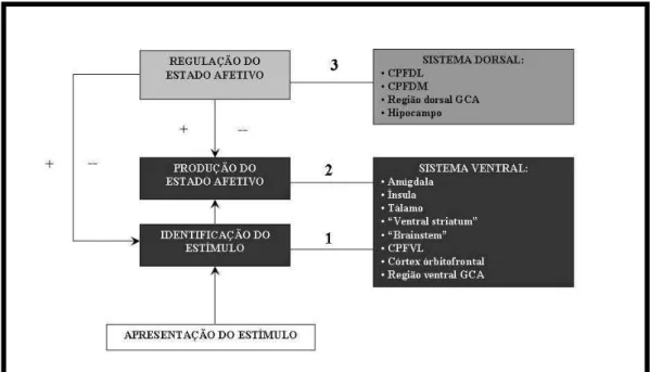
The neurophysiological model describes several feedback loops in the perception of tinnitus. The ‘lower loop’ describes a feedback system between the detection of tinnitus, limbic and ANS activity, and it is suggested that this system operates at a subconscious level without the need for conscious awareness. The ‘upper loop’ describes a feedback system again involving detection, limbic, and ANS activity but also the conscious evaluation of tinnitus (i.e. beliefs about the significance of the noise; Jastreboff, 1990). It was suggested by Jastreboff (1999) that the lower loop is dominant in the majority of tinnitus patients, and stresses the importance of unconscious conditioning over conscious cognitive processes (Mckenna, 2004).
In most of the his published work addressing the neurophysiological models, Jastreboff cited the participation of prefrontal and other cortical areas besides limbic system in the ‘upper loop’, but he did not specify which cortical areas. In a book chapter published in 2004, we find a more elaborate description of the upper loop, in which he cited that “when the tinnitus signal becomes highly significant, brain centers involved in attention are involved besides the limbic system. Others centers, such as the prefrontal cortex (tendency to perseveration, problems with task switching), and the cerebellum (e.g, multisensory integration, interaction with somatosensory system) must be considered in clinically significant tinnitus” (Jastreboff, 2004).
functions of the brain of particular relevance to cognition and behavior, helping the exacerbated perception of tinnitus. In addition, due to the fact that the lateral aspect of anterior lobe of the cerebellum showed the highest BOLD activity in tinnitus, it is possible to speculate that the patients with tinnitus are acquiring the unpleasant sounds more significantly than the healthy controls. The lateral cerebellum has been associated with the acquisition and discrimination of sensory information (Gao et al., 1996). Patients with tinnitus may be highly processing the unpleasant sounds: thus, they may have a link to unpleasant emotions, as suggested by the models.
Both models of developing tinnitus have much in common, but differ in the emphasis of the role of cognition and classical conditioning within the process of tinnitus perception, suffering and treatment. While the neurophysiological model includes a cognitive component (‘upper loop’), but asserts that this component is not critical, and that the ‘lower loop’ is dominant in most tinnitus patients, the psychological model gives a central role to the beliefs and their manipulation. Our findings related to the cerebellum suggest that cognition processes play an important role in the perception of the tinnitus. Besides the different emphasis on cognition between the models, both therapies pay attention to cognitive processes, indicating patient-oriented counseling as an important step. Others have demonstrated the importance of counseling for the treatment of tinnitus (Tyler, 2006).
information as it develops. Thus, the cerebellum, which is described as part of the ‘upper loop’, may be activated in early stages of tinnitus, and not only in highly annoyed patients, as our patients only comprised slight, mild and moderately handicapped patients. We also believe that the role of the prefrontal area in the ‘upper loop’ should be better defined, using paradigms to test those areas and functions that the model predicts to be impaired. The prefrontal area comprises a large extension anterior to the frontal lobes of the brain, lying in front of the motor and premotor areas, and comprises many important parts (such as orbitofrontal cortex, dorsolateral prefrontal cortex, and anterior cingulated cortex, and others) with distinct functions that have been studied extensively with functional imaging (Kandel et al., 1991). The prefrontal cortex includes the IFG that, in our study, showed significantly lower activation in tinnitus patients (p<0.05; figure 5, table 5). Thus, this aspect represents an open field for further research, using functional imaging modalities to study the role of attention, mediated by specific prefrontal areas, in tinnitus perception. Other paradigms may be more appropriate to study prefrontal activity. Some authors recently described the use of paradigms that elicit fear to activate prefrontal cortical areas (Mobbs et al., 2008).
activation in patients with tinnitus may be related to a lack of regulation of the affective response to unpleasant sounds. However, these areas did not arise when the groups were compared (p>0.05; table 5), showing that there is not a significant difference upon the activation of limbic structures between individuals with and without tinnitus.
In addition, functional neuroimaging studies have implicated the insula during the anticipation of an aversive stimulus (Phelps et al., 2001), suggesting its role in conveying the representation of aversive sensory information to the amygdala (Phillips et al., 2003a). In addition, we expected to find the amygdala activation in the TG, as it has been suggested that it is the primary area of activation involved in tinnitus-related suffering (Carpenter and Williams, 2006; Tyler, 2006). However, we did not find any activation of the amygdala in both groups.
The amygdala has been related to the processing of fear (Phillips et al., 2003a). Some authors hypothesized that fear conditioning plays a mayor role in sustaining the tinnitus perception (Levine, 1994). This concept is appealing because fear conditioning has strong underpinnings in psychological theory and is linked to a wide range of affective conditions known to have an effect on individuals with tinnitus (Hallam et al., 1984). The neurophysiologic model also considers fear, when cites that the tinnitus problems occurs in situations of dealing of with threat (Jastreboff, 1990; Mckenna, 2004). The absence of the amydala activation in both groups suggests that our paradigm may not the best one to study the perception of fear. A paradigm that elicits fear may yield information regarding the specific activation of the prefrontal area in tinnitus.
Traditionally, thecerebellumhas been considered to have a primary role ofin the coordination of voluntary movement, gait, posture, speech and motor functions. However, cumulative evidence shows that the cerebellum play a role in cognition and behavior (Rapoport et al., 2000). The cerebellum is largely connected to the brain, via the thalamus, to many brain areas relevant to cognition and behavior, including the dorsolateral prefrontal cortex, the medial frontal cortex, the parietal and superior temporal areas, the anterior cingulated, and the posterior hypothalamus (Middleton and Strick, 1997; Dolan, 1998). That connection between cerebellum and superior temporal area was identified in our TG, and may be described as part of the ‘upper loop’ cited in the Jastreboff’s model.
Caution must be taken in interpreting the role of the cerebellum in tinnitus. Experience warns that is highly unlikely that a single and specific area of the brain causes cognitive and emotional changes, as well as their disorders, since mental functions tend to be widely distributed in various brain circuits (Alexander et al., 1986; Rapoport, 2000). In functional neuroimaging studies, the cerebellar effects do not occur in isolation and are rarely the areas of the most robust change, suggesting that cortical areas mediate the cognitive changes implied by the cerebellum, which has an effect on integrating the information (Rapoport, 2000). Similarly, in our study the cerebellum did not show the most robust BOLD activity in either the control or tinnitus patients; however, the cerebellum arose as the most significant activated brain area when the two groups were compared. This suggests signalizing that the cerebellum represented an integrative area for cognition in tinnitus patients.
activity in the hippocampus in the tinnitus patients, which may also be related to the cognition, could also suggest that the cerebellum arose in patients with tinnitus to compensate the improper function of the hippocampus.
Considerations about BA 22 and BA 44
The STG [Brodmann area (BA) 22] contains several important structures of the brain, including: 1) primary auditory cortex (BA 41 and 42), which is the cortical region responsible for the sensation of sound; and 2) Wernicke's area (posterior BA 22), which is an important region for the processing of speech (Kandel et al., 1991).
We found a significantly low BOLD activity in the left STG in tinnitus patients (p<0.05, figure 5, table 5). Several authors described abnormal BOLD changes (increase or decrease) in the left primary auditory cortex in tinnitus patients during different PET and fMRI paradigms (Arnold et al., 1996; Oestreicher et al., 1999; Melcher et al., 2000), suggesting that TRA may be occurring in the BA 41 and 42. We agree with Melcher’s idea (2000) that “the tinnitus perception corresponds to abnormally increased neural activity that results in abnormally low sound-evoked activation (e.g., saturation or physiological masking)”. In other words, in our study the tinnitus patients may have high neural activity in the primary auditory cortex, and the unpleasant sounds evoked low activation of the BA 41 and 42 because the auditory pathways may be saturated by the TRA.
Broca’s area (ITG, BA 44) by a neural pathway called the arcuate fasciculus, and both areas are related mostly to language processing (Kandel et al., 1991). The BA 22p and BA44 also have connections to the primary auditory cortex (BA 41 and 42), especially the left side, because they play a role in the comprehension of the words (Kandel et al., 1991). Therefore, the left STG (BA 22) and the left ITG (BA 44) in our study showed a similar pattern of BOLD activity (i.e., significantly lower activation in tinnitus patients than the healthy controls). The effect on the ITG (BA 44) may be created by the activity in the STG (BA 22) because these areas very closed connected.
prefrontal activity in patients highly annoyed by tinnitus; however, the left inferior frontal gyrus in the patients with tinnitus showed lower neural activity than the healthy participants. We recommend additional fMRI studies involving paradigms that elicit fear to more properly unveil the prefrontal involvement in tinnitus-related annoyance.
ACKNOWLEDGMENTS
We thank the kind assistance of João Ricardo Sato for the statistical analysis. The State of São Paulo Research Foundation financially supported the present study (FAPESP; grant n° 2004/08579-9).
REFERENCES
1. Alexander, G.E., DeLong, M.R., Strick, P.L., 1986. Parallel organization of functionally segregated circuits linking basal ganglia and cortex. Annu. Rev. Neurosci. 9, 347-381.
2. Amaro, E.J., Williams, S.C.R., Shergill, S.S., FU, C.H.Y., Macsweeney, M., Picchioni, M.M., 2002. Acoustic noise and functional magnetic resonance imaging: current strategies and future prospects. J. Magn. Reson. Imaging. 16(2) 497-510.
3. Arnold, W., Bartenstein, P., Oestreicher, E., 1996. Focal metabolic activation in the predominant left auditory cortex in patients suffering from tinnitus: a PET study with (18F) deoxyglucose. J. Oto-Rhino-Laryngol. Relat. Spec. 58, 195-199. 4. Beck, A.T., Ward, C.H., Mendelson, M., Mock, J., Erbaugh, G., 1961. An
5. Beck, A.T., Steer, R.A., Garbin, M.G., 1998. Psychometric properties of the Beck depression inventory: twenty-five years of evaluation. Clin. Psychol. Rev. 8, 77-100.
6. Bradley, M.M, Lang, P.J., 1994. Measuring emotion: the self-assessment manikin and the semantic differential. J. Beh. Ther. Exp. Psychiatry. 25, 49-59. 7. Bradley, M.M., Lang, P.J., 1999. International affective digitized sounds (IADS):
stimuli, instruction manual and affective ratings. Technical Report B-2, The Center for Research in Psychophysiology, University of Florida, Gainesville. 8. Bradley, M.M., Lang, P.J., 2000. Affective reactions to acoustic stimuli.
Psychophysiology. 37, 204-215.
9. Brammer, M., Bulmore, E., 1996. XBAM_v.3.4, Brain Image Analysis Unit – BIAU, Center for Neuroimaging Sciences, Institute of Psychiatry, London (available on http:// www.iop. kcl.ac.uk /iop/ Departments/BioComp/BIAU/xbam_v3.shtml.
10.Cacace, A.T., Cousins, J., Moonen, C.W.T., 1995. In-vivo localization of phantom auditory perceptions during functional magnetic resonance imaging of the human brain. In: Proceedings of 5th International Tinnitus Seminar, Portland, USA, pp.397-401.
11.Cacace, A.T., 1997. Imaging tinnitus with fMRI. JARO 20, 7.
13.Cacace, A.T., 1999. Delineating tinnitus-related activity in the nervous system: Application of functional imaging at the fin de siècle. In: Proceedings of 6th Internacional Tinnitus Seminar, Cambridge,UK, pp.39-44.
14.Cacace, A.T., 2003. Expanding the biological basis of tinnitus: crossmodal origins and the role of neuroplasticity. Hear. Res. 175(1-2) 112-132.
15.Carpenter, M., Williams, M., 2006. Temperament measures for the assessment of
tinnitus-related suffering. ASHA (available on http://www.docstoc.com/docs/522608/Temperament-Measures-for-the
Assessment-of-Tinnitus-Related-Suffering.
16.Coelho, C.C.B., Sanchez, T.G., Bento, R.F., 2004. Características do zumbido em pacientes atendidos em serviço de referência. International Arquives of Otorhinolaryngology 8(3) 216-224.
17.Dolan, R.J., 1998. A cognitive affective role for the cerebellum. Brain. 121, 545-546.
18.Domènech, J., Casellas, S., Traserra, J., Falcó, C., Berenguer, J., Pujol, T., 2005. Changes in brain activity in patients with tinnitus studied with functional magnetic resonance imaging. In: Proceedings of 8th International Tinnitus Seminar, Pau, France, p.44.
19.Doeller, C., Opitz, B., Mecklinger, A., Krick, C., Reith, W., Schöger, E., 2003. Prefrontal cortex involvement in preattentive auditory deviance detection: neuroimaging and electrophysiological evidence. NeuroImage. 20, 1270-1282. 20.Ferreira, P.E.A., Cunha, F., Onishi, E.T., Branco-Barreiro, F.C.A., Ganança, F.F.,
21.Folmer, R.L., Stevens, A.A., Martin, W.H, Honey, R.E., Thraves, L.L., 2002. fMRI of brain activity associated with tinnitus severity and residual inhibition. In: Proceedings of 7th International Tinnitus Seminar, Perth, Australia, p.131-135.
22.Gao, J.H., Parsons, L.M., Bower, J.M., et al., 1996. Cerebellum implicated in sensory acquisition and discrimination rather than motor control. Science. 272, 545-547.
23.Ghez, C., 1991. The cerebellum. In: Kandel, E. R., Schwartz, J.H., Jessel, T.M. (Eds.), Principles of neural science, 3rd edition, Appleton&Lange, Norwalk, USA, pp. 626-645.
24.Giraud, A.L., Chéry-Croze, S., Fischer, G., 1999. A selective imaging tinnitus. Neuro. Report. 10, 1-5.
25.Guimarães, A.R., Melcher, J.R., Talavage, T.M., 1998. Imaging subcortical auditory activity in humans. Hum. Brain. Mapp. 6, 33-41.
26.Gorestein, C., Andrade, L., 1998. Inventário de depressão de Beck: propriedades psicométricas da versão em português. Rev. Psiq. Clin. 25(5) 245-250.
27.Hallam, R.S., Rachman, S., Hinchcliffe, R., 1984. Psychological aspects of tinnitus. In: Rachmann, S. (Eds.), Contributions to Medial Psychology 3, Pergamon, Oxford.
28.Henry, J.A., Meikle, M.B., 2000. Psychoacustic measures of tinnitus. J. Am. Acad. Audiol. 11 (3), 138-55.
30.Jastreboff, P.J., 1990. Phantom auditory perception (tinnitus): mechanisms of generation and perception. Neurosci. Res. 8, 221-254.
31.Jastreboff, P.J., Hazell, J.W.P., Graham, R.L., 1994. Neurophysiological model of tinnitus. Dependence of the minimal masking level on treatment outcome. Hear. Res. 80, 216-232.
32.Jastreboff, M., 1999. Controversies between cognitive therapies and TRT counseling. In: Hazell, J. (Eds.), Proceedings of 6th International Tinnitus Seminar, Cambridge, UK.
33.Jastreboff, P.J., 2004. The neurophysiological model of tinnitus. In: Snow, J.B. (Eds.), Tinnitus: theory and management, Chapter 3, BC Decker, Hamilton, Canada, pp.96-107.
34.Kandel, E.R., 1991. Brain and behavior. In: Kandel, E. R., Schwartz, J.H., Jessel, T.M. (Eds), Principles of neural science, 3rd edition, Appleton&Lange, Norwalk, USA.
35.Knecht, S., Dräger, B., Deppe, M., Bobe, L., Lohmann, H., Flöel, A., Ringelstein, E.B., Henningsen, H., 2000. Handedness and hemispheric language dominance in healthy humans. Brain. 123, 2512-2518.
36.Kovacs, S., Peeters, R., Smits, M., De Ridder, D., Van Hecke, P., Sunaert, S., 2005. Activation of cortical and subcortical auditory structures at 3T by means of an fMRI paradigm suitable for clinical use. In: Proceedings of 8th International Tinnitus Seminar, Pau, France, p.41.
Technology in mental health care delivery systems, Ablex, Norwood, pp.199-237.
38.Levine, R.A., 1994. Tinnitus. Curr. Opin. Otolaryngol. Head. Neck. Surg. 2, 171-176.
39.Levine, R.A., Benson, R.R., Talavage, T.M., Melcher, J.R., Rosen, B.R., 1997. Functional magnetic resonance imaging and tinnitus: preliminary results. JARO 20, 65.
40.Levine, R.A., Melcher, J.R., Sigalovsky, I., 1998. Abnormal inferior colliculus activation in subjects with lateralized tinnitus. Ann. Neurol. 44, 441.
41.Lima, A.S., 2005. Efeito da melhora da audição sobre o zumbido em pacientes com hipoacusia condutiva submetidos à timpanoplastia e estapedectomia [thesis, in Portuguese]. School of Medicine University of São Paulo, São Paulo, Brazil. 42.Lima, A.S., Sanchez, T.G., Moraes, M.F.B., Batezati-Alves, S.C., Bento, R.F.,
2007. The effect of timpanoplasty on tinnitus in patients with conductive hearing loss: a six-month follow-up. Rev. Bras. Otorrinolaringol. 73(3) 384-389.
43.Lockwood, A.H., Salvi, R.J., Coad, M.L., 1998. The functional neuroanatomy of tinnitus: Evidence for limbic system links and neural plasticity. Neurol. 50, 114-120.
44.McCombe, A., Bagueley, D., Coles, R., Mckenna, L., Mckinney, C., Windle-Taylor, P., 2001. Guidelines for the grading of tinnitus severity. Clin. Otolaryngol. 26, pp.388-393.
46.Mckenna, L., 2004. Models of tinnitus suffering and treatment compared and contrasted. Audiological Medicine. 2, 1-14.
47.Melcher, J.R., Sigalovsky, I.S., Guinan, J.J., Levine, R.A., 2000. Lateralized tinnitus studied with functional magnetic resonance imaging: Abnormal inferior colliculus activation. Journal of Neurophisiology (Bethesda), 1058-1072.
48.Melcher, J.R., Sigalovsky, I.S., Levine, R.A., 2002. Tinnitus-related fMRI activation patterns in human auditory nuclei. In: Proceedings of 7th International Tinnitus Seminar, Perth, Australia, pp.166-70.
49.Melcher, J.R., Levine, R.A., Norris, B., Bergevin, C., 2005. Abnormal fMRI activation in the inferior colliculi (IC) of tinnitus subjects. In: Proceedings of 8th International Tinnitus Seminar, Pau, France, p.42.
50.Middleton, F.A.; Strick, P.L., 1997. Cerebellar output channels. Int. Rev. Neurobiol. 41, 61-82.
51.Middleton, F.A.; Strick, P.L., 1998. The cerebellum: an overview. Neuroscience. 21(9) 367-369.
52.Mirz, F., Brahe, P.C., Koichi, I., Peter, J., 1999. Positron emisson tomography of cortical centers of tinnitus. Hear. Res., 133-144.
53.Mobbs, D., Petrovic, P., Marchant, J., Hassabis, D., Weiskopf, N., Seymour, B., Dolan, R.J., Frith, C.D., 2007. When fear is near: threat imminence elicits prefrontal-periaqueductal gray shifts in humans. Science. 317(5814), 1079-1083. 54.Moore, D.R., 2005. Effects of long-term unilateral hearing loss on the
55.Newman, C.W., Jacobson, G.P., Spitzer, J.B., 1996. Development of tinnitus handicap inventory. Arch. Otolaryngol. Head. Neck. Surg. 122, 143-48.
56.Olfield, R.C., 1971. The assessment and analysis of handedness: the Edinburgh Inventory. Neuropsychology. 9(1) 97-113.
57.Oestreicher, E., Willoch, F., Lamm, K., 1999. Changes in metabolic glucose rate in the central nervous induced tinnitus. JARO. 22, 42.
58.Phillips, M.L., Drevets, W.C., Rauch, S.L., Lane, R., 2003a. Neurobiology of emotion perception I: the neural basis of normal emotion perception. Biol. Psychiatry. 54, 504-514.
59.Rapoport, M., Van Reekum, R., Mayberg, H., 2000. The role of the cerebellum in cognition and behavior: a selective review. J. Neuropsychiatry. Clin. Neurosci. 12, 193-198.
60.Sanchez, T.G., 1997a. Zumbido: Estudo da correlação entre limiar tonal e eletrofisiológico e das respostas elétricas do tronco cerebral [thesis, in Portuguese], University of São Paulo School of Medicine, São Paulo, Brazil. 61.Sanchez, T.G., Bento, R.F., Miniti, A., Câmara, J., 1997b. Zumbido:
características e epidemiologia. Experiência do Hospital das Clínicas da Faculdade de Medicina da Universidade de São Paulo. Rev. Bras. Otorhinolaryngol. 63(3) 229-235.
62.Sanchez, T.G., 1998. Zumbido. In: Bento, R.F., Miniti, A., Marone, S.A.M. (Eds.), Tratado de Otologia, Editora USP, São Paulo, pp.322-30.
64.Sanchez, T.G., Medeiros, I.R.T., Levy, C.P.D., Ramalho, J.R.O., Bento, R.F., 2005. Tinnitus in normally hearing patients: clinical aspects and repercussions. Rev. Bras. Otorrinolaryngol. 71(4) 427-431.
65.Sasaki, C.T., Kauer, J.S., Babitz, L., 1980. Differential (14 C) 2-deoxyglucose uptake after deafferentation of the mammalian auditory pathway – a model for examining tinnitus. Brain. Res. 194, 511-16.
66.Savastano, M., 2008. Tinnitus with or without hearing loss: are its characteristics different? Eur. Arch. Otorhinolaryngol. Mar 4, (available online first: http: // www. Springerlink .com /content /l1x 1675n52058676/)
67.Sigalovsky, I.S., Melcher, J.R., Levine, R.A., 2002. Growth of fMRI activation with stimulus level in the inferior colliculi: Implications for understanding tinnitus-related abnormalities. In: Proceedings of 7th International Tinnitus Seminar, Perth, Australia, pp.317-22.
68.Smits, M., Kovacs, S., De Ridder, D., Peeters, R., Van Hecke, P., Sunaert, S., 2005. Lateralization of signal change in the auditory pathway in patients with lateralized tinnitus studied with functional Magnetic Resonance Imaging (fMRI) In: Proceedings of 8th International Tinnitus Seminar, Pau, France, p.45.
69.Talairach, J., Tournoux, P., 1988. Co-planar stereotaxic atlas of the human brain: 3-dimensional proportional system: an approach to cerebral imaging. Thieme Medical Pub, New York.
71.Zhang, Y., Geng, Z., Zhang, Q., Li, W., Zhang, J., 2006. Auditory cortical responses evoked by pure tones in healthy and sensorineural hearing loss subjects: functional MRI and magnetoencephalography. Chin. Med. J. 119(18), 1548-1554.
LIST OF TABLES
Table 1 - Age, Beck and Edinburgh inventories for control and tinnitus groups
Table 2 - Clinical characteristics of tinnitus
Table 3 – Tinnitus group, activated cerebral regions for unpleasant IADS sounds
Table 4 – Control group, activated cerebral regions for unpleasant IADS sounds
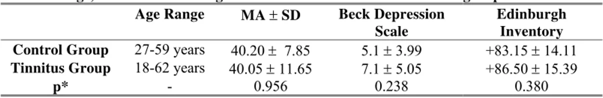
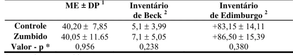
Table 1: Age, Beck and Edinburgh inventories for control and tinnitus groups Age Range MA ± SD Beck Depression
Scale Edinburgh Inventory Control Group 27-59 years 40.20 ± 7.85 5.1 ± 3.99 +83.15 ± 14.11
Tinnitus Group 18-62 years 40.05 ± 11.65 7.1 ± 5.05 +86.50 ± 15.39
p* - 0.956 0.238 0.380
Table 2 - Clinical characteristics of tinnitus
Patient Laterality Tinnitus Description Duration Tinnitus Perception THI
1 L Buzzing 2 years Intermitent 54
2 L Whooshing 1 year Intermitent 34
3 Head Whooshing 10 months Intermitent 18
4 Bilateral L>R Ocean waves 4 years Intermitent 32
5 L Crickets 5 years Continuous 30
6 L Whooshing 3 years Intermitent 34
7 Bilateral L=R Whooshing 2 ½ years Continuous 54 8 Bilateral L<R Whistle 2 years Intermitent 14
9 Head Whistle 11 years Continuous 20
10 R Whooshing 2 years Intermitent 22
11 Bilateral L=R Whistle 10 years Continuous 42 12 Bilateral L=R Whooshing 2 years Intermitent 14
13 L Whooshing 1 year intermitent 40
14 Head Whooshing 5 months Intermitent 16
15 Bilateral L>R Buzzing 2 years Continuous 28
L=left ear, R=right ear
Table 3 – Tinnitus group, activated cerebral regions for unpleasant IADS sounds
Size 1 Tal [x,y,z] p value * % BOLD effect BA Brain Region 2
399 [-33,-11,53] 0.005 0.05 72 Left Insula
77 [40,-7,42] 0.01 0.02 72 Right Insula
44 [47,15,-7] 0.01 0.04 72 Right Insula
67 [-61,-30,9] 0.01 0.03 22 Left Superior Temporal Gyrus
53 [0,-4,48] 0.01 0.02 67 Thalamus
47 [61,-11,4] 0.02 0.04 22 Right Superior Temporal Gyrus
38 [-43,-56,-29] 0.02 0.01 71 Left Cerebellum
40 [14,-78,26] 0.02 0.01 18 Right Primary Visual Cortex
28 [-25,-81,-18] 0.03 0.01 18 Left Primary Visual Cortex
14 [22,-78,-46] 0.03 0.009 71 Right Cerebellum
13 [58,-37,-2] 0.03 0.01 21 Right Middle Temporal Gyrus
20 [29,-63,26] 0.03 0.01 37 Right Infero-posterior
Temporal Lobe
11 [7,-74,-29] 0.04 0.01 71 Right Cerebellum
8 [-36,15,-7] 0.04 0.01 72 Left Insula
8 [-7,-11,4] 0.04 0.01 67 Left Thalamus
* p<0.05, corrected, GBAM
Tal = Talairach coordinates, BOLD = blood oxygenation level dependent BA= Brodmann area
(1)
Table 4 – Control group, activated cerebral regions for unpleasant IADS sounds
Size 1 Tal [x,y,z] p value * % BOLD effect BA Brain Region 2
438 [-36,-44,42] 0.0005 0.03 66 Left Hippocampus
185 [33,-70,-18] 0.004 0.02 19 Right Primary Visual Cortex 80 [58,-22,10] 0.007 0.02 22 Right Superior Temporal Gyrus
54 [40,-48,-35] 0.007 0.02 71 Right Cerebellum
71 [-36,-52,-40] 0.007 0.01 71 Left Cerebellum
47 [47,4,42] 0.008 0.02 22 Right Superior Temporal Gyrus
62 [4,-7,59] 0.009 0.009 67 Right Thalamus
36 [51,11,4] 0.01 0.01 45 Right Inferior Frontal Gyrus
31 [-7,-59,-46] 0.01 0.009 71 Left Cerebellum
15 [51,-52,-13] 0.02 0.008 71 Right Cerebellum
17 [40,-48,42] 0.02 0.006 66 Right Hippocampus
17 [-29,-59,37] 0.02 0.008 37 Left Inf-Posterior Temporal Lobe
13 [-47,-44,-7] 0.03 0.005 71 Left Cerebellum
13 [54,-44,-2] 0.03 0.006 71 Right Cerebellum
12 [-47,-19,-2] 0.03 0.005 20 Left Inferior Temporal Gyrus
10 [29,-56,26] 0.03 0.006 66 Right Hippocampus
17 [11,0,20] 0.03 0.004 69 Right Putamen
13 [36,-15,59] 0.05 0.006 72 Right Insula
6 [4,-74,26] 0.05 0.005 18 Right Primary Visual Cortex * p<0.05, corrected, GBAM
Tal = Talairach coordinates, BOLD = blood oxygenation level dependent BA= Brodmann area
(1)

![Table 3 – Tinnitus group, activated cerebral regions for unpleasant IADS sounds Size 1 Tal [x,y,z] p value * % BOLD effect BA Brain Region 2](https://thumb-eu.123doks.com/thumbv2/123dok_br/16680620.212589/59.892.164.749.322.727/table-tinnitus-activated-cerebral-regions-unpleasant-brain-region.webp)
![Table 4 – Control group, activated cerebral regions for unpleasant IADS sounds Size 1 Tal [x,y,z] p value * % BOLD effect BA Brain Region 2 438 [-36,-44,42] 0.0005 0.03 66 Left Hippocampus 185 [33,-70,-18] 0.004 0.02 19 Right Primary Visual C](https://thumb-eu.123doks.com/thumbv2/123dok_br/16680620.212589/60.892.163.724.322.796/control-activated-cerebral-unpleasant-region-hippocampus-primary-visual.webp)

![Figura 3. Escala SAM [67, 68] modificada para o ambiente de RMf, utilizada na classificação emocional do estímulo (A= valência emocional, B= estímulo)](https://thumb-eu.123doks.com/thumbv2/123dok_br/16680620.212589/84.892.211.779.600.1081/modificada-utilizada-classificação-emocional-estímulo-valência-emocional-estímulo.webp)
