FACULDADE DE ODONTOLOGIA
PROGRAMA DE PÓS-GRADUAÇÃO EM ODONTOLOGIA MESTRADO EM ENDODONTIA
MARÍLIA OSTERMANN BÜRGEL
AVALIAÇÃO DA CENTRALIZAÇÃO DO PREPARO DO CANAL RADICULAR E DA FADIGA DO INSTRUMENTO COMPARANDO TRÊS SISTEMAS ROTATÓRIOS DE NÍQUEL-TITÂNIO
AVALIAÇÃO DA CENTRALIZAÇÃO DO PREPARO DO CANAL RADICULAR E DA FADIGA DO INSTRUMENTO COMPARANDO TRÊS SISTEMAS ROTATÓRIOS DE NÍQUEL-TITÂNIO
Dissertação apresentada como parte dos requisitos obrigatórios para obtenção do título de Mestre em Endodontia, pelo Programa de Pós-Graduação da Faculdade de Odontologia da Pontifícia Universidade Católica do Rio Grande do Sul.
Orientadora: Profa. Dr. Fabiana Vieira Vier Pelisser Co-orientadora: Profa. Dr. Patricia Maria Poli Kopper Móra
AVALIAÇÃO DA CENTRALIZAÇÃO DO PREPARO DO CANAL RADICULAR E DA FADIGA DO INSTRUMENTO COMPARANDO TRÊS SISTEMAS ROTATÓRIOS DE NÍQUEL-TITÂNIO
Dissertação apresentada como parte dos requisitos obrigatórios para obtenção do título de Mestre em Endodontia pelo Programa de Pós-Graduação da Faculdade de Odontologia da Pontifícia Universidade Católica do Rio Grande do Sul.
Aprovada em ____ de _____________ de 2012.
BANCA EXAMINADORA:
________________________________________ Profª. Dr. Simone Bonato Luisi
________________________________________ Dr. Roberta Kochenborger Scarparo ________________________________________
Profa. Dr. Fabiana Vieira Vier Pelisser (orientadora) Porto Alegre
Agradeço primeiramente a Deus, por guiar meus passos nessa trajetória;
À minha família, especialmente aos meus pais, Luciane e Luiz Claudio, e ao
meu marido, Caio, por estarem sempre presentes e serem tão compreensivos
nessa etapa;
Especialmente às minhas professoras Dr. Fabiana Vieira Vier Pelisser e Dra.
Patrícia Poli Kopper Móra, por estarem sempre dispostas a orientar. Agradeço
pelo exemplo pessoal e profissional que vocês me passaram, mostrando que
as dificuldades existem, mas que podem ser superadas com entusiasmo e
dedicação.
Aos demais professores desse curso, em especial ao Prof. Dr. José Antônio
Poli de Figueiredo, pelo exemplo profissional, pelo auxílio nessa pesquisa e por
tudo que me ensinou nesses anos.
À Profa. Dr. Vânia Fontanella por seus ensinamentos fundamentais para que
essa pesquisa fosse concluída.
Ao Dr. Vinícius Dutra, pela sua colaboração.
Ao Michel Klymus, pela doação das limas Wizard CD Plus e Wizard Navigator.
Ao pessoal do centro de microscopia da PUCRS, em especial à Mirian e
à Jennifer, pela ajuda na aquisição das imagens.
Aos colegas de profissão, especialmente à Clínica BellDent, à Claudia Aline,
aos colegas Eduardo Dreher, Luiz Britto, Magno Rigo, Ricardo Meneguzzi e
Rolf Muner Filho, pela parceria.
À Odete, que esteve sempre do meu lado, me apoiando em cada etapa.
Aos pacientes, pela compreensão devido à minha ausência.
Aos colegas de curso pelo apoio e amizade, em especial às amigas Deborah
Cogo, Fernanda López e Renata Morgental, e ao meu professor e colega
Alexandre Ghisi.
Aos professores e colegas de graduação e especialização que sempre me
incentivaram, em especial às profa. Dr. Maristela Gutiérrez de Borba e profa.
Dr. Marcia Rejane Brücker, Michele Dias, Renata de Azevedo, Roberta
Chazan, Roberta Terme, Carina Follmann e Roberta Dresch.
RESUMO
Introdução: Este estudo in vitro avaliou a capacidade de centralização do preparo, bem como o desgaste distorção e a fratura de três tipos de instrumentos rotatórios de níquel-titânio (Ni-Ti) – Wizard CD Plus (WP), Wizard Navigator (WN) e BioRace(BR). Metodologia: Foram utilizadas 90 raízes mésio-vestibulares (MV) de primeiros molares superiores (1ºs MS) e 10 conjuntos de cada tipo de instrumento rotatório. A sobreposição de imagens tomográficas de feixe cônico pré e pós-instrumentação avaliaram a capacidade de centralização do preparo, a 2,0, 4,0, 6,0 e 8,0mm do ápice. Os instrumentos foram avaliados em microscopia eletrônica de varredura (MEV), antes e após até o terceiro uso, na sua ponta e a 5mm desta. Os dados foram analisados pelo Teste de Kruskal-Wallis, complementado pelo Teste de Tukey (P<0.05). Resultados: Houve transporte do canal radicular em todos os grupos, não havendo diferença estatística entre os mesmos (P>0.05). Não houve fratura e distorção a 5mm da ponta nos instrumentos analisados. O grupo BR apresentou mais distorção na ponta do instrumento do que o grupo WP (P=0,011). Houve mais desgaste do instrumento no grupo WP do que no BR (P<0,001). Ocorreu aumento progressivo da distorção na ponta do instrumento e do desgaste em relação ao uso no grupo BR (distorção P=0,026 e desgaste P<0,001), assim como do desgaste no grupo WP (P<0,001). Conclusão: Nenhum dos sistemas rotatórios empregados foi capaz de proporcionar preparos centralizados e o uso progressivo dos instrumentos aumenta a ocorrência de distorção e desgastes em sua topografia.
ABSTRACT
Introduction: This in vitro study evaluated the capacity of centralization of the preparation, as well as the wear, distortion and fracture of three rotary nickel-titanium (Ni-Ti) systems – Wizard CD Plus (WP), Wizard Navigator (WN) and BioRace(BR). Methodology: Were used 90 mesiobuccal roots (MV) of first upper molars (MS). The overlap of tomographic images before and after instrumentation was used to evaluate the transport of the preparation, at 2.0, 4.0, 6.0 and 8.0 mm from the apex. The instruments were observed by SEM before and after the third use, at its tip and at 5 mm of it. The data were analyzed by Kruskal-Wallis, complemented by Tukey test (P<0.05). Results: There was transport of the root canal in all groups, with no statistical difference between them (P> 0.05). There was no fracture and distortion at 5mm from the tip of the instruments. The group BR exhibited more distortion at the tip than the group WP (P = 0.011). There was more wear of the instrument in the group WP than in the BR (P <0.001). There was a progressive increase in distortion at the tip (P = 0.026) and of the wear (P <0.001) compared to the use in the group BR, as well as the wear in the group WP (P<0.001). Conclusion: None of the rotary systems employed was able to provide centralized preparation and the progressive use of the instruments did not favor the occurrence of fracture despite having increased the occurrence of distortion and wear.
1. INTRODUÇÃO ... 10
2. ARTIGO 1 ... 13
3. ARTIGO 2 ... 25
4.DISCUSSÃO ... ... 41
4. REFERÊNCIAS ... 47
ANEXOS ... 53
1 INTRODUÇÃO
A instrumentação mecânica do sistema de canais radiculares é uma
importante fase do preparo do canal radicular (Auerbach, 1948; Schilder, 1974),
pois designa espaço para a ação de soluções irrigadoras e medicações
intracanal a fim de combater bactérias e seus subprodutos. Entretanto, essa
constitui a etapa com maior dificuldade da terapia endodôntica (Hulsmann,
Peters et al., 2005). Sugere-se que quanto mais alargado for o preparo do canal radicular (tão largo quanto a anatomia radicular permitir), somado ao uso
frequente e abundante de soluções irrigadoras antimicrobianas, mais eficaz
será o preparo químico-mecânico (Siqueira, Rocas et al., 2002).
Mesmo a partir da década de 80, após o advento dos instrumentos de
níquel-titânio (Ni-Ti) na Endodontia, a busca por técnicas e sistemas rotatórios
que propiciem um preparo ideal permanece um desafio. Diferentes sistemas
rotatórios vêm sendo propostos, os quais são utilizados em baixa-rotação em
contra-ângulos acoplados a motores elétricos ou pneumáticos, esses sistemas
buscam realizar preparos centralizados que proporcionem a manutenção do
formato original do canal radicular e conicidade, sem a formação de degraus,
zips, transportes e desvios (Hulsmann, Peters et al., 2005; Hartmann, Barletta
et al., 2007; Lopez, Fachin et al., 2008; Gergi, Rjeily et al., 2010).
Durante o preparo do canal radicular, a remoção excessiva de dentina
em uma única direção causa transporte do mesmo (Hartmann, Barletta et al., 2007), o que compromete sua limpeza e aumenta os riscos de acidentes como
capacidade maior de manter a centralização do canal radicular, resultando em
um preparo mais satisfatório para canais radiculares com curvaturas
acentuadas quando comparados com preparos feitos por instrumentos
manuais. (Schafer, Tepel et al., 1995; Chan e Cheung, 1996; Lopez, Fachin et
al., 2008).
Por outro lado, o uso continuado dos instrumentos de NiTi pode causar
deformações na estrutura metálica da liga (Troian, So et al., 2006), o que pode contribuir para um preparo inadequado, resultando em uma limpeza e
modelagem insuficientes para a sanificação do sistema de canal radicular.
Além disso, defeitos na superfície do instrumento endodôntico são capazes de
favorecer a fratura dos mesmos (Kuhn, Tavernier et al., 2001), o que diminui a segurança dos profissionais para a sua utilização (Plotino, Grande et al., 2009).
Apesar da grande quantidade de estudos encontrados na literatura a
respeito dos instrumentos de NiTi, constata-se que a relação entre possíveis
alterações na topografia do instrumento, causadas pelo uso repetido, com a
sua capacidade de proporcionar preparos centralizados dos canais não foi
investigada. Além disso, os instrumentos Wizard Navigator e Wizard Plus, que
apresentam secção transversal triangular e não possuem qualquer tipo de
tratamento de superfície, não possuem investigações prévias quanto à
centralização do canal e a possíveis alterações de sua superfície após o uso.
Em contrapartida, os instrumentos BioRace, que possuem secção transversal
al., 2009). Além disso, demonstraram, por meio de microscópio de força atômica (AFM), alteração na topografia do seu terço cervical, após doze
usos(Yamazaki-Arasaki, Cabrales et al., 2011).
Assim, o presente estudo teve como objetivo avaliar a centralização do
preparo do canal, mediante o primeiro uso dos sistemas Wizard CD Plus
(Medin, Nové Město na Moravě, Czech Republic), Wizard Navigator (Medin, Nové Město na Moravě, Czech Republic) e BioRace (FKG Dentaire, Les Chaux-de-Fonds, Suíça), assim como a distorção de espiras, desgaste
2 ARTIGO 1
Formatado conforme diretrizes do Journal of Endodontics (J Endod)- Fator de impacto: 3.291
Canal centering ability and stress suffered by Ni-Ti instruments: a comparison among Wizard CD Plus, Wizard Navigator and BioRace. Marília Ostermann BürgelaBDS; Patrícia Poli Kopper MorabBDS, MSc, PhD; Vania Fontanellac BDS, MSc, PhD; José Antonio Poli de Figueiredoa BDS, MSc, PhD; Fabiana Viera Vier-PelisseraBDS, MSc, PhD.
a Department of Endodontics, Graduate Program, School of Dentistry, Pontifical Catholic University of Rio Grande do Sul (PUCRS), Porto Alegre, Brazil;
b Department of Endodontics, Graduate Program, School of Dentistry, Federal University of Rio Grande do Sul (UFRGS), Porto Alegre, Brazil;
c Department of Oral Radiology, Graduate Program, School of Dentistry, Brazilian Lutheran University (ULBRA), Canoas, RS, Brazil;
Address correspondence to: Fabiana Vieira Vier Pelisser Graduate Program in Dentistry
Pontifical Catholic University of Rio Grande do Sul – PUCRS, Porto Alegre, Brazil Corresponding address:
Av. Ipiranga 6681 Prédio 6 sala 206 CEP 90619-900
Canal centering ability and stress suffered by Ni-Ti instruments: a comparison among Wizard CD Plus, Wizard Navigator and BioRace.
ABSTRACT
Introduction: This in vitro study evaluated the canal centering ability as well as the wear, distortion and fracture of three rotary nickel-titanium (Ni-Ti) systems – Wizard CD Plus (WP), Wizard Navigator (WN) and BioRace(BR). Methodology: 90 mesiobuccal roots (MB) of first upper molars (UM) were used. Canal transportation was evaluated at 2.0, 4.0 and 8.0mm from the apex, by means of subtracting tomographic images taken before and after preparation. The instruments were observed by SEM before and after the third use, at its tip and at 5 mm of it. The data were analyzed by Kruskal-Wallis, complemented by Tukey test (P<0.05). Results: Canal transportation occurred in all groups, and significant differences were not detected (P> 0.05). There was no fracture and distortion at 5mm from the tip of the instruments. The group BR exhibited more distortion at the tip than the group WP (P = 0.011). There was more wear of the instrument in the group WP than in the BR (P <0.001). In BR instruments, surface wear (P<0.001) and distortion of its tip (P=0.026) increased according to the number of uses, while for WP instruments only surface wear was affect (P<0.001). Conclusion: None of the rotary systems employed was able to provide centralized preparation and the progressive use of the instruments did not favor the occurrence of fracture until third use despite having increased the occurrence of distortion and wear.
INTRODUCTION
Appropriate cleaning and shaping are essential for a successful root canal therapy (1-2). On this regard, a centralized enlargement allows the maintenance of root canal original shape and taper, provides more efficient disinfection and avoid root perforations (3-6). As a matter of fact, as of the 80’s. the introduction of nickel-titanium (Ni-Ti) instruments opened new perspectives in Endodontics, since their superelasticity helps on centering ability (4, 7-8). On the other hand, ideal instruments are still desired, and a wide range of rotary systems are constantly developed.
The instruments Wizard Navigator and Wizard Plus, which have triangular cross-section and do not have any surface treatment, have no prior investigations of canal centering ability and the possible changes in its surface after use. In contrast, BioRace instruments, which have triangular cross-section and electropolished surface (9-11), are more resistant to cyclic fatigue (10) and have satisfactory results in the preparation of curved root canals (11). Also, they demonstrated by means of an atomic force microscope (AFM), change in the topography of their cervical third after twelve uses (9).
On the other hand, the continuous use of Ni-Ti instruments may cause deformations in the metal structure of the alloy (12), which can contribute to an inadequate preparation, resulting in an insufficient cleaning and shaping for the disinfection of the root system. Moreover, defects in the surface of the endodontic instrument are capable of favoring their fracture (13), which reduces its security (14).
Despite the vast amount of research found in the literature regarding the NiTi tools, it is noted that the correlation between possible changes in the topography of the instrument caused by the repeated use, with its ability to provide centralized preparation of the canals has not been investigated.
MATERIALS AND METHODS
This study was approved by the Pontifical Catholic University of Rio Grande do Sul (PUCRS) institutional Ethics and research committees. (CEP 11/05622).
Ninety mesiobuccal roots were used (MB) of human first upper molars (UP) extracted, provided by PUCRS institutional bank of teeth.
The roots were radiographed with the direct digital system CCD Cygnus Ray MPS (Progeny - Buffalo Grove - IL-USA) and the device Gnatus 70X (Gnatus Medical and Dental Equipment Ltd - Ribeirão Preto - SP) in order to provide the stratified randomization of the sample, the degree of root canal curvature, measured as established by Schneider (15) and the distance from the beginning of the curve in relation to the root apex (16), were considered. Operated at 10 mA, 70 kVp, and with exposure time of 0.4 seconds. Teeth presenting previous endodontic manipulation, incomplete root formation, calcifications and/or root resorption were excluded.
Preparation of the canals
A single operator, trained for the use of rotary systems, performed the canals instrumentation. After the endodontic access and irrigation of the canal with NaOCl 1% (Liquid from Milton, Biodynamic, Ibiporã, Brazil), the working length (CT) was established retreating 1mm of the measurement obtained after the Instrument # 10 (Dentsply-Maillefer , Ballaigues, Switzerland) be juxtaposed to the apical foramen. In all cases the CT was between 16 and 18mm.
For the preparation of the root canals 10 sets of each rotatory system of Ni-Ti: Wizard CD Plus (WP); Wizard Navigator (WN) and BioRaCe (BR), were used. Each set of instruments was used to prepare three root canals.
the maximum torque for BR. At each change of instrument, the canals were irrigated with 2 ml of the same solution.
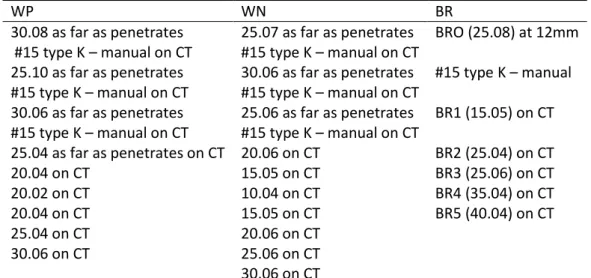
The sequence of instruments used in each of the systems can be found in Table 1.
Tomography
To perform the scans before and after root canal preparation, the teeth were fixed in a rectangular acrylic platform previously perforated (17) with the buccal surface toward its front end, and scanned (l-Cat cone beam, Imaging Sciences International, Hatfield, PA, USA, with 120,000 kV and 46.72 mA, acquisition time of 40 seconds, FOV = 6.0 cm, matrix 800/800 pixels and voxel of 0.2 mm). The raw scanning data were analyzed by the software Xoran-Cat (Imaging Sciences International, Hatfield, PA, EUA).
Evaluation of the centralization of preparation
The tomographic images before and after preparation were acquired through the DICOM files (17) obtained through the program Efilm (Merge HealthCare) and transferred to the Adobe Photoshop program (version CS3, Adobe Systems Inc, San Jose, CA, USA). In the axial sections corresponding to 2, 4, 6 and 8 mm from the apex, a point was marked at the center of the root canal (Fig. 1b-c).
The images before and after preparation were overlapped by the subtraction technique (Fig 1a). The distance between the marked points was measured, in pixels, in the directions buccal-palatal (BP) and mesiodistal (MD) (Fig. 1d). The closer to zero was the obtained measure, more centered was the canal preparation (6).
SEM of the instruments
Mourão, Brazil), packed in surgical grade paper (Descapark, Sao Paulo, Brazil), and sterilized by autoclaving (Dabi Atlante, Ribeirão Preto, Brazil).
Evaluation of the instruments
The images were classified by a blinded calibrated investigator (Spearman Correlation , P <0.05) as to the distortion of the spirals (unwinding, reverse winding or shortening of the spirals along the surface examined), the superficial wear (surface defects of the instrument), and the fracture of the instrument, as described by Troian et al. (12) with some modifications (Figure 2).
Statistical analysis
The data regarding the centralization of the preparation of the canal and of the instruments were analyzed by the Kruskal-Wallis test, complemented by the Tukey test when necessary (P<0.05).
RESULTS
All techniques produced root canal transport, with no statistically significant difference among groups, both in the direction B-P (P = 0.093) and in the direction M-D (P = 0.063), regardless of the location of observation (2, 4, 6 and 8mm.) The results are shown in the graph of Figure 1E.
There was no fracture and distortion at 5 mm from the tip of the instruments analyzed. However, in relation to the distortion of spirals at the tip of the instrument, the group BR distorted more than the group WP (P = 0.011).
As to the wear on the tip and at 5 mm, there was statistically significant difference among the three groups. The WP group showed the greatest wear in both assessments and the group BR the smallest (P <0.001).
DISCUSSION
Even with the increasing evolution of rotary instruments in endodontics, there is still a constant search for centralized preparation, made by instruments that suffer the minimum degree of distortion, wear and fracture. The present research showed that there was transport of the root canal, regardless of the rotary system employed, of the sections and the senses analyzed, which agrees with the findings of other authors (6, 17, 18). However, even being the apical transport still frequent with the use of these instruments, it appears to be less frequent compared to manual techniques for preparation of the canal (19). As to the direction of transport, our results differ from those found by Oliveira et al (17), who demonstrated a greater tendency toward mesial transport.
Several studies, using simulated canals (9, 12 ,20) and extracted human teeth (3, 6, 18, 21-24) have been conducted to evaluate the centralization and transport of the preparation. To this end, methods of scanning of cross (18) and longitudinal (4) sections, photographic (25) and radiographic (23, 26) and, more recently, computed tomography (3, 6, 17) and microtomography (27-28) have been used. Extracted human molars were preferred in this study, rather than resin simulated canals due to the anatomical complexity (29-30) and because they require of the instrument a smaller number of revolutions to complete the preparation of the canal (31). Moreover, the resin is not as hard as the dentine, and the resin chips generated are larger, causing frequent blockages of the apical third (5).
In this study, no instruments ended up fractured, reinforcing the findings of Kawakami and Gavini (32) which showed that the use of instruments for up to seven times does not seem to influence the occurrence of fracture. However, in another study, the repeated use of Profile instruments significantly reduced the torque and the angle of rotation necessary to occur the fracture of these instruments (33).
BR, there was a perception of active cutting of the instrument, which was not perceived with the use of WP and WN.
The WP instruments presented higher superficial wear at the tip as well as at about 5 mm from it, followed by WN and BR instruments, which demonstrated the lowest scores. The surface treatment by electropolishing may have influenced these results.
The increase in the number of use of the instruments seems to have a direct effect on the distortion of spirals and on superficial wear of the instruments of the BR group, a fact also mentioned in other studies with respect to the instruments Race (12) and Profile (34). In this experiment, maximum speed and torque were used as recommended by the manufacturer, which may have favored a higher occurrence of distortion of spirals. In a recent study it was found that the torque had affected the resistance to the cyclic flexural fatigue of the instruments utilized, a factor that may also suggest a change in the morphology of the instrument (32). Also the WP instruments had a progressive increase of wear, with respect to use, which did not occur in the WN. The latter have instruments with the tip diameter of 0.10 mm and 0.15 mm, and may not have acted on the walls of the canal in apical, since, anatomically, the apical diameter of the MV root of the upper molars is of 0.24 mm, 0.28 mm and 0.31 mm in patients up to 24 years, among 25 and 40 years and over 40 years, respectively (35).
Further studies in the same research line, are necessary to better elucidate the relationship among the occurrence of apical transport and the presence of changes in the topography of the instruments.
REFERENCES
1. Auerbach MB. Factors for successful pulp canal therapy. N Y Univ J Dent 1948;6(3):213-216.
2. Schilder H. Cleaning and shaping the root canal. Dent Clin North Am 1974;18(2):269-296.
3. Gergi R, Rjeily JA, Sader J, Naaman A. Comparison of canal transportation and centering ability of twisted files, Pathfile-ProTaper system, and stainless steel hand K-files by using computed tomography. J Endod 2010;36(5):904-907.
4. Lopez FU, Fachin EV, Camargo Fontanella VR, Barletta FB, So MV, Grecca FS. Apical transportation: a comparative evaluation of three root canal instrumentation techniques with three different apical diameters. J Endod 2008;34(12):1545-1548.
5. Hulsmann M, Peters OA, Dummer PMH. Mechanical preparation of root canals: shaping goals, techniques and means. Endodontic Topics 2005;10:30-76.
6. Hartmann MS, Barletta FB, Camargo Fontanella VR, Vanni JR. Canal transportation after root canal instrumentation: a comparative study with computed tomography. J Endod 2007;33(8):962-965.
7. Schafer E, Tepel J, Hoppe W. Properties of endodontic hand instruments used in rotary motion. Part 2. Instrumentation of curved canals. J Endod 1995;21(10):493-497.
8. Chan AW, Cheung GS. A comparison of stainless steel and nickel-titanium K-files in curved root canals. Int Endod J 1996;29(6):370-375.
9. Yamazaki-Arasaki A, Cabrales R, Santos M, Kleine B, Prokopowitsch I. Topography of four different endodontic rotary systems, before and after being used for the 12th time. Microsc Res Tech 2011;75(1):97-102.
10. Lopes HP, Elias CN, Vieira VT, Moreira EJ, Marques RV, de Oliveira JC, et al. Effects of electropolishing surface treatment on the cyclic fatigue resistance of BioRace nickel-titanium rotary instruments. J Endod 2010;36(10):1653-1657.
11. Bonaccorso A, Cantatore G, Condorelli GG, Schafer E, Tripi TR. Shaping ability of four nickel-titanium rotary instruments in simulated S-shaped canals. J Endod 2009;35(6):883-886. 12. Troian CH, So MV, Figueiredo JA, Oliveira EP. Deformation and fracture of RaCe and K3 endodontic instruments according to the number of uses. Int Endod J 2006;39(8):616-625. 13. Kuhn G, Tavernier B, Jordan L. Influence of structure on nickel-titanium endodontic instruments failure. J Endod 2001;27(8):516-520.
14. Plotino G, Grande NM, Cordaro M, Testarelli L, Gambarini G. A review of cyclic fatigue testing of nickel-titanium rotary instruments. J Endod 2009;35(11):1469-1476.
15. Schneider SW. A comparison of canal preparations in straight and curved root canals. Oral Surg Oral Med Oral Pathol 1971;32(2):271-275.
16. Dummer PM, Alodeh MH, Doller R. Shaping of simulated root canals in resin blocks using files activated by a sonic handpiece. Int Endod J 1989;22(5):211-215.
17. Oliveira CA, Meurer MI, Pascoalato C, Silva SR. Cone-beam computed tomography analysis of the apical third of curved roots after mechanical preparation with different automated systems. Braz Dent J 2009;20(5):376-381.
18. Al-Sudani D, Al-Shahrani S. A comparison of the canal centering ability of ProFile, K3, and RaCe Nickel Titanium rotary systems. J Endod 2006;32(12):1198-1201.
19. Schafer E, Lohmann D. Efficiency of rotary nickel-titanium FlexMaster instruments compared with stainless steel hand K-Flexofile--Part 1. Shaping ability in simulated curved canals. Int Endod J 2002;35(6):505-513.
21. Cheung GS, Liu CS. A retrospective study of endodontic treatment outcome between nickel-titanium rotary and stainless steel hand filing techniques. J Endod 2009;35(7):938-943. 22. Ankrum MT, Hartwell GR, Truitt JE. K3 Endo, ProTaper, and ProFile systems: breakage and distortion in severely curved roots of molars. J Endod 2004;30(4):234-237.
23. Goldberg F, Araujo JA. Comparison of three instruments in the preparation of curved root canals. Endod Dent Traumatol 1997;13(6):265-268.
24. Sattapan B, Nervo GJ, Palamara JE, Messer HH. Defects in rotary nickel-titanium files after clinical use. J Endod 2000;26(3):161-165.
25. Ersev H, Yilmaz B, Ciftcioglu E, Ozkarsli SF. A comparison of the shaping effects of 5 nickel-titanium rotary instruments in simulated S-shaped canals. Oral Surg Oral Med Oral Pathol Oral Radiol Endod 2010;109(5):e86-93.
26. Schafer E, Vlassis M. Comparative investigation of two rotary nickel-titanium instruments: ProTaper versus RaCe. Part 1. Shaping ability in simulated curved canals. Int Endod J 2004;37(4):229-238.
27. Peters OA, Boessler C, Paque F. Root canal preparation with a novel nickel-titanium instrument evaluated with micro-computed tomography: canal surface preparation over time. J Endod 2010;36(6):1068-1072.
28. Peters OA, Paque F. Root canal preparation of maxillary molars with the self-adjusting file: a micro-computed tomography study. J Endod 2011;37(1):53-57.
29. Somma F, Leoni D, Plotino G, Grande NM, Plasschaert A. Root canal morphology of the mesiobuccal root of maxillary first molars: a micro-computed tomographic analysis. Int Endod J 2009;42(2):165-174.
30. Degerness RA, Bowles WR. Dimension, anatomy and morphology of the mesiobuccal root canal system in maxillary molars. J Endod 2010;36(6):985-989.
31. Peters OA, Barbakow F. Dynamic torque and apical forces of ProFile.04 rotary instruments during preparation of curved canals. Int Endod J 2002;35(4):379-389.
32. Kawakami DAS, Gavini G. Flexural cyclic fatigue resistance of Ni-Ti ro tary instruments due to number of uses and torque. Revista de Odontologia da Universidade Cidade de São Paulo 2007;19(3):300-311.
33. Yared G. In vitro study of the torsional properties of new and used ProFile nickel titanium rotary files. J Endod 2004;30(6):410-412.
34. Zuolo ML, Walton RE. Instrument deterioration with usage: nickel-titanium versus stainless steel. Quintessence Int 1997;28(6):397-402.
LIST OF FIGURES
FIGURE 1 – Sequence of the overlapping of images (final over the initial) until approximation to the outer contour of the tooth (a-b), the center of the canal was marked with a red dot (1 pixel) (initial) and a yellow dot (1 pixel) (final) (c) and the transport of the canal was measured in the direction B-P (vertical) and MD (horizontal) - arrows (d). Chart showing median (med) and IIQ of the shifts of the different sections (expressed in mm). FIGURE 2 - New WP, WN and BR Instruments (0), after the second (2) and third (3) use. Images obtained from the tip of the instrument (a) and 5mm from the tip (b); whitish areas on the surface of the WP and WN instrument corresponds to the surface wear (¤); is observed wear of the edge of the WN instrument after the second use and BR instrument after second and third use(*); instrument BR presented distortion in the spiral of the image of the tip of the instrument after the second and third use (# ); graph expressing the mean of scores according to the number of uses. SCORES: Spiral Distortion – (0) No unwinding, reverse winding or shortening of spirals along the shaft examined, (1) Unwinding, reverse winding or shortening of only one spiral along the shaft examined, (2) Unwinding, reverse winding or shortening of more than one spiral along the shaft examined; Surface Wear – (0) No wear along the shaft examined, (1) Small amount of wear: one to three areas with defects along the shaft examined; (2) Moderate wear: four to five areas with defects along the shaft examined; (3) Severe wear: more than five areas with defects along the shaft examined.
LIST OF TABLES
TABLE 1 – Sequence of the instruments utilized
WP WN BR
30.08 as far as penetrates 25.07 as far as penetrates BRO (25.08) at 12mm #15 type K – manual on CT #15 type K – manual on CT
25.10 as far as penetrates 30.06 as far as penetrates #15 type K – manual #15 type K – manual on CT #15 type K – manual on CT
30.06 as far as penetrates 25.06 as far as penetrates BR1 (15.05) on CT #15 type K – manual on CT #15 type K – manual on CT
25.04 as far as penetrates on CT 20.06 on CT BR2 (25.04) on CT
20.04 on CT 15.05 on CT BR3 (25.06) on CT
20.02 on CT 10.04 on CT BR4 (35.04) on CT
20.04 on CT 15.05 on CT BR5 (40.04) on CT
25.04 on CT 20.06 on CT
30.06 on CT 25.06 on CT
FIGURE 1
3 ARTIGO 2
Submetido ao Journal of Endodontics (J Endod)- Fator de impacto: 3.291
Canal centering ability and design of Ni-Ti instruments: a comparison among Wizard CD Plus, Wizard Navigator and BioRace.
Marília Ostermann Bürgela DDS, MsC; Patrícia Poli Kopper DDS, MsC, PhDb; Vânia Fontanellac DDS, MsC, PhD; Roberta Kochenborger Scarparoa DDS, MsC, PhD; José Antonio Poli de Figueiredoa DDS, MsC, PhD; Fabiana Vieira Vier-Pelissera DDS, MsC, PhD.
a Department of Endodontics, Graduate Program, School of Dentistry, Pontifical Catholic University of Rio Grande do Sul (PUCRS), Porto Alegre, Brazil;
b Department of Endodontics, Graduate Program, School of Dentistry, Federal University of Rio Grande do Sul (UFRGS), Porto Alegre, Brazil;
c Department of Oral Radiology, Graduate Program, School of Dentistry, Brazilian Lutheran University (ULBRA), Canoas, RS, Brazil;
Corresponding author
Fabiana Vieira Vier-Pelisser
Pontifical Catholic University of Rio Grande do Sul – PUCRS, Porto Alegre, Brazil Av. Ipiranga 6681
Prédio 6 sala 206 CEP 90619-900
Porto Alegre – RS – Brazil Phone: 55 51 3320 3500
ABSTRACT
Aim: To evaluate the canal centering ability of BioRace (BR), Wizard CD
Plus (WP) and Wizard Navigator (WN), associating it with the instruments
designs. Methodology: The instruments surface and cross sections were
observed by means of SEM and described. Additionally, upper first molars
mesiobuccal roots were selected, morphologically balanced and divided into
three groups (n=10 per group), according to the rotary system used for
instrumentation. Canal transportation was evaluated at 2, 4, 6 and 8 mm from
the apex, by means of subtracting tomographic images taken before and after
preparation. The center of root canal pre- and postpreparation was marked, and
the distance between these points was measured, in the buccal-palatal (BP)
and mesio-distal (MD) directions. Data were analyzed by two-way ANOVA and
Bonferroni post-hoc (P<0.05). Results: Only BR instruments showed a polished and regular surface. Canal transportation occurred in all groups, and in both
directions. Significant differences were not detected among the groups,
regardless the distance from root apex. Although all systems demonstrated
triangular cross sections, BR files showed sharper angles. Conclusion: None
of the rotary instruments presented ideal centering ability. Differences were
found amongst instruments regarding quality of finishing and polishing, with
better features for BR instruments.
Key words: centering ability; rotary instrumentation; cone beam; BioRace;
INTRODUCTION
Appropriate cleaning and shaping are essential for a successful root
canal therapy (1-2). In this regard, a centralized enlargement allows the
maintenance of root canal original shaping, provides more efficient disinfection
and avoid root perforations (3-6). The introduction of nickel-titanium (Ni-Ti)
instruments opened new perspectives in Endodontics, since their superelasticity
contributes with the instrumentation centering ability (4,7-8). On the other hand,
ideal instruments are still desired, and a wide range of rotary systems are
constantly developed.
Among them, BioRace (BR) (FKG Dentaire, Les Chaux-de-Fonds,
Switzerland), Wizard CD Plus (WP) (Medin, NovéMěsto at Moravě, Czech Republic) and Wizard Navigator (WN) (Medin, NovéMěsto at Moravě, Czech Republic) were recently introduced. These three rotary systems present
instruments with iso-sized tips (9). Nevertheless, each one of them offer
sequences of files with different tapers, i.e .02 to .08 for BR, .02 to .14 for WP
and .04 to .07 for WN instruments.
Moreover, these instruments present differences regarding their design
and surface treatment. According to the manufacture, WP instruments show
grooves and discontinuous cutting edges, aiming at reducing the risk of
torsional fracture (10). BR instruments show an electropolished surface
(10,11-13), intending to improve its resistance to cyclic fadigue (13). These features, as
well as other non-investigated aspects related to the files design, could affect
instruments avoiding canal transportation (13), whereas WP and WN systems
have yet to be studied in this regard.
Thus, the present study aimed at evaluating the canal centering ability of
BR, WP and WN instruments, while associating it with the files features
observed by means of SEM.
MATERIALS AND METHODS
This study was sanctioned by Pontifical Catholic University of Rio Grande
do Sul (PUCRS) institutional ethics and research committees (11/05622). Thirty
upper first molars, provided by PUCRS institutional bank of teeth, were
selected. The molars were radiographed using a direct digital system (CCD
Cygnus Ray MPS - Progeny - Buffalo Grove - USA). Teeth presenting
endodontic manipulation, incomplete root formation, calcifications and/or root
resorption were excluded. The maximum degree of mesiobuccal canal
curvature and its position were determined as previously reported (14-15). In
most of the samples, the canals showed curvatures between 25o and 35o
degrees (ranging from 0.5o to 58.1o), and its beginning was observed between 6
and 7 mm from the apex (apical distance ranging from 3.96 to 14.35 mm). The
selected samples were divided into three groups balanced for variations in
canal anatomy, according to the rotary systems employed for preparation:
BioRaCe (BR), Wizard CD Plus (WP) or Wizard Navigator (WN). Ten sets of
each system were used, thus establishing a single use for the Ni-Ti files. Prior
junction, using a high-speed diamond bur (KG Sorensen, Cotia, Brazil) under
water-cooling, and the instruments were analyzed by SEM.
SEM analysis of instruments designs
Surface features and the design of BR, WP and WN instruments were
examined under SEM (Philips XL-30, Lichtenstein, Netherland) operating at
20kV under magnifications of 100x. The samples were mounted on a stub, in a
standardized position so that the files shaft could be observed.
Additionally, one set of each rotary system, not employed for canals
preparation, was used to assess the instruments cross section. The files were
sectioned at its apical, middle and cervical portions, using a carburundum disc
(Talmax, Curitiba, Brazil) under water cooling. Representative images were
recorded in a TIFF format at a resolution of 300 dpi. Descriptive analysis was
performed.
Canals Preparation
The working length (WL) was visually established, being set, in all samples,
in 17 mm. A single trained operator performed the canals instrumentation. For
all groups, #10 and #15 files (Dentsply-Maillefer, Ballaigues, Switzerland) were
used throughout the WL. Then, the rotary systems were used with pecking
motion. At each change of instrument, the canals were irrigated with 2 ml of 1%
NaOCl (Biodynamic, Ibiporã, Brazil). The sequences of instruments, speed and
Cone Beam (CBCT) Imaging
Canal transportation was evaluated by means of subtracting CBCT
images obtained before and after instrumentation. The specimens were fitted
into a Fox scale (Bio Art Equipamentos Odontológicos, São Carlos, Brazil) and
adapted to a Cone Beam I - Cat tomograph (Imaging Sciences International,
Hatfield, USA), operated at 120000 kV and 46.72 mA. The images were
captured in a small field of view (6 cm) using exposure time of 40 s, a matrix of
800 x 800 pixels and voxel of 0.2 mm. Xoran- Cat software (Imaging Sciences
International, Hatfield, USA) was used for image reconstruction. Slices in the
axial direction generated DICOM format archives (16).
Measurement of canal transportation
Centering ability analysis was performed by a single blinded, calibrated
examiner. Images of the axial sections corresponding to 2, 4, 6 and 8 mm from
the apex were transferred to the Adobe Photoshop program (version CS3,
Adobe Systems Inc, San Jose, USA). At each section, images corresponding to
the center of the canals pre and post-preparation were marked. These images
were overlapped by the subtraction technique (Fig 1a), and the distance
between the two points was measured in both BP and MD directions (Fig. 1b-d).
The closer to zero was the obtained measure, the more centered was the canal
Statistical analysis
Measures of centering ability were analyzed using two-way ANOVA and
Bonferroni post-hoc (P<0.05).
RESULTS
Instruments Designs
SEM analysis of BR instruments showed few irregularities and a
polished surface. The files edges and tip presented well-defined contours.
Nevertheless, both WP and WN instruments presented surface defects and
irregular edges. Although all systems demonstrated triangular cross sections,
BR files showed sharper angles (Figure 2).
Centering ability
Canal transportation occurred in all groups. Significant differences were
not detected (Figure 1 e-f), regardless the apical distance (2, 4, 6 and 8 mm)
and canal direction (P<0.05).
DISCUSSION
The present study analyzed the centering ability of three recently
introduced rotary systems. The results observed herein showed that, generally,
BR, WP and WN instruments produced similar canal transportation among
them, which was also comparable with the amount of deviation produced by
from others showed that Ni-Ti files did not avoid the occurrence of apical
transportation (6,16,18), even if reducing it compared to hand techniques (3-5,
19).
Safe limits for canal transportation must be based on dental anatomical
features. Thus, the measures of canal wall thickness at different apical
distances are of paramount importance when determining the preparation
security. Dental walls appear to be thicker toward the cervical portion.
Regarding mesiobuccal canals of maxillary molars, approximately 2 mm from
the apex, dental walls thickness are set between 0.84 and 2.15 mm, whilst, at
an apical distance of about 8 mm, this measures are comprised between 1.23
and 1.58 mm (20). The correlation between these data and the present
outcomes suggests that all groups promoted tolerable canal transportation in
most of the samples. Meanwhile, particularly for BR and WN groups, and at 2
mm from apex, canal transportation was critical in some of the specimens if
considering the possibility of iatrogenic complications. At this point, although not
statistically significant, a clinically relevant advantage was detected for WP
centering ability. This fact should be explained by the WP files greater tapers,
which were designed intending to provide further elimination of cervical
interferences (10).
A number of study models have been used to compare canal shape
before and after instrumentation (3-4,6,8,13,16,18,21-22). However, taken into
account the importance of dental tissues hardness and of the morphological
Moreover, to avoid the influence of confounding factors, the groups were
balanced for variations in canal anatomy as recommended (14).
As well as previous studies (3,6,16,18), this investigation used CBCT
images to assess the preparation centering ability. Besides being a
non-destructive and more accurate method, this technique allows the observation in
both MD and BP directions, which is not possible when applying conventional
radiographs (25). The endodontic literature has yet to clearly established an
association between canal curvature and transportation directions. Some
studies using mesiobuccal canals showed a tendency toward mesial
transportation (16,21), which has been attributed to the fact that curvatures
were frequently observed in a distal position. Meanwhile, other authors state
that the direction of canal curvature has no effect on the direction of
transportation (26). One of the possible explanations for conflicting results is
that the methods employed no longer evaluated the discrepancies between MD
and BP transportation. The methodology adopted herein allowed this approach,
showing similar canal transportation in both directions.
On the other hand, especially for WP and WN systems, the apical
distance appears to have an influence on the discrepancies between MD and
BP deviations. The greatest differences were observed 6 mm from the apex,
showing a tendency toward BP transportation. This section, as showed herein,
coincides with the beginning of canals curvatures in most of the samples, which
could have affected the direction of enlargement. Besides, the lower amount of
discrepancies observed for the BR group at this point was probably influenced
Unlike BR instrumentation, in which a regular and polished surface was
observed, WP and WN instruments showed irregular edges. These features
were tactile perceived by the friction during instrumentation, and should have
affected the obtainment of circular and uniform preparations, thus producing
greater transportations in one of the directions. Besides, files irregularities may
have contributed with a poor standardization of centering ability, which was
corroborated by the high standard deviations observed. Considering that in the
present study a single use was established for the instruments, further studies
are warranted to assess the influence of repeated uses on centering ability.
Although the quality of surface finishing did not promote statistically
significant differences for the preparations centering ability, it is important to
point out that BR instrumentation showed similar results compared to the other
groups, regardless the use of a larger-sized master apical file, which could
influence the maintenance of canals original position (4).
Conclusion
Taken into account the dimensions of root canal walls, it can be
concluded that none of the rotary instruments presented ideal centering ability.
Differences were found amongst instruments regarding quality of finishing and
polishing, with better features for BR instruments.
REFERENCES
1. Auerbach MB. Factors for successful pulp canal therapy. N Y Univ J Dent
2. Schilder H. Cleaning and shaping the root canal. Dent Clin North Am
1974;18(2):269-296.
3. Gergi R, Rjeily JA, Sader J, Naaman A. Comparison of canal
transportation and centering ability of twisted files, Pathfile-ProTaper system,
and stainless steel hand K-files by using computed tomography. J Endod
2010;36(5):904-907.
4. Lopez FU, Fachin EV, Camargo Fontanella VR, Barletta FB, So MV,
Grecca FS. Apical transportation: a comparative evaluation of three root canal
instrumentation techniques with three different apical diameters. J Endod
2008;34(12):1545-1548.
5. Hulsmann M, Peters OA, Dummer PMH. Mechanical preparation of root
canals: shaping goals, techniques and means. Endodontic Topics
2005;10:30-76.
6. Hartmann MS, Barletta FB, Camargo Fontanella VR, Vanni JR. Canal
transportation after root canal instrumentation: a comparative study with
computed tomography. J Endod 2007;33(8):962-965.
7. Schafer E, Tepel J, Hoppe W. Properties of endodontic hand instruments
used in rotary motion. Part 2. Instrumentation of curved canals. J Endod
1995;21(10):493-497.
8. Chan AW, Cheung GS. A comparison of stainless steel and
nickel-titanium K-files in curved root canals. Int Endod J 1996;29(6):370-375.
9. International organization of Standardization. Dentistry – Root canal instruments – Part 1: General requirements and test methods. ISO 3630-1:2008
http://www.iso.org/iso/iso_catalogue/catalogue_tc/catalogue_detail.htm?csnum
ber=37702
10. Medin as. Rotary instruments for dentistry. 2011[cited 2012 16 May];42].
Available from
http://www.medin.eu/upload/catalogs/catalog-of-rotary-dental-instruments-complete.pdf.
11. Yamazaki-Arasaki A, Cabrales R, Santos M, Kleine B, Prokopowitsch I.
Topography of four different endodontic rotary systems, before and after being
used for the 12th time. Microsc Res Tech 2011;75(1):97-102.
12. Lopes HP, Elias CN, Vieira VT, Moreira EJ, Marques RV, de Oliveira JC,
et al. Effects of electropolishing surface treatment on the cyclic fatigue
resistance of BioRace nickel-titanium rotary instruments. J Endod
2010;36(10):1653-1657.
13. Bonaccorso A, Cantatore G, Condorelli GG, Schafer E, Tripi TR. Shaping
ability of four nickel-titanium rotary instruments in simulated S-shaped canals. J
Endod 2009;35(6):883-886.
14. Schneider SW. A comparison of canal preparations in straight and
curved root canals. Oral Surg Oral Med Oral Pathol 1971;32(2):271-275.
15. Dummer PM, Alodeh MH, Doller R. Shaping of simulated root canals in
resin blocks using files activated by a sonic handpiece. Int Endod J
1989;22(5):211-215.
16. Oliveira CA, Meurer MI, Pascoalato C, Silva SR. Cone-beam computed
tomography analysis of the apical third of curved roots after mechanical
17. Baumann MA, Roth A. Effect of experience on quality of canal
preparation with rotary nickel-titanium files. Oral Surg Oral Med Oral Pathol Oral
Radiol Endod 1999;88(6):714-718.
18. Tasdemir T, Aydemir H, Inan U, Unal O. Canal preparation with Hero 642
rotary Ni-Ti instruments compared with stainless steel hand K-file assessed
using computed tomography. Int Endod J 2005;38(6):402-408.
19. Schafer E, Lohmann D. Efficiency of rotary nickel-titanium FlexMaster
instruments compared with stainless steel hand K-Flexofile--Part 1. Shaping
ability in simulated curved canals. Int Endod J 2002;35(6):505-513.
20. Degerness RA, Bowles WR. Dimension, anatomy and morphology of the
mesiobuccal root canal system in maxillary molars. J Endod
2010;36(6):985-989.
21. Al-Sudani D, Al-Shahrani S. A comparison of the canal centering ability
of ProFile, K3, and RaCe Nickel Titanium rotary systems. J Endod
2006;32(12):1198-1201.
22. Schafer E, Vlassis M. Comparative investigation of two rotary
nickel-titanium instruments: ProTaper versus RaCe. Part 1. Shaping ability in
simulated curved canals. Int Endod J 2004;37(4):229-238.
23. Somma F, Leoni D, Plotino G, Grande NM, Plasschaert A. Root canal
morphology of the mesiobuccal root of maxillary first molars: a micro-computed
tomographic analysis. Int Endod J 2009;42(2):165-174.
24. Dummer PM, Kelly T, Meghji A, Sheikh I, Vanitchai JT. An in vitro study
of the quality of root fillings in teeth obturated by lateral condensation of
25. Goldberg F, Araujo JA. Comparison of three instruments in the
preparation of curved root canals. Endod Dent Traumatol 1997;13(6):265-268.
26. Kosa DA, Marshall G, Baumgartner JC. An analysis of canal centering
using mechanical instrumentation techniques. J Endod 1999;25(6):441-445.
FIGURE LEGENDS
FIGURE 1 – Methodology used for measuring centering ability and the obtained results. (a) images pre and post-instrumentation were overlapped by the
subtraction technique; (b-c) the central position of root canal was marker prior
(red dot) and post-preparation (yellow dot); (d) canal transportation was
measured in buccalpalatal (BP) and mesiodistal (MD) directions (arrows); (e-f)
The resulting outcomes of canal transportation did not show statistically
significant differences in both directions (P>0.05).
FIGURE 2 - Instrument surface and cross-sectional features under SEM.
(A) 25.08 BR instrument showing round tip and transitional angle. 30.08 WP file
presenting a transitional angle, although the tip had a grosser outline. 25.07 WN
showing an abrupt transition from the instrument shank to its tip (arrows);
Surface finishing was irregular for WP and WN files, and polished for BR
instruments (*). (B) WP instrument flutes presented flattened cutting edges ( )
compared to BR and WN files, in which a sharp angle could be detected. (C) All
rotary systems demonstrated triangular cross sections, in spite of the BR files
sharper angles. (D) Intentional groove defects were found at the flutes of 30.08
instrument differing from 25.07 WN and 25.08 BR instruments. BR files
presented a well-defined and polished outline.
TABLE LEGEND
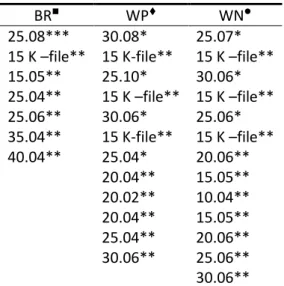
TABLE 1 - Sequence of instruments, speed and torque used for canals
preparation
TABLE 1 – Sequence of instruments, speed and torque used for canals preparation
BR WP WN
25.08*** 30.08* 25.07* 15 K –file** 15 K-file** 15 K –file** 15.05** 25.10* 30.06* 25.04** 15 K –file** 15 K –file** 25.06** 30.06* 25.06* 35.04** 15 K-file** 15 K –file** 40.04** 25.04* 20.06**
20.04** 15.05** 20.02** 10.04** 20.04** 15.05** 25.04** 20.06** 30.06** 25.06** 30.06**
*as far as penetrate; ** at the WL; ***at 12mm.
600 RPM / maximum torque;250 RPM / 0.2 -3 N.cm;
FIGURE1
4 DISCUSSÃO
Apesar do avanço tecnológico na Endodontia, ainda há uma constante
busca por preparos centralizados, realizados por instrumentos que sofram o
mínimo grau de distorção, desgaste e fratura.
Vários estudos, empregando canais simulados (Troian, So et al., 2006; Schafer e Oitzinger, 2008; Yamazaki-Arasaki, Cabrales et al., 2011) e dentes humanos extraídos (Goldberg e Araujo, 1997; Sattapan, Nervo et al., 2000; Ankrum, Hartwell et al., 2004; Al-Sudani e Al-Shahrani, 2006; Hartmann, Barletta et al., 2007; Cheung e Liu, 2009; Gergi, Rjeily et al., 2010) vêm sendo conduzidos para avaliar a centralização dos preparos. Com este objetivo,
métodos de scaneamento de seccções transversais (Al-Sudani e Al-Shahrani,
2006) e longitudinais (Lopez, Fachin et al., 2008), fotográficos (Ersev, Yilmaz et
al., 2010) e radiográficos (Goldberg e Araujo, 1997; Schafer e Vlassis, 2004) e, mais recentemente, tomografias computadorizadas (Hartmann, Barletta et al., 2007; Oliveira, Meurer et al., 2009; Gergi, Rjeily et al., 2010) e microtomografias (Peters, Boessler et al., 2010; Peters e Paque, 2011) têm sido utilizados. (International, 2008)
Por apresentarem uma anatomia complexa, as raízes mésio-vestibulares
(MV) de molares superiores (MS) são capazes de reproduzir uma condição
clínica adversa, que constitui um desafio para os profissionais. Isto por que a
incidência, em relação ao número de canais na raiz MV, foi de 20%, 79,8%,
1,1% para um, dois e três, respectivamente (Degerness e Bowles, 2010). Além
(3,6mm) e a presença de istmos é menos freqüente próximo do ápice (Somma,
Leoni et al., 2009).
Este estudo foi realizado com dentes humanos extraídos para que
houvesse uma aproximação com a realidade clínica. Peters e Barbakow
(Peters e Barbakow, 2002) avaliaram o torque gerado pelo instrumento
endodôntico quando canais simulados curvos e estreitos são preparados
comparando-os com o preparo em dentes humanos extraídos. Foi verificado
que um maior número de rotações é necessário para completar o preparo do
canal radicular quando são utilizados blocos de resina. Além disso, o tamanho
de raspas de resina e dentina geradas durante o preparo biomecânico não são
idênticas, resultando em frequentes bloqueios do terço apical e dificuldades
para remover debris em canais simulados. Em contrapartida, a microdureza da
dentina varia de 35-40Kg/mm2 próximo ao espaço pulpar, enquanto a da resina
utilizada em canais simulados varia de 20-22Kg/mm2. Para tanto para remover
dentina natural o dobro de força seria necessário(Hulsmann, Peters et al., 2005).
Para análise do preparo do transporte do canal radicular com os
diferentes instrumentos foi utilizada a sobreposição de imagens obtidas por
meio da tomografia de feixe cônico. Outros autores (Hartmann, Barletta et al., 2007; Oliveira, Meurer et al., 2009; Gergi, Rjeily et al., 2010) também utilizaram tomografia para análise da qualidade do preparo do canal radicular.
A presente pesquisa demonstrou que houve transporte do canal
radicular, independente do sistema rotatório empregado, das secções e dos
(Al-Sudani e Al-Shahrani, 2006; Hartmann, Barletta et al., 2007; Oliveira, Meurer et
al., 2009). No entanto, mesmo o transporte apical sendo ainda frequente com o uso desses instrumentos, parece ser menos frequente quando comparado com
técnicas manuais de preparo do canal (Schafer e Lohmann, 2002). Quanto ao
sentido do transporte, nossos resultados diferem dos encontrados por Oliveira
et al (Oliveira, Meurer et al., 2009), que demonstraram uma grande tendência de transporte na direção mesial, porém está de acordo com investigações
anteriores que mostram que a direção do transporte do canal radicular não é
influenciada pela direção da curvatura radicular (Kosa, Marshall et al., 1999). Em contrapartida, nos grupos WP e WN a distância em relação ao ápice
parece ter influência na diferença entre o desvio MD e VP, pois principalmente
a 6mm do ápice houve a maior diferença entre as duas direções, essa região
corresponde ao início da curvatura dos canais na maioria das amostras.
Provavelmente, a menor diferença entre as direções MD e VP observada no
grupo BR foi devido à secção transversal e ao desenho do instrumento. Tanto
os instrumentos WP quanto os instrumentos WN apresentam diferentes
secções transversais ao longo do instrumento, fator que pode ter influenciado
um maior alargamento a 6mm do ápice. Além disso, os instrumentos WN e WP
apresentaram arestas irregulares, o que pode ter produzido maior transporte
em uma das direções observadas. Os instrumentos BR apresentaram
superfície polida e arestas regulares.
As características anatômicas dentárias devem basear os limites de
segurança para o transporte do canal radicular. No entanto, as medidas de
importância para determinar a segurança do preparo. As paredes dentárias
parecem ser mais espessa na porção cervical. Em relação aos canais
mésio-vestibulares de molares superiores, a 2 mm do ápice, a espessura das paredes
dental pode ser determinada entre 0,84 e 2,15 mm, enquanto que, a uma
distância apical de cerca de 8 mm, essas medidas são compreendidas entre
1,23 e 1,58 mm (Degerness e Bowles, 2010). A correlação entre estes dados e
os resultados deste estudo sugerem que todos os sistemas rotatórios testados
promovem transporte do canal tolerável na maioria das amostras. Enquanto
isso, particularmente para BR e grupos WN, e a 2 mm do ápice, o transporte do
seria crítico em alguns dos espécimes se considerar a possibilidade de
iatrogenias. Neste ponto, embora não estatisticamente significativa, uma
vantagem clinicamente relevante, foi constatada para o grupo WP na
capacidade de centralização. Esse fato pode ser explicado pois os
instrumentos WP apresentam taper maiores, o que pode causar uma maior
eliminação de interferências cervicais (Medin, 2011).
Os instrumentos WP e WN novos apresentaram bordos irregulares,
diferentemente do grupo BR, em que uma superfície regular e polida foi
observada. Porém a qualidade de acabamento superficial não promoveu
diferenças estatisticamente significativas para a capacidade de centralização
dos preparos, mas é importante ressaltar que o grupo BR apresentou
resultados semelhantes aos demais grupos, independente do uso de um
instrumento mestre maior porte apical, o que poderia influenciar a manutenção
No presente estudo, nenhum instrumento fraturou reforçando os
achados de Kawakami e Gavini (Kawakami e Gavini, 2007) que evidenciaram
que a utilização dos instrumentos por até sete vezes parece não influenciar na
ocorrência de fratura. Em contrapartida, em outra investigação, o uso repetido
de instrumentos Profile reduziu significativamente o torque e o ângulo de
rotação necessários para que ocorresse a fratura desses instrumentos (Yared,
2004).
A análise dos instrumentos em relação ao desgaste e distorção de
espiras foi realizada por meio da MEV. Esse método foi utilizado em outros
estudos (Zuolo e Walton, 1997; Troian, So et al., 2006) e parece ser um método apropriado para avaliação dos instrumentos. O grupo BR apresentou
maior distorção na ponta que os WP. Talvez este fato possa estar relacionado
com o diâmetro de alargamento apical, que foram 0,40mm e 0,30mm,
respectivamente. Apesar de não ter havido diferença entre os instrumentos WN
e BR neste quesito, durante o preparo do canal com BR, havia uma percepção
de corte ativo do instrumento, o que não foi percebido com o uso do WP e WN.
Os instrumentos WP apresentaram desgaste superficial maior, tanto na
ponta quanto a 5 mm desta, seguidos pelos instrumentos WN e BR, que
demonstraram os menores escores. O tratamento de superfície por
eletropolimento pode ter influenciado estes resultados. (Dummer, Kelly et al.,
1993; Baumann e Roth, 1999; Tasdemir, Aydemir et al., 2005)
O aumento do número de uso dos instrumentos parece ter efeito direto
na distorção de espiras e no desgaste superficial dos instrumentos do grupo
BR, fato também mencionado em outros estudos, com relação aos
presente experimento, velocidade e torques máximos foram empregados,
conforme recomendado pelo fabricante, o que pode ter favorecido a maior
ocorrência de distorções de espiras. Em recente estudo, verificou-se que o
torque afetou a resistência à fadiga cíclica flexural dos instrumentos utilizados,
fator que pode também sugerir uma alteração na morfologia do instrumento
(Kawakami e Gavini, 2007). Também os instrumentos WP tiveram um aumento
progressivo do desgaste, em relação ao uso, fato que não ocorreu nos WN.
Esses últimos possuem instrumentos com diâmetro na ponta de 0,10mm e
0,15mm, podendo não ter atuado nas paredes do canal em apical, uma vez
que anatomicamente o diâmetro apical da raiz MV de MS é de 0,24mm,
0,28mm e 0,31mm em pacientes com até 24 anos, entre 25 e 40 anos e com
mais de 40 anos, respectivamente (Vier, Tochetto et al., 2004).
Novas investigações, nesta mesma linha de pesquisa, são necessárias
para melhor elucidar a relação entre a ocorrência de transporte apical com a
presença de alterações na topografia dos instrumentos.
Dentro das limitações desse estudo, pode-se concluir que nenhum dos
sistemas rotatórios empregados foi capaz de proporcionar preparos
centralizados e o uso progressivo dos instrumentos até o terceiro uso não
favoreceu a ocorrência de fratura apesar de ter aumentado a ocorrência de
4 REFERÊNCIAS
AL-SUDANI, D.; AL-SHAHRANI, S. A comparison of the canal centering ability of ProFile, K3, and RaCe Nickel Titanium rotary systems. J Endod, v. 32, n. 12, p. 1198-201, Dec 2006. ISSN 0099-2399 (Print)
0099-2399 (Linking). Disponível em: <
http://www.ncbi.nlm.nih.gov/entrez/query.fcgi?cmd=Retrieve&db=PubMed&dopt=Citation&li st_uids=17174683 >.
ANKRUM, M. T.; HARTWELL, G. R.; TRUITT, J. E. K3 Endo, ProTaper, and ProFile systems: breakage and distortion in severely curved roots of molars. J Endod, v. 30, n. 4, p. 234-7, Apr 2004. ISSN 0099-2399 (Print)
0099-2399 (Linking). Disponível em: <
http://www.ncbi.nlm.nih.gov/entrez/query.fcgi?cmd=Retrieve&db=PubMed&dopt=Citation&li st_uids=15085054 >.
AUERBACH, M. B. Factors for successful pulp canal therapy. N Y Univ J Dent, v. 6, n. 3, p. 213-6, Apr 1948. ISSN 0028-7865 (Print)
0028-7865 (Linking). Disponível em: <
http://www.ncbi.nlm.nih.gov/entrez/query.fcgi?cmd=Retrieve&db=PubMed&dopt=Citation&li st_uids=18915848 >.
BAUMANN, M. A.; ROTH, A. Effect of experience on quality of canal preparation with rotary nickel-titanium files. Oral Surg Oral Med Oral Pathol Oral Radiol Endod, v. 88, n. 6, p. 714-8, Dec 1999. ISSN 1079-2104 (Print)
1079-2104 (Linking). Disponível em: <
http://www.ncbi.nlm.nih.gov/entrez/query.fcgi?cmd=Retrieve&db=PubMed&dopt=Citation&li st_uids=10625855 >.
BONACCORSO, A. et al. Shaping ability of four nickel-titanium rotary instruments in simulated S-shaped canals. J Endod, v. 35, n. 6, p. 883-6, Jun 2009. ISSN 1878-3554 (Electronic)
0099-2399 (Linking). Disponível em: <
http://www.ncbi.nlm.nih.gov/entrez/query.fcgi?cmd=Retrieve&db=PubMed&dopt=Citation&li st_uids=19482191 >.
CHAN, A. W.; CHEUNG, G. S. A comparison of stainless steel and nickel-titanium K-files in curved root canals. Int Endod J, v. 29, n. 6, p. 370-5, Nov 1996. ISSN 0143-2885 (Print)
0143-2885 (Linking). Disponível em: <
http://www.ncbi.nlm.nih.gov/entrez/query.fcgi?cmd=Retrieve&db=PubMed&dopt=Citation&li st_uids=10332236 >.
CHEUNG, G. S.; LIU, C. S. A retrospective study of endodontic treatment outcome between nickel-titanium rotary and stainless steel hand filing techniques. J Endod, v. 35, n. 7, p. 938-43, Jul 2009. ISSN 1878-3554 (Electronic)
0099-2399 (Linking). Disponível em: <