Original Research
Evaluation of Different Dentin Bonding Agents Accompanied with
Composite Coronal Barrier
Zahed Mohammadi
1, Loghman Rezaei-Soufi
2, Tahereh Omidipoor
3,
Massoud Felegary
4, Farshid Vahdatinia
51
Endodentist, Hamadan, Iran.
2
Dental Research Center and Department of Operative Dentistry, Dental School, Hamadan
University of Medical Sciences, Hamadan, Iran
3
Department of Operative Dentistry, Dental School, Hamadan University of Medical Sciences,
Hamadan, Iran
4
Dentist, Ghorveh, Iran
5
Dental School, Hamadan University of Medical Sciences, Hamadan, Iran
Received 5 December 2014 and Accepted 2 February 2015
Abstract
Introduction: The purpose of this study was to evaluate the sealing ability of dentin bonding agents in root canals obturated with gutta-percha and MTA. Methods: Forty-five single rooted human premolar teeth were decoronated so that remaining root portions were 12 mm in length. The samples were divided randomly into three experimental (n=13) and two control groups (n=3). All teeth were instrumented up to #40 K-file using step-back technique. The roots were obturated with gutta-percha/AH26 and MTA for 5 and 3 mm, respectively. Excite, Clearfil SE Bond, and iBond were applied for experimental groups and then 2mm was filled with composite Filtek Z250. The roots in the controls were merely instrumented and obturated. Two coats of nail varnish were applied on the surface of the teeth in the experimental and positive groups, except 2 mm around the apical foramen and coronal surfaces. In the negative control, the surfaces were completely covered by two layers of nail varnish. After thermocycling, the roots mounted in plastic caps of tubes containing BHI medium and inoculated coronally with Enterococcus faecalis. The data were statistically analyzed using Fisher's exact and Kaplan-Meier survival Analysis. Results: There were no statistically significant differences between three experimental groups regarding the leakage rate (P=0.738). Conclusion: Within the limitations of this study, it was observed that the adhesive systems in alliance with gutta-percha and MTA obturation could not entirely
prevent bacterical leakage, and their sealing abilities were not statistically different.
Key words: Bacterial leakage, adhesives, Enterococcus faecalis
---
Mohammadi Z, Rezaei-Soufi L, Omidipoor T, Felegary M, Vahdatinia F. Evaluation of Different Dentin Bonding Agents Accompanied with Composite Coronal Barrier. J Dent Mater Tech 2015; 4(2): 73-80.Introduction
when teeth were restored with composite resin in combination with dentin bonding systems (6, 7). Essentially the performance of adhesive systems on the enamel surface is appropriate (8), while the bonding to dentin is barely possible (9). Optimizing bond strength between resin and dentin and resin and root canal posts is of crucial importance. The inadequate bond strength of adhesive systems used for cementing root canal posts can lead to failure of the restoration and an increased possibility of subsequent microleakage and failure of the endodontic treatment (10). In endodontically treated teeth, the higher amount of composite material may increase polymerization shrinkage stress (11). Other problem is the root perforation during the canal preparation (12). The use of Mineral Trioxide Aggregate (MTA) as a canal filling material can reduce these two problems. MTA powder consists of fine hydrophilic particles. The principle compounds present in this material are tricalcium silicate, tricalcium aluminate, tricalcium oxide, and silicate oxide. In addition, small amounts of other mineral oxides exist that are responsible for the chemical and physical properties of this aggregate. The biocompatibility of gray MTA has been reported using cell culture techniques and connective tissue reactions (13). Sarkar et al. reported the propensity of MTA to release Ca and its ability to form hydroxyapatite and concluded that the sealing ability, biocompatibility and dentinogenic activity of MTA is attributed to these physicochemical reactions (14). Mineral trioxide aggregate (MTA) has been regarded as an ideal material for perforation repair, retrograde filling, pulp capping, and apexification since its introduction in 1993 (15, 16).The repair capacity of MTA can in turn be attributed to the antimicrobial properties and high pH (12.5) of MTA, these characteristic of MTA promote growth of the cementum and formation of bone (17, 18).
Korasli et al evaluated the coronal marginal leakage of endodontically treated teeth bonded with four self-etching adhesives and one total-etch adhesive system (19). None of the tested self-etch adhesives completely eliminated microleakage. Dye leakage was restricted to the coronal cavity walls it did not migrate toward the pulp chamber or toward the root canal. Single Bond and Clearfil SE Bond showed significantly lower dye penetration values at occlusal and gingival margins. Er et al compared the sealing ability of different dentin bonding adhesive systems in root-end cavities by the bacterial leakage method (20). They found that there were no statistically significant differences in rate of bacterial leakage among the experimental test materials at 1-4 weeks. Gonzalez-Castillo et al evaluate the
capability of Cavit™ G to seal the pulp chamber, used
alone, with Clearfil™ S3 Bond or Prime & Bond NT
adhesive systems, at 24 hours, 7, 30 or 45 days (4). They concluded that the adhesive systems did not
improve the capability of Cavit™ G to seal the pulp
chamber over time.
Root perforation is one of the probable problems that may occur during the root canal therapy. Selection of a proper dentin bonding agent for composite filling of such teeth can overcome the leakage problems after restoration. Therefore, the purpose of this study was the evaluation of different dentin bonding agents accompanied with composite coronal barrier
Materials and Methods
A whole number of 45 noncarious human premolar extracted teeth with straight, single root canals were selected for this study. Soft tissues were mechanically removed from the root surfaces. The teeth were placed in a disinfectant (10% formalin) for 48 h in and then stored in normal saline at room temperature until ready to be used. The teeth were decoronated by diamond disk so that the remaining root portion were12 mm in length. The working length was measured by deducting 1 mm from the length recorded when tip of a #10 file (Dentsply/ Maillefer Instruments SA , Ballaigues, Switzerland) was visible at the apical foramen. All teeth were instrumented up to #40 K-file using step-back technique. Irrigation was carried out using 5 mL of a 5.25% NaOCl solution between files. The canals were then dried with sterile paper points. The roots were obturated with gutta-percha (Aria Dent, Tehran, Iran) and AH26 sealer (Dentsply, DeTrey, Konstanz, Germany) by using lateral condensation technique. Then the coronal part of the gutta-percha has been removed by pizorimer No III (Mani, Germany) so that 5mm of apical gutta percha remains within the root canal. Afterward, 3mm of roots were filled with MTA (Dentsply, Tulsa Dental, Tulsa, OK, USA) coronal to gutta-percha. The samples were placed in 100% humidity at 37°C for 24 h to allow the MTA to set.
The samples were divided randomly into three experimental (n=13) groups and two positive and negative control groups (n=3). Excite (2 step etch & rinse), Clearfil SE Bond (2 step self-etch), and iBond (1 step self-etch) adhesive systems were applied for
experimental groups according to the manufacturers’
instructions (Table 1). After that, 2mm of canal above the MTA was filled with composite Filtek Z250 (3M ESPE, St. Paul, MN, USA) in two 1mm layers. Each layer has been polymerized for 20 second with a
light-curing unit (Hilux LEDMax, Benlioğlu Dental, Ankara,
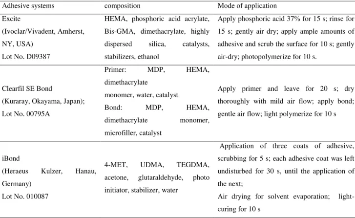
Table 1. Adhesive Systems, Batch Number, Composition, and Application Mode Adhesive systems composition Mode of application
Excite
(Ivoclar/Vivadent, Amherst,
NY, USA)
Lot No. D09387
HEMA, phosphoric acid acrylate,
Bis-GMA, dimethacrylate, highly
dispersed silica, catalysts,
stabilizers, ethanol
Apply phosphoric acid 37% for 15 s; rinse for
15 s; gently air dry; apply ample amounts of
adhesive and scrub the surface for 10 s; gently
air-dry; photopolymerize for 10 s.
Clearfil SE Bond
(Kuraray, Okayama, Japan);
Lot No. 00795A
Primer: MDP, HEMA,
dimethacrylate
monomer, water, catalyst
Bond: MDP, HEMA,
dimethacrylate monomer,
microfiller, catalyst
Apply primer and leave for 20 s; dry
thoroughly with mild air flow; apply bond;
gentle air flow; light polymerize for 10 s
iBond
(Heraeus Kulzer, Hanau,
Germany)
Lot No. 010087
4-MET, UDMA, TEGDMA,
acetone, glutaraldehyde, photo
initiator, stabilizer, water
Application of three coats of adhesive,
scrubbing for 5 s; each adhesive coat was left
undisturbed for 30 s, until the application of
the next;
Air drying for solvent evaporation;
light-curing for 10 s
Two coats of nail varnish were applied on the surface of the teeth in the experimental and positive groups, except 2 mm around the apical foramen and coronal surfaces. In the negative control, the surfaces of the specimens were completely covered by two layers of nail varnish. All groups were thermocycled for 500 cycles between 5 °C and 55 °C with a dwell time of 15 seconds.
Then, the roots mounted in plastic caps of glass tubes containing Brain Heart Infusion (BHI) broth. A hole was made through the centre of each cap and the tooth was placed into the hole to the cementoenamel junction. The gap between the tooth and the hole was filled with sticky wax (Fig. 1). The completed apparatus was then sterilized with ethylene dioxide. A 24-h broth
Figure 1. Schematic diagram of the experimental model
Results
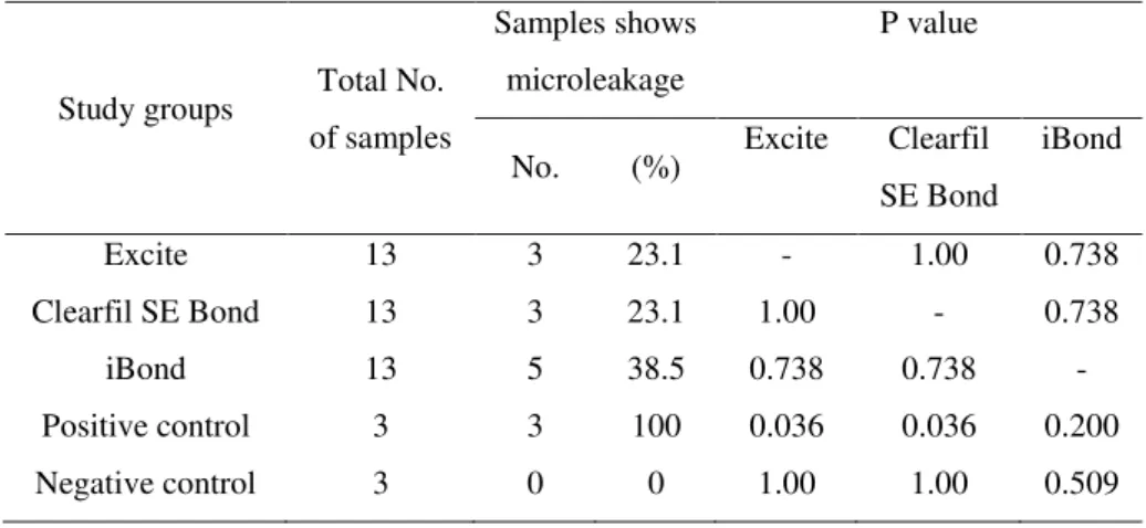
During the experiments all positive controls leaked, based on the results of Fisher's exact test, the positive control group showed significant differences with Excite and Clearfil SE Bond (P=0.036) and no significant differences with iBond (P=0.2). The negative controls do not leak. Based on the results of Fisher's exact test, the negative control group shows no significant differences with Excite and Clearfil SE Bond (P=1.00) as well as iBond (P=0.509). Table 2 illustrates that bacterial microleakage were detected in the test
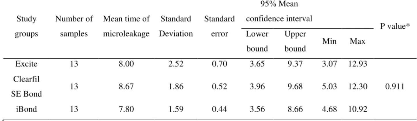
groups: Excite (23.1%), Clearfil SE Bond (23.1%), and iBond (38.5%). Based on the results of Fisher's exact test, there were no significant differences of microleakage among the experimental groups (P=0.738). The estimated mean times of bacterial microleakage were 8 days in Excite group, 8.67 days in Clearfil SE Bond group, and 7.80 days in iBond group (Table 3). Kaplan-Meier Survival analysis showed no statistically significant difference among experimental groups (P=0.911).
Table 2. Frequencydistribution of the samples shows the bacterial microleakage from Fisher's Exact for study groups during 40 days
Study groups Total No. of samples
Samples shows
microleakage
P value
No. (%) Excite Clearfil SE Bond
iBond
Excite 13 3 23.1 - 1.00 0.738
Clearfil SE Bond 13 3 23.1 1.00 - 0.738
iBond 13 5 38.5 0.738 0.738 -
Positive control 3 3 100 0.036 0.036 0.200
Table 3. Mean time and confidence interval of bacterial microleakage from Kaplan-Meier for study groups during 40 days
Study
groups
Number of
samples
Mean time of
microleakage
Standard
Deviation
Standard
error
95% Mean
confidence interval
P value* Lower
bound
Upper
bound Min Max
Excite 13 8.00 2.52 0.70 3.65 9.37 3.07 12.93
0.911 Clearfil
SE Bond 13 8.67 1.86 0.52 3.96 9.68 5.03 12.30
iBond 13 7.80 1.59 0.44 3.56 8.66 4.68 10.92
*
log-rank (Mantel Cox)
Discussion
In the endodontic treatments for filling and sealing of the root canal system, generally gutta-pecha and sometimes newly presented resilon with a sealer has been used. If perforation occur during the root canal therapy (especially in the middle of the canal), MTA has been proposed for sealing the canal after sealing the canal apical end with the common obturation materials (15). Nevertheless, the ability of MTA in preventing the microleakage is doubtful (21, 22). Therefore, filling the coronal part of the canal by the material which provides acceptable coronal sealing is a proper technique. So in the present study, the sealing ability of canal coronal end filling has been evaluated by a composite bonded with three different bonding systems in canals filled with gutta-percha and MTA. For this purpose MTA might be an ideal material, because it induces, by its high pH an effect on dentin, a release of wound healing signals (growth factors) and a chemical bond between MTA and dentin (a layer of hydroxyapatite) that prevents or reduces oral bacterial penetration to the pulp amputation site (23).
In the present study the sealing ability of the adhesives has been evaluated by using bacterial penetration method. The microleakage evaluation technique used in the current study identified only the bacterial penetration, whereas the penetration of endo-toxin also plays an important role in the failure of endodontic treatments and periapical diseases. But the study method used in this work could not evaluate these factors. In the present study the light cure composite (Filtek Z250) has been used. The polymerization shrinkage of this type of composite has been reported in the range of 1.6% to 2% (24). This is important, because
the root canal has an inappropriate geometry for bonding of resin materials (25). Therefore, the selection of material such as Filtek Z250 with acceptable volumetric change is necessary to prevent the gap formation between the filling material and canal wall.
In the present study three adhesive systems (5th, 6th and 7th generations) have been used to bond the composite to the root canal wall. Although the application of the more recent composite generations is easier and faster (with eliminating one step of the procedure), but little is known regarding their performance in prevention of bacterial penetration. Therefore the present study may be useful for determining the performance of these adhesive systems in the root canal dentin. According to the results of the current study, the leakage of bacteria was not seen in any sample of negative control group. This shows that an appropriate sealing quality obtained at the space between the tooth and the plastic cap by using the sticky wax. In the other word easily prepared and inexpensive assembly used in the present study can reliably evaluate the bacterial microleakage.
In the positive control group the bacterial microleakage has been shown after 15 day. However, this has been observed only in less than 48 hours in the same studies (26, 27). This difference may be related to the positive control group preparation method and the better sealing ability obtained from gutta-percha obturation and MTA filling.
significant differences in rate of bacterial leakage among the experimental test materials. This finding has also been reported by Er et al, which demonstrated that the type of dentin bonding gents had no influence in the bacterial leakage of the root-end fillings (20). Our finding also shows that the there were no significant differences in rate of bacterial leakage between 2-step etch & rinse and self etch adhesive systems. We can justify this result by relating this finding to dentin bond strength, because Lopes et al depict that there were no significant differences between dentin bond strength of 2-step etch & rinse and self etch adhesive systems (29). In contrast to the present study, Almeida et al reported that the etch-and-rinse adhesive system had a better performance compared to the self-etching adhesive system on marginal microleakage of dentin (30). The least desirable results for self-etching adhesive systems may be related to the little acidity, which provide a lower degree of demineralization and further infiltrates the enamel surface in a shallow etching depth when compared to phosphoric acid conditioning, then reducing close contact with substrates (30, 31). Although in the present study radicular dentin surfaces have been prepared by Clearfil SE Bond (pH= 2) which has less acidity than 35% phosphoric acid (pH=0.65), but there is no significant difference between the microleakage of self-etch and 2-step etch & rinse groups. Therefore, the Almeida et al (30) and Khosravi et al (31) explanations based on the acidity of conditioning agent cannot justify the findings of the present study. The Inconsistency between the results of the present study and Almeida et al (30) and Khosravi et al (31) can be related to different study methods.
The results of the present study show that there are no significant differences between the microleakage in all experimental groups. Therefore, in coronal sealing of endodontically treated tooth, the use of the latest adhesive generations has been recommended because of their simpler application procedure.
Conclusion
Within the limitations of the present study, it was observed that the adhesive systems in alliance to gutta-percha and MTA obturation could not entirely prevent the bacterial leakage, and their sealing abilities were not statistically different.
References
1. Medić V, Obradović-Djuricić K, Dodić S, Petrović
R. In vitro evaluation of microleakage of various
types of dental cements. Srp Arh Celok Lek 2010;
138:143-9.
2. Zoletti GO, Siqueira JF Jr, Santos KR.
Identification of Enterococcus faecalis in root-filled
teeth with or without periradicular lesions by
culture-dependent and-independent approaches. J
Endod 2006;32:722-6.
3. Veloso HH, Estrela CRA, Decurcio DA, Alves D,
Estrela C. Microbial microleakage in temporary
restorative after post space preparation. Odonto
Ciência 2008;23:187-91.
4. González-Castillo S1, Bailón-Sánchez ME,
González-Rodríguez MP, Poyatos-Martínez R,
Ferrer-Luque CM. An in vitro evaluation of two
dentine adhesive systems to seal the pulp chamber
using a glucose penetration model. Med Oral Patol
Oral Cir Bucal 2011;16:e556-60.
5. Cobankara FK, Unlu N, Cetin AR, Ozkan HB. The
effect of different restoration techniques on the
fracture resistance of endodontically-treated molars.
Oper Dent 2008;33:526-33.
6. Ausiello P, De Gee AJ, Rengo S, Davidson CL.
Fracture resistance of endodontically treated
premolars adhesively restored. Am J Dent 1997;
10:237-41.
7. Hernandez R, Bader S, Boston D, Trope M.
Resistance to fracture of endodontically treated
premolars restored with new generation dentine
bonding systems. Int Endod J 1994;27:281-4.
8. Yazici AR, Baseren M, Dayangac B. The effect of
current-generation bonding systems on
microleakage of resin composite restorations.
Quintessence Int 2002;33:763-9.
9. Pashley DH. Dentin: a dynamic susbstrate. Scan
Microsc 1989;3:161-76.
10. Gogos C, Stavrianos C, Kolokouris I, Economides
N, Papadoyannis I, Shear bond strength of two
resin cements to human root dentin using three
dentin bonding agents. Oper Dent 2007;32:31-6.
11. Zivković S. Quality assessment of marginal sealing
using 7 dentin adhesive systems. Quintessence Int
12. Arias VG, Campos IT, Pimenta LA. Microleakage
study of three adhesive systems. Braz Dent J 2004;
15:194-8.
13. Shahi S, Rahimi S, Lotfi M, Yavari H, Gaderian A.
A comparative study of the biocompatibility of
three root-end filling materials in rat connective
tissue. J Endod 2006;32:776-80.
14. Sarkar NK, Caicedo R, Ritwik P, Moiseyeva R,
Kavashima I. Physicochemical basis of the biologic
properties of mineral trioxide aggragate. J Endod
2005;31:97-100.
15. Lee SJ, Monsef M, Torabinejad M. Sealing ability
of a mineral trioxide aggregate for repair of lateral
root perforations. J Endod 1993;19:541-4.
16. Osorio RM, Hefti A, Vertucci FJ, Shawley AL.
Cytotoxicity of endodontic materials. J Endod
1998; 24:91-6.
17. Sakaue H, Komatsu K, Yoshioka T, Ishimura H,
Ebihara A, Suda H. Evaluation of coronal leakage
and pathway of dye leakage after obturation with
various materials for open apical foramina. Dent
Mater J 2013;32:130-7.
18. Unal GC, Maden M, Isidan T. Repair of Furcal
Iatrogenic Perforation with Mineral Trioxide
Aggregate: Two Years Follow-up of Two Cases.
Eur J Dent 2010;4:475-81.
19. Korasli D, Ziraman F, Ozyurt P, Cehreli SB.
Microleakage of self-etch primer / adhesives in
endodontically treated teeth. J Am Dent Assoc
2007;138:634-40.
20. Er K, Taşdemir T, Bayramoğlu G, Siso SH.
Comparison of the sealing of different dentin
bonding adhesives in root-end cavities: a bacterial
leakage study. Oral Surg Oral Med Oral Pathol Oral
Radiol Endod 2008;106:152-8.
21. Vizgirda PJ, Liewehr FR, Patton WR, McPherson
JC, Buxton TB. A comparison of laterally
condensed percha, thermoplasticized
gutta-percha, and mineral trioxide aggregate as root canal
filling materials. J Endod 2004;30:103-6.
22. Al-Hezaimi K, Naghshbandi J, Oglesby S, Simon
JHS, Rotstein I. Human saliva penetration of root
canals obturated with two types of mineral trioxide
aggregate cements. J Endod 2005;31:453-6.
23. Bakland, L. K. and J. O. Andreasen. Will mineral
trioxide aggregate replace calcium hydroxide in
treating pulpal and periodontal healing
complications subsequent to dental trauma? A
review." Dent Traumatol 2012;28:25-32.
24. Jeong TS, Kang HS, Kim SK, Kim S, Kim HI,
Kwon YH. The effect of resin shades on
microhardness, polymerization shrinkage, and color
change of dental composite resins. Dent Mater J
2009;28:438-45.
25. Tay FR, Loushine RJ, Lambrechts P, Weller RN,
Pashley DH. Geometric factors affecting dentin
bonding in root canals: a theoretical modeling
approach. J Endod 2005;31:584-9.
26. Grecca FS, Rosa AR, Gomes MS, Parolo CF,
Bemfica JR, Frasca LC, Maltz M. Effect of timing
and method of post space preparation on sealing
ability of remaining root filling material: in vitro
microbiological study. J Can Dent Assoc
2009;75:583.
27. Parirokh M, Askarifard S, Mansouri S, Haghdoost
AA, Raoof M, Torabinejad M. Effect of phosphate
buffer saline on coronal leakage of mineral trioxide
aggregate. J Oral Sci 2009;51:187-91.
28. Hilton TJ. Can modern restorative procedures and
materials reliably seal cavities? In vitro
investigations: part 2. Am J Dent 2002;15:279-89.
29. Lopes MB, Sinhoreti MA, CorrerSobrinho L,
Consani S. Comparative study of the dental
substrate used in shear bond strength tests. Pesqui
Odontol Bras 2003;17:171-5.
30. Almeida KG, Scheibe KG, Oliveira AE, Alves CM,
on the microleakage of two adhesive systems. J
Appl Oral Sci 2009;17:92-6.
31. Khosravi K, Ataei E, Mousavi M, Khodaeian N.
Effect of phosphoric acid etching of enamel
margins on the microleakage of a simplified
all-in-one and a self-etch adhesive system. Oper Dent
2009;34:531-6.
Corresponding Author: Tahereh Omidipoor
Hamadan University of Medical Sciences, Hamadan, Iran. Tel: 09163673904