Braz Dent J 19(3) 2008
228 P.T. Silva et al.
Correspondence: Dra. Fernanda Geraldes Pappen, Rua Sete de Setembro 43/1101, 96015-300 Pelotas, RS, Brasil. Tel/Fax: +55-53-3222-9760. e-mail: [email protected]
ISSN 0103-6440
Cytotoxicity Evaluation of Four Endodontic Sealers
Paulo Tadeu da SILVA1 Fernanda Geraldes PAPPEN1
Erick Miranda SOUZA1 João Eduardo DIAS2 Idomeo BONETTI FILHO1 Iracilda Zepponi CARLOS3 Renato Toledo LEONARDO1
1Department of Restorative Dentistry, Dental School of Araraquara,
State University of São Paulo, Araraquara, SP, Brazil
2Discipline of Endodontics, Dental School of Caparica, Portugal 3Discipline of Immunology, School of Pharmaceutical Sciences of Araraquara,
State University of São Paulo, Araraquara, SP, Brazil
This study evaluated in vitro the cytotoxicity of four root canal sealers (Topseal, EndoRez, TubliSeal and Kerr Pulp Canal Sealer E.W.T.) and their effects on reactive oxygen/nitrogen intermediate induction by mouse peritoneal macrophages. Thioglycollate-induced cells were obtained from Swiss mice by peritoneal lavage with 5 mL 10 mM phosphate-buffered saline, washed twice and resuspended (106 cells/mL) in appropriate medium for each test. Cytotoxicity was determined by the presence of hydrogen peroxide (H
2O2) and nitric oxide (NO) by the peroxidase-dependent oxidation of phenol red and Griess reaction, respectively. Sealer suspensions were obtained in two different concentrations from each material: 18 mg/mL and 9 mg/mL, established according to compatibility parameters following MTT assay. Comparing the sealers, H2O2 release at concentrations of 9 mg/mL and 18 mg/mL was similar: Topseal > positive control (medium + cells + 5 mg/mL zimozan solution) > EndoRez > TubliSeal > Kerr Pulp E.W.T. > negative control (medium + cells). NO release at concentration of 9 mg/mL was: positive control (medium + cells + 10 µg/mL LPS solution) > Topseal > Kerr Pulp E.W.T. > TubliSeal = EndoRez > negative control (medium + cells); at concentration of 18 mg/mL was: positive control > Topseal > Kerr Pulp E.W.T > TubliSeal > EndoRez > negative control. Based on the results, it may be concluded that Topseal presented the highest cytotoxicity among the tested sealers, releasing higher concentrations of NO and H2O2 in macrophage culture.
Key Words: cytotoxicity, root canal sealer, macrophages, nitric oxide, hydrogen peroxide.
INTRODUCTION
The goal of a root canal filling is to prevent coronal re-infection and entomb remaining bacteria (1) since the absence of root filling or presence of poor fillings may provide pathways for bacteria and toxins to the periapex. Additionally, root canal sealers must be biocompatible because they may be in direct contact with the apical connective tissues for a long period and might cause inflammatory degeneration of part of these tissues, delaying the periapical healing (2,3). Sealing and biological properties of endodontic sealers are thus essential for the clinical success of endodontic therapy.
Root canal sealers can have different bases, and epoxy resins, calcium hydroxide, zinc-oxide eugenol (ZOE) and silicone present different biological charac-teristics. ZOE-based sealers have a long history of use, with good mechanical properties, but known cytotoxic effects due to eugenol release (4). Resin-based sealers are becoming more popular, but studies have indicated some toxic and mutagenic effects (5,6).
Macrophages are widely distributed in different tissues and play an essential role in the development of the specific and nonspecific immune response. These cells can be activated by bacterial components, cytokines and chemicals. When activated, macrophages produce Braz Dent J (2008) 19(3): 228-231
Braz Dent J 19(3) 2008
Cytotoxicity of root canal sealers 229
and release numerous secretory products like cytokines, inorganic reactive radicals, reactive oxygen intermedi-ates (ROI) and reactive nitrogen intermediintermedi-ates (RNI), all having biological activity (7). Hydrogen peroxide (H2O2) and nitric oxide (NO) are important in cell signaling and are effective molecules for the microbicidal and cyto-toxic response of macrophages after stimulation (8). ROI and RNI can be considered as beneficial interme-diates for presenting microbiocidal and tumoricidal activities. Otherwise, they can also be destructive for the host tissue under certain conditions (9).
This study evaluated in vitro the cytotoxicity of different endodontic sealers by the analysis of NO and H2O2 production by mouse peritoneal macrophages.
MATERIAL AND METHODS
Eleven Swiss mice weighting 18-25 g from the Animal House of the School of Pharmaceutical Sciences of Araraquara, Brazil were used in all experiments. All procedures were undertaken in accordance with the institutional Research Ethics Committee.
Thioglycollate-elicited peritoneal exudate cells (PECs) were obtained from Swiss mice following intraperitoneal injection of 3 mL of thioglycollate medium and rinsing of the peritoneal cavity with 5.0 mL of 10 mM phosphate buffered saline (PBS; pH 7.4; Sigma-Aldrich Brasil, São Paulo, SP, Brazil). The cells were washed twice with PBS by centrifugation at 200 x g for 5 min at 4° C and resuspended in appropriate medium for each test. Macrophages (2 x 10⁶ cells/mL) were suspended in RPMI-1640 containing 5% (v/v) heat-inactivated fetal calf serum (FCS; Cutilab, Campinas, SP, Brazil), 100 IU/mL penicillin (Sigma-Aldrich Brasil), 100 µg/mL streptomycin (Sigma-Aldrich Brasil) and 50 mM 2-mercaptoethanol (Sigma-Aldrich Brasil).
Four endodontic sealers were evaluated: Topseal (epoxy-based; Dentsply De Trey, Konztanz, Germany), EndoRez (methacrylate-based; Ultradent Products, South Jordan, UT, USA), TubliSeal (ZOE-based; SybronEndo, Orange, CA, USA) and Kerr Pulp Canal Sealer E.W.T. (ZOE-based; Kerr Hawe, Bioggio, Switzerland). Materials were prepared as per manufacturers’ instructions under aseptic conditions to prevent biological contamination during the cytotoxicity test. Sealer sus-pensions were made in polyethylene glycol 400 in two concentrations per material: 18 mg/mL and 9 mg/mL. The suspensions were autoclaved for 20 min at 121 ºC.
These concentrations were established according to compatibility parameters following the MTT assay.
Analysis of Hydrogen Peroxide
H2O2 was measured by the horseradish peroxi-dase (HRP)-dependent oxidation of phenol red (10). Macrophages (2 x 10⁶ cells/mL) were suspended in 10 mM potassium phosphate buffer containing 140 mM NaCl, 5.5 mM dextrose, 0.56 mM phenol red, and 0.01 mg/mL type II HRP, pH 7.4. 100 µL of this suspension were added to each well of a 96-well tissue culture plate and exposed to 50 µL of sealer suspension for 1 h at 37°C, 5% CO2 atmosphere. The reaction was inacti-vated with 10 µL of 4 N NaOH. The absorbances were read at 620 nm. Results were expressed as nmol H2O2 per 2 x 10⁵ cells as calibrated against solutions of known H2O2 concentration. The experiments were performed in triplicate. Each experiment was accompanied by a positive control (medium + cells + 5 mg/mL zimozan solution) and a negative control (medium + cells).
Analysis of Nitric Oxide
NO synthesis was determined by measuring the accumulation of nitrite (NO2-), a stable metabolite of NO, in culture supernatants using the Griess reaction (11). Macrophages at 5 x 10⁶ cells/mL in RPMI-1640 (Sigma, St. Louis, MO, USA) containing 5% heat-inactivated FCS, 100 IU/penicillin, 100 µg/mL strepto-mycin and 50 mM 2-mercaptoethanol were added to each well of a 96-well tissue culture dish with 100 µL of the sealer solution. The cells were incubated for 24 h (time for NO assay) at 37oC in a 5% CO
2 atmosphere. After incubation, 50 µL aliquots of culture supernatant were mixed with 50 µL of Griess reagent (1% w/v sulphanylamide, 0.1% w/v naphthylethylenediamine and 3% H3PO4), and incubated at room temperature for 10 min. Absorbance at 540 nm was measured using a microplate reader. NO2- concentration (µmol/L) was calculated from a NaNO2 standard curve. Each experi-ment was accompanied by a positive control (medium + cells + 10 µg/mL LPS solution) and a negative control (medium + cells). The experiments were done in triplicate.
Statistical Analysis
Results are representative of three independent
Braz Dent J 19(3) 2008
230 P.T. Silva et al.
experiments and were presented as means ± SEM of quadruplicate observations (n=12). Data were analyzed statistically by the Student’s t-test using the Origin software (OriginLab Corp., Northamptom, MA, USA).
RESULTS
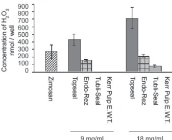
The means were compared in pairs and analyzed by the Student’s t-test. The statistical results of the mean release is summarized in the following lines.H2O2 release at concentrations of 9 mg/mL and 18 mg/mL was similar: Topseal > positive control > EndoRez > TubliSeal > Kerr Pulp E.W.T. > negative control. NO release at concentration of 9 mg/mL was: positive control > Topseal > Kerr Pulp E.W.T. > TubliSeal = EndoRez > negative control; NO release at concentration of 18 mg/ mL was: positive control > Topseal > Kerr Pulp E.W.T > TubliSeal > EndoRez > negative control. (Figs. 1 and 2).
DISCUSSION
Macrophages have been used for immunocytotoxicity testing because they permit the measurement of the cytotoxic response directly in the cell culture and have the ability to maintain immunological functions in the presence of many different chemical agents (12). The effect of endodontic sealers on macrophage activity is of interest since these cells play a key role on innate and acquired immune defenses and on the pathogenesis of inflammation (13).
In response to antigens or inflammatory signals generated at sites of tissue injury, macrophages un-dergo a process of cellular “activation” which is asso-ciated with morphological, functional, and biochemical changes in the cells. One prominent characteristic of activated macrophages is their increased capacity to release pro-inflammatory and cytotoxic mediators, which aid in antigen destruction (14).
In the present study, the determination of sub-toxic concentrations of the sealers using the MTT assay permitted the measurement of the parameters and the exclusion of cell mortality as a variable.
The measurement of H2O2 and NO levels by colorimetric analysis allows, associated with other parameters, the understanding of the compatibility of the material with the host tissues (2). Under physiological conditions, H2O2 is generated in low levels and is quickly metabolized or degraded, but long exposures and high concentrations of H2O2 can destroy biological structures and lead to irreversible cell damage (15).
Both resin-based sealers (Topseal and EndoRez) required more energy to be phagocytized, what was expressed by the amount of H2O2 released. This indi-cates that these sealers are more cytotoxic than TubliSeal and Kerr Pulp Canal Sealer E.W.T., respectively. Topseal induced higher levels of NO release than Kerr Pulp Canal Sealer E.W.T., TubliSeal and EndoRez, respectively. NO affects virtually every step of the development of inflammation. Large amounts of NO can be toxic and pro-inflammatory. NO is not a strong oxidant. However,
Figure 1. Graphic presentation of mean hydrogen peroxide release and the respective 95% range for the means.
Figure 2. Graphic presentation of mean nitric oxide release and the respective 95% range for the means.
60.9
Braz Dent J 19(3) 2008
Cytotoxicity of root canal sealers 231
it reacts at a nearly diffusion limited rate with superoxide to form peroxynitrite, which is a strong oxidant. Peroxynitrite is formed by activated inflammatory cells and agonist-stimulated endothelial cells, and has been found to oxidize several biological molecules and to nitrate free or protein tyrosine residues and other phenolics (16,17). Thus, NO may become cytotoxic or cytostatic. The higher cytotoxicity of resin-based sealers observed in this study confirms the findings of previous investigations (5,6,18,19). The cytotoxicity of TopSeal can be due to minimum amounts of formaldehyde contained in this sealer or to the release of the amine and epoxy resin components from this material, as previously suggested (19). AHPlus has the same formulation of TopSeal. The urethane dimethacrylate (UDMA) in the structure of EndoREZ could be the responsible for its cytotoxic effect, as reported elsewhere (20).
In conclusion, Topseal presented the highest cytotoxicity among the tested sealers, releasing higher concentrations of NO and H2O2 in macrophage culture.
RESUMO
Este estudo avaliou in vitro a citotoxicidade de quatro cimentos obturadores (Topseal, EndoRez, TubliSeal e Kerr Pulp Canal Sealer E.W.T) e seus efeitos na liberação de reativos intermediários do oxigênio e do nitrogênio em cultura de macrófagos peritoniais de ratos.Tioglicolato foi utlizado para se obter células peritoneias de camundongos. A cavidade peritoneal foi irrigada com 5 mL de solução salina 10 mM. As células foram lavadas duas vezes e foi feita uma suspensão (106 células/mL) em meio apropriado para cada um dos testes. A citotoxicidade dos cimentos foi determinada pela presença de peróxido de hidrogênio (H2O2) e óxido nítrico (NO) pela oxidação peroxidase-dependente do vermelho fenol e pela reação de Griess, respectivamente. Suspensões de cimento foram obtidas em duas diferentes concentrações para cada mate-rial: 18 mg/mL e 9 mg/mL, estabelecidas previamente pelo teste de viabilidade celular MTT. Comparando os cimentos, a liberação de H2O2 foi similar nas duas concentrações: Topseal > controle positivo (meio + células + Zimozan a 5mg/mL ) > EndoRez > TubliSeal > Kerr Pulp E.W.T. > controle negativo (meio + células). A liberação de NO na concentração de 9 mg/mL foi: de 9 mg/mL foi: controle positivo (meio + células + solução de LPS a 10 ¼g/mL) > Topseal > Kerr Pulp E.W.T. > TubliSeal = EndoRez > controle negativo (meio + células); e na concentração de 18 mg/mL; e na concentração de 18 mg/mL: controle positivo > Topseal > Kerr Pulp E.W.T > TubliSeal > EndoRez > controle negativo. Baseado nos resultados, pode-se concluir que o Topseal apresentou a maior citotoxicidade dentre os cimentos avaliados, liberando as mais altas concentrações de NO e H2O2 em cultura de macrófagos.
REFERENCES
1 . Torabinejad M, Ung B, Kettering JD. In vitro bacterial
pen-etration of coronally unsealed endodontically treated teeth. J Endod 1990;16:566-569.
2 . Williams DF. Biocompatibility: an overview. In: Encyclopaedia of Medical and Dental Materials. Williams DF, Oxford: Pergamon; 1990. p.51-59.
3 . Geurtsen W, Leyhausen G. Biological aspects of root canal filling materials. Histocompatibility, cytotoxicity and mu-tagenicity. Clin Oral Investig 1997;1:5-11.
4 . Schmaltz G, Hoffmann M, Weis K, Schweikl H. Influence of albumin and collagen on the cell mortality evoked by zinc oxide-eugenol in vitro. J Endod 2000;26:284-287.
5 . Schweikl H, Schmalz G, Federlin M. Mutagenicity of the root canal sealer AHPlus in the Ames test. Clin Oral Invest 1998;2:125-129.
6 . Eldeniz AU, Mustafa K, Orstavik D, Dahl JE. Cytotoxicity of new resin-, calcium hydroxide- and silicone-based root canal sealers on fibroblasts derived from human gingiva and L929 cell lines. Int Endod J 2007;40:329-337.
7 . Forman HJ, Torres M. Redox signaling in macrophages. Mo-lecular aspects of Medicine 2001;22:189-216.
8 . Kayser O, Kolodziej H, Kiderlen AF. Immunomodulatory principles of Pelargonium sidoides. Phytother Res 2000;15:122-126.
9 . McBride AG, Borutaité V, Brown GC. Superoxide dismutase and hydrogen peroxide cause rapid nitric oxide breakdown, peroxynitrite production and subsequent cell death. Biochim Biophys Acta 1999;1454:275-288.
10. Pick E, Mizel D. Rapid microassay of measurement of superox-ide and hydrogen products. J Immun Meth 1981;46:211-226. 11. Green LC, Wagner DA, Glogowski J, Skipper PL, Wishnok JS,
Tannenbaum SR. Analysis of nitrate, nitrite, and [15N] ni-trate in biological fluids. Anal Biochem 1982;126:131-138. 12. Barile FA, Dierickx PJ, Kristen U. In vitro cytotoxicy testing
for prediction of acute human toxicity. Cell BiolToxicol 1994;10:155-162.
13. Unanue ER. The regulation of lymphocyte functions by macrophage. Immunol Rev 1978;40:228-235.
14. Laskin DL, Laskin JD. Role of macrophages and inflamma-tory mediators in chemically induced toxicity. Toxicology 2001;160:111-118.
15. Ramasarma T. H2O2 has a role in cellular regulation. Indian J Biochem Biophys 1990;27:269-274.
16. Ischiropoulos H, Nelson J, Duran D, Al-Medhdi A. Reactions of nitric oxide and peroxynitrite with organic molecules and ferrihorseradish peroxidase: interference with the determina-tion of hydrogen peroxide. Free Radic Biol Med 1996;20:373-381. 17. Linares E, Giorgio S, Mortara RA, Santos CXC, Yamada AT, Augusto O. Role of peroxynitrite in macrophage microbicidal mechanisms in vivo revealed by protein nitration and hy-droxylation. Free Radic Biol Med 2001;30:1234-1242. 18. Bouillaguet S, Wataha JC, Lockwood PE, Galgano C, Golay A,
Krejci I. Cytotoxicity and sealing properties of four classes of endodontic sealers evaluated by succinic dehydrogenase activ-ity and confocal laser scanning microscopy. Eur J Oral Sci 2004;112:182-187.
19. Cohen BI, Pagnillo MK, Musikant BL, Deutsch AS. Formalde-hyde from endodontic materials. Oral Health 1998;88:37-39. 20. Hikage S, Sato A, Suzuki S, Cox CF, Sakaguchi K. Cytotoxic-ity of dental resin monomers in the presence of S9 mix enzymes. Dent Mater J 1999;18:76-86.
Accepted March 31, 2008