Assessment of the chemopreventive effect of casearin B, a clerodane diterpene
extracted from
Casearia sylvestris
(Salicaceae)
Aline M. Prieto
a, André G. dos Santos
b, Ana Paula S. Oliveira
c, Alberto J. Cavalheiro
d, Dulce H.S. Silva
d,
Vanderlan S. Bolzani
d, Eliana Ap. Varanda
c, Christiane P. Soares
a,⇑
aUNESP – Univ. Estadual Paulista, Araraquara, School of Pharmaceutical Sciences, Department of Clinical Analysis, Rua Expedicionários do Brasil 1621, Araraquara, SP, Brazil bUNESP – Univ. Estadual Paulista, Araraquara, School of Pharmaceutical Sciences, Department of Natural Principles and Toxicology, Rodovia Araraquara-Jaú km 01, Araraquara,
SP, Brazil
cUNESP – Univ. Estadual Paulista, Araraquara, School of Pharmaceutical Sciences, Department of Biological Sciences, Rodovia Araraquara-Jaú km 1, Araraquara, SP, Brazil dUNESP – Univ. Estadual Paulista, Araraquara, Núcleo de Bioensaios, Biossíntese e Ecofisiologia de Produtos Naturais, Chemistry Institute, Rua Prof. Francisco Degni, s/n, Araraquara,
SP, Brazil
a r t i c l e
i n f o
Article history:
Received 12 September 2012 Accepted 17 November 2012 Available online 28 November 2012
Keywords: Casearia sylvestris
Ames test Comet assay Casearin B Antioxidant DCFDA
a b s t r a c t
Studies have shown thatCasearia sylvestriscompounds protect DNA from damage both in vitroand in vivo. Complementarily, the aim of the present study was to assess the chemopreventive effect of case-arin B (CASB) against DNA damage using the Ames test, the comet assay and the DCFDA antioxidant assay. The genotoxicity was assessed by the comet assay in HepG2 cells. CASB was genotoxic at concen-trations higher than 0.30
l
M when incubated with the FPG (formamidopyrimidine-DNA glycosylase) enzyme. For the antigenotoxicity comet assay, CASB protected the DNA from damage caused by H2O2in the HepG2 cell line in concentrations above 0.04
l
M with post-treatment, and above 0.08l
M with pre-treatment. CASB was not mutagenic (Ames test) in TA 98 and TA 102. In the antimutagenicity assays, the compound was a strong inhibitor against aflatoxin B1 (AFB) in TA 98 (>88.8%), whereas it was mod-erate (42.7–59.4%) inhibitor against mytomicin C (MMC) in TA 102. Additionally, in the antioxidant assay using DCFDA, CASB reduced reactive oxygen species (ROS) generated by H2O2. In conclusion, CASB wasgenotoxic to HepG2 cells at high concentrations; was protective of DNA at low concentrations, as shown by the Ames test and comet assay; and was also antioxidant.
Ó2012 Elsevier Ltd.
1. Introduction
Casearia sylvestris
is a plant commonly known in Brazil as
‘‘guaçatonga,’’ and its leaves are widely used for their
anti-inflam-matory and anti-ulcerative activities (
Basile et al., 1990; Borges
et al., 2000; Sertié et al., 2000
). In addition, the bark of
C. sylvestris
has been used to treat fever symptoms and for treatment of herpes
virus and diarrhea (
Esteves et al., 2005
). Phytochemical
investiga-tions revealed that the major compounds isolated from
C. sylvestris
(clerodane diterpenes) exhibited anti-fungal activities (
Oberlies
et al., 2002
).
Extracts and chemical components of
C. sylvestris
, particularly
clerodane diterpenes, showed cytotoxic activity against tumor
cells. Specifically to the casearin B (CASB), there is only one
pub-lished study concerning its biological activity, which accessed the
cytotoxicity of the sample against human cell lines using MTT
as-say. The study presents the following CASB cytotoxicity results:
MOLT-4 (leukemia; IC
50= 0.22
l
M), MDA-MB-435 (melanoma;
IC
50= 0.35
l
M), HCT-8 (colon cancer; IC
50= 0.97
l
M), and SF-295
(glioblastoma; IC
50= 0.43
l
M), whereas CASB was less cytotoxic
to L-929 cells (normal fibroblasts; IC
50= 1.52
l
M) demonstrating
a more selective cytotoxic response to tumor cell lines (
dos Santos
et al., 2010
). However, there are no further mechanistic
investiga-tions concerning these results.
Regarding the effects of
C. sylvestris
compounds on DNA, studies
have reported the absence of genotoxic activity of an ethanolic
extract from the leaves of
C. sylvestris
in HTC (hepatoma) and
V79 cells (Chinese hamster lung cells) using comet assays (
Maistro
et al., 2004
). Additionally, the essential oil of the leaves was
anti-clastogenic when evaluated by observing chromosome aberrations
in HTC cells (
Sousa et al., 2007
). Recently, a study using mouse
blood cells showed that an ethanolic extract and caseargrewiin F
0278-6915Ó2012 Elsevier Ltd.
http://dx.doi.org/10.1016/j.fct.2012.11.029
Abbreviations:2-AF, 2-aminofluorene; 2-AN, 2-anthramine; AFB, aflatoxin B1; CASB, casearin B; DCFDA, dichlorodihydrofluorescein diacetate; FPG, formamido-pyrimidine-DNA glycosylase; HPLC-UV, High-performance liquid chromatography-ultraviolet; IR, infrared; LPCC, low-pressure column chromatography; MMC, mytomicin C; MS, mass spectrometry; NMR, nuclear magnetic resonance; NPD, 4-nitro-o-phenylenediamine; SPE, solid-phase extraction; TLC, thin layer chromatography.
⇑
Corresponding author. Address: UNESP, School of Pharmaceutical Sciences, Rua Expedicionários do Brasil 1621, 14 801 902 Araraquara, São Paulo, Brazil. Tel.: +55 16 33015706; fax: +55 16 33220073.E-mail address:soarescp@hotmail.com(C.P. Soares).
Contents lists available at
SciVerse ScienceDirect
Food and Chemical Toxicology
j o u r n a l h o m e p a g e : w w w . e l s e v i e r . c o m / l o c a t e / f o o d c h e m t o x
Open access under the Elsevier OA license.
protected DNA from damage in low concentrations and were
geno-toxic and mutagenic in high concentrations (
de Oliveira et al.,
2009
).
In fact, currently, there are a range of studies that investigate
compounds isolated from plants that protect DNA from damage,
elucidating the possible mechanisms by which these reductions
in DNA damage occur. To complement the previous studies
per-formed with
C. sylvestris
compounds, the aim of the present study
was to assess the chemopreventive effect of CASB using the comet
assay, the Ames test and an antioxidant assay.
2. Materials and methods
2.1. Chemicals
Dulbecco’s Modified Eagle Medium (DMEM), antibiotic–antimycotic solution, kanamycin sulfate, 3-[4,5-dimethylthiazol-2-yl]-2,5-diphenyltetrazolium bromide (MTT) (CAS # 298-93-1), dimethylsulfoxide (DMSO) (CAS # 67-68-5), formaldehyde (CAS # 50-00-0), mitomycin C (MMC) (CAS # 50-07-7), 2-aminofluorene (2-AF) (CAS # 153-78-6), 4-nitro-o-phenylenediamine (NPD) (CAS # 99-56-9), 2-anthra-mine (2-AN) (CAS # 613-13-8) and aflatoxin B1 (AFB) were purchased from Sig-ma–AldrichÒ
(St. Louis, Missouri, USA). Fetal calf serum (FCS) was obtained from CultilabÒ
(Campinas, São Paulo, BRA). Sterile H2O2(CAS # 7722-84-1),ethanol (CAS # 64-17-5), silica (CAS # 7631-86-9), hexane (CAS # 110-82-7), ethyl acetate (CAS # 141-78-6) and isopropanol (CAS # 67-63-0) were purchased from MerckÒ
(Darmstadt, Hessen, DEU). Doxorubicin (Dox) (CAS # 23214-92-8) was purchased from EurofarmaÒ
(São Paulo, BRA). FPG endonuclease was purchased from New England BiolabsÒ
(Ipswich, MA, USA). S9 mixture was obtained from MoltoxÒ
(Boone, NC, USA). Activated charcoal was purchased from SynthÒ
(Diadema, São Paulo, BRA).
2.2. Preparation of extract, and purification and structure determination of CASB
Dried and powdered leaves ofC. sylvestris(20.5 kg) were extracted with ethanol in a stainless steel extractor with solvent re-circulation for 24 h at 40°C. The crude extract was concentrated under reduced pressure to yield 1540.0 g of dry residue. A portion (473.6 g) of the residue was separated by SPE (solid-phase extraction) from silica (60–200
l
m)/activated charcoal (1:1, w/w) by elution with hexane/ ethyl ace-tate (95:5, v/v), ethyl aceace-tate, and methanol to afford three fractions (SPE1-SPE3). SPE2 (16.0 g) was submitted to normal-phase LPCC (low-pressure column chroma-tography) over silica (40–63l
m) eluted with a gradient of hexane/ethyl acetate/ isopropanol of increasing polarity (78:20.5:1.5 to 60:37.3:2.7, v/v): 45 fractions were collected and monitored by TLC (thin layer chromatography), HPLC-UV (High-performance liquid chromatography-ultraviolet), and1H NMR (nuclear mag-netic resonance). Fractions 15–19 were submitted to preparative reversed-phase HPLC (C18 column; 25050 mm; 12l
m) with methanol/ water mixture (67:33or 75:25, v/v) as the mobile phase to yield CASB (250.6 mg),Fig. 1. The compound was identified by spectrometric analysis using NMR, MS (mass spectrometry), IR (infrared) and UV, as described bydos Santos et al. (2010).
2.3. Cell culture
The HepG2 cell line was purchased from the Rio de Janeiro Cell Bank, Brazil. Be-cause this cell line was obtained from a human hepatocellular carcinoma, there are studies showing that it can express hepatic enzymes such as lipase, reductase and catalase (Busch et al., 1990; Cuthbert et al., 1997), leading to a protective effect against promutagens (Wilkening et al., 2003), making it a good model to test anti-oxidant properties. Additionally, HepG2 cell line showed to be highly sensitive to-ward several genotoxicants that give negative results in otherin vitroassays such as mycotoxins (Knasmüller et al., 2004a), becoming a very suitable tool for genotoxi-city testing (Knasmüller et al., 2004b). Additionally, there are no studies concerning if CASB is a phase I/II inductor, which makes HepG2 more suitable to test CASB since there are studies that show that HepG2 cell line is sensitive to both phase I and II inductors genotoxicants (Majer et al., 2004). The cells were grown in 75 cm3flasks (Techno Plastic ProductsÒ
, Trasadingen, CHE) with loosened caps, containing DMEM supplemented with 10% FCS (v/v), antibiotic–antimycotic Solution (1000 U of pen-icillin, 100
l
g/mL of streptomycin sulfate and 0.25l
g/mL amphotericin B), and kanamycin sulfate (100l
g/mL) at 37°C in a CO2incubator (Thermo Fisher Scientific Inc.Ò, MA, USA) under a humidified atmosphere composed of 5% CO2in 95% air.
2.4. Viability
As proposed byTice et al. (2000), the comet assay must be performed in condi-tions of low cytotoxicity; consequently the genotoxicity and antigenotoxicity of the HepG2 cells were assessed under the IC20obtained from the MTT assay. The anti-mutagenicity was assessed under non-cytotoxic conditions using the Ames test, as previously performed inSalmonella typhimuriumstrains.
2.4.1. MTT assay
For the MTT assay, 104cells/well were seeded in a 96 well plate, then the cells were exposed to at least five different concentrations of CASB compounds for a per-iod of 24 h in triplicate, for three independent experiments. The treatments were: (1) negative control (DMEM), (2) positive control (Dox at 15
l
g/mL), (3) vehicle control (DMEM plus DMSO 0.05% (v/v)), (4) five different concentrations of CASB (0.65–10.00l
M). After the treatments, the MTT assay was performed as described byMosmann (1983). Finally, the viability was obtained using the following for-mula: viability (%) = (treatments100)/(negative control).2.4.2. Viability in
S. typhimuriumFirst, the culture medium, bacteria (TA98 or TA102), sterile physiological solution and the compound to be tested were homogenized by vor-texing. Subsequently, the mixture was added to tubes with 9 mL of sterile physio-logical solution to obtain a dilution of 1:10, which was homogenized, and a final volume of 1 mL was transferred to another tube with the same amount of physio-logical solution; this procedure was repeated to obtain a dilution of 105. From the tube containing a dilution of 105, 0.1 mL was seeded on nutrient agar plates, then incubated for 24 h at 37°C. After this period, the colonies were counted and the col-ony-forming units/mL was calculated (CFU = coloniesdilution10). The percent-age of viability for each treatment was compared to the number of colonies from the negative control. According toVargas et al. (1993), cytotoxicity was considered for samples with a percentage of viability below 60%.
2.5. Comet assay
2.5.1. Genotoxicity
HepG2 cells were seeded in 24-well plates (5104cells/well) and treated for 24 h with five concentrations of CASB using 1:2 dilution ratio. The treatments for the comet assay were performed to allow the cells to complete at least one entire cell cycle (24 h); in this way, it was possible to assess the damages that the cells were not able to repair. The CASB concentrations tested were: 0.15, 0.30, 0.60, 1.20 and 2.30
l
M. DMEM plus 0.05% DMSO and 0.5% Hank’s solution (v:v) was used as vehicle control (VC); for the negative control (NC), the cells were treated only with DMEM; and for the positive control, the cells were treated with 1 mM H2O2 for 10 min. The concentration of hydrogen peroxide was obtained by absorbance as proposed byBrestel (1985). After the treatments, the cells were detached, re-sus-pended and homogenized with low-melting-point agarose, spread on a microscope slide pre-coated with normal-melting-point agarose and covered with a coverslip. Then, the slides were incubated for 30 min with 90l
L of FPG enzyme (1:3000, v:v) at 37°C, to specifically assess the oxidative damage in the DNA (Collins et al., 2008). Additionally, an experiment without FPG incubation was also per-formed. The alkaline version of the comet assay (single cell gel electrophoresis) was performed as described bySingh et al. (1988). Duplicate slides were prepared and stained with ethidium bromide, and 50 cells were screened per sample with a fluorescent microscope (ZEISSÒDNA damage was assessed by an image analysis system (TriTek CometScoreÒ
1.5, 2006, Sumerduck, VA, USA), and the DNA percent in the tail was obtained for each treatment. The groups treated with the compounds were compared with the vehicle control using the Kruskal–Wallis test and the associated Dunn post-test using the OriginPro 8.0 software (OriginLabÒ
, Northampton, MA, USA).
2.5.2. Antigenotoxicity
HepG2 cells were pre-treated with CASB and were then exposed to 1 mM of H2O2for 10 min, or the cells were exposed to H2O21 mM for 10 min and post-trea-ted with the CASB. Five concentrations were tespost-trea-ted: 0.02, 0.04, 0.08, 0.15 and 0.30
l
M. The positive control consisted of treating with H2O2(1 mM) for 10 min, and the cells were pre- or post-treated with DMEM, negative control (DMEM) and vehicle control (cells pre- or post-treated with 0.05% DMSO plus Hank’s 0.5% in DMEM plus H2O2(1 mM)). Subsequently, the comet assay was performed as pre-viously described for the genotoxicity assay, incubating the slides with FPG enzyme (1:3000, v:v) because the mutagen used in this test was H2O2. Statistical analysis was performed using the Kruskal–Wallis test and the associated Dunn post-test to compare the treated groups with the vehicle control group using the OriginPro 8.0 software (OriginLabÒ, Northampton, MA, USA). The damage inhibition percent (I) was calculated as proposed byHosseinimehr and Karami (2005): I = [(Positive control)(Test)]100/(Positive control). Additionally, the percentage of inhibi-tion was classified as strong (higher than 60%), moderate (60–41%), weak (40– 21%) and negligible (20–0%) (Calomme et al., 1996).
2.6. Ames test
2.6.1. Mutagenicity
Mutagenicity was evaluated by theSalmonella/microsome assay that is based on the plate-incorporation method proposed byMaron and Ames (1983), usingS. typhimuriumtest strains TA98 and TA102, provided by Dr. B.N. Ames (Berkeley, CA, USA), both with and without metabolization by the S9 mixture. The test strains were obtained from frozen cultures and were grown overnight for 12–14 h at 37°C in oxoid nutrient broth No. 2. The CASB was dissolved in DMSO and added to 2 mL of top agar mixed with 100
l
L of bacterial culture (1–2108cells/mL) and were then added to a plate with minimal agar. Then, the plates were incubated at 37°C for 48 h and the number of His+ revertant colonies was manually counted. Additionally, the influence of metabolic activation was assessed by adding 500l
L of S9 mixture (S9 at a concentration of 0.04 mL/mL of mix). All experiments were analyzed in triplicate. The standard mutagens used as positive controls in each experiment were: TA 102 (S9) MMC (0.5l
g/plate), TA 102 (+S9) 2-AF (10l
g/ plate), TA 98 (S9) NPD (10l
g/plate), TA98 (+S9) 2-AN (1.25l
g/plate). DMSO was used as the negative (vehicle) control (100l
L – maximum volume that was used in the assay). We selected the concentrations based on the toxicity. In a pre-liminary test, we considered toxicity to be when we observed either a reduction in the number of his+ revertants, or an alteration in the auxotrophic background (i.e., background lawn). In all subsequent assays, the upper limit of the dose range tested was the highest nontoxic dose or the lowest toxic dose determined in this assay. In the mutagenicity assay, we used the following concentrations: 0.03; 0.05; 0.10, 0.15 and 0.21 mg/plate. Statistical analysis was performed using the Sal-anal software (Integrate Laboratory Service, NC, USA, 1992), adopting theBernstein et al. (1982)model. The mutagenic index (MI) was also calculated for each dose; that is, the average number of revertants per plate divided by the average number of revertants per plate in the negative (solvent) control. A sample was considered positive when MIP2 for at least one of the tested doses, and there was a reproduc-ible dose–response curve (Varella et al., 2004).2.6.2. Antimutagenicity
Before assessing the antimutagenicity, the cytotoxicity of the highest dose of the compounds associated with the mutagen was tested. Based on the results of this preliminary assay, theC. sylvestriscomponents were tested in non-cytotoxic doses for their antimutagenic properties. The procedures for the antimutagenicity assays were similar to those described for the mutagenicity assays, except that in each tube of top agar containing the bacterial strain and the CASB, the mutagenic agent was also added. One of the mutagens tested was a direct-acting genotoxic com-pound, MMC in TA 102 (4107mg/plate); while the other was the indirect-acting promutagen AFB in TA 98 (5107mg/plate). In addition to AFB, the promutagen-activating fraction S9 was added. This fraction was prepared and used following
Maron and Ames (1983). Toxicity to the various bacteria was also tested, and the sample concentrations employed for the antimutagenic test were all found to be non-toxic. The following CASB concentrations were used in the antimutagenicity assay: 0.02; 0.03; 0.05; 0.10 mg/plate. The concentration of CASB that inhibits 50% of mutagenicity (MID50) was obtained using regression analysis (OriginPro 8.0 software – OriginLabÒ
, Northampton, MA, USA). The calculation of the percentage of mutagenic inhibition (I) was carried out as described byTachino et al. (1994). We also classified the mutagenic inhibition as described in antigeno-toxicity assay.
I¼ ½1 ðrevertants per plate with inhibitor=revertants per plate without inhibitorÞ 100
2.7. Antioxidant assay
The intracellular reactive oxygen species (ROS) were measured using 20,70 -dichlorodihydrofluorescein diacetate (DCFDA). Inside the cell, DCFDA (inactive ROS) is converted to DCFH (active ROS) by an esterase. H2O2and other ROS oxidize intracellular DCFH (non-fluorescent) to DCF (fluorescent). HepG2 cells (104cells/ well) were seeded in sterile black plates. Then, the cells were treated with CASB in three concentrations: 0.04, 0.08 and 0.15 mM for 24 h with their respective con-trols. The concentrations of this compound were chosen based on previously ob-tained results of cytotoxicity and genotoxicity assays. Additionally, the following controls were used for the experiment: (1) negative control (NC) cells (cells main-tained in culture medium), (2) vehicle control (VC) (cells treated with 0.05% DMSO plus Hank’s 0.5% in DMEM) and (3) quercetin (a natural antioxidant) control (Q) (40 mM). After treatment, the wells were washed twice with Hank’s solution. Next, 100
l
L of a solution of 5 mM DCFDA diluted in Hank’s was added and incubated for 30 min at 37°C while protected from light. Thereafter, 1 mM hydrogen peroxide was added for 10 min, after which time the hydrogen peroxide was removed from the plate by aspiration. The wells were washed twice with Hank’s, and 100l
L of Hank’s solution was added to each well for reading with a fluorimeter. Reading was performed every minute for 30 min, at excitation: k= 485 nm, emission:k= 528 nm. A curve of fluorescence intensityversustime was plotted, and from this curve, the total fluorescence intensity for each treatment was estimated by obtain-ing the integral of each curve (area under the curve) usobtain-ing the OriginPro 8.0 soft-ware (OriginLabÒ
, Northampton, MA, USA). Three independent experiments were performed in triplicate. Next, a one-way analysis of variance (ANOVA) was per-formed with the Dunnett post-test to compare the total intensities obtained from the treatmentsversusthe vehicle control (VC) (Nakajima et al., 2009).
3. Results
3.1. Viability and genotoxicity
Before the effect of the compounds on the DNA in the HepG2
cell line was assessed, the MTT assay was performed. The assays
were conducted under the IC
20,2.3
l
M (
Fig. 2
). Complementarily,
the results, shown in
Fig. 2
, demonstrate that this compound was
genotoxic to HepG2 cells at concentrations above 0.30
l
M when
incubated with FPG enzyme, whereas without the enzyme
incuba-tion, CASB begins to show genotoxicity at 1.15
l
M (
Fig. 3
).
3.2. Antigenotoxicity
To assess antigenotoxicity, concentrations that slightly affected
the cell viability and that were not genotoxic were chosen. CASB
significantly reduced the DNA damage caused by H
2O
2in HepG2
cells in both pre- (0.08 and 0.15
l
M) and post-treatments (0.04,
0.08 and 0.15
l
M) (
Fig. 4
A). With regard to the percent inhibition
of DNA damage (
Fig. 4
B), the post-treatment was the most
Fig. 2.MTT viability assay of CASB in the HepG2 cell line treated with the compound for 24 h. IC20= 1.15
l
M.efficient, presenting strong inhibition (70.1%) in the higher
concen-tration tested (0.15
l
M). At the same concentration, the
pre-treat-ment exhibited moderate inhibition (47.8%).
3.3. Mutagenicity
Table 1
shows that CASB was not mutagenic to
S. typhimurium
strains TA 98 and TA 102 with and without metabolization. That
the mutagenic index was <2 demonstrates that the compound
tested could not induce an increase in the number of revertants.
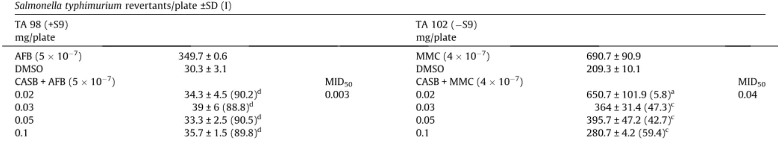
3.4. Antimutagenicity
The results of the antimutagenicity assay for CASB are shown in
Table 2
. The compound was used in association with MMC and AFB
in the strain TA 98 under metabolization (+S9) and in the strain TA
102 without metabolization (
S9). The combination of CASB
(0.10 mg/plate) with the mutagens AFB and MMC resulted in a
via-bility of 100% and 69%, respectively, which are acceptable to
per-form the antimutagenicity assay. Under metabolization using the
strain TA 98, CASB was a strong inhibitor (approximately 90%)
against DNA damage induced by AFB. For the strain TA 102 without
metabolization, CASB was a weak (40–21%) or moderate (41–60%)
inhibitor against the mutagenicity induced by MMC.
3.5. Antioxidant assay
The results of the antioxidant assay (
Fig. 5
) show that CASB was
able to significantly reduce ROS generated by H
2O
2at the two
high-er concentrations tested (0.08 and 0.15
l
M) compared with the
vehicle control (
Fig. 5
B). These results corroborate the H
2O
2DNA
damage reduction shown in the post-treatment with CASB
as-sessed by the comet assay, as shown in
Fig. 4
A.
4. Discussion
The present study assessed the mutagenic/genotoxic,
antimuta-genic/antigenotoxic and antioxidant activities of CASB in
S.
typhimurium
and HepG2 cell lines. Recently, natural products have
been suggested to possess DNA protective and antioxidant
proper-ties (
Dellai et al., 2009; Gupta et al., 2009
), which can be useful to
prevent diseases such as cancer.
The
S. typhimurium
strains TA 98 and TA 102 were used in the
Ames test. The TA 98 strain has a specific deletion in the plasmid
pKM 101 that makes this strain reversible in frameshift events
(
Maron and Ames, 1983
).
S. typhimurium
TA 102 is susceptible to
oxidative damage due to a mutation in the gene
G428
(
Levin
et al., 1982
).
CASB was not mutagenic to the strains TA 98 and TA 102. As
de-scribed previously, these results indicate that the compound did
not induce significant frameshift events or oxidative damage at
the concentrations tested.
However, CASB was genotoxic at concentrations below the IC
20to the HepG2 cells. Interestingly, when the enzyme FPG was used,
the genotoxicity increased significantly compared to the assay
Fig. 3.Genotoxicity in the HepG2 cell line exposed to CASB for 24 h assessed by thecomet assay, with and without incubation with FPG enzyme. NC = negative control (cells in cell culture medium, DMEM); VC = vehicle control (cells treated with DMSO 0.05%, Hank’s 0, 5% in DMEM). Kruskal–Wallis with Dunn post-test VCversus
treatments:⁄p< 0.05;⁄⁄p< 0.01;⁄⁄⁄p< 0.001. Mann–Whitney FPG(+)versusFPG(): cp< 0.001.
without enzyme incubation (
Fig. 3
). Thus, CASB itself caused
oxida-tive damage to DNA with a concentration–response profile.
Although this mechanism has not been elucidated for CASB,
de
Carvalho et al. (1998)
demonstrated that two different diterpene
clerodanes extracted from
C. sylvestris
, known as casearins S and
T, caused acetylation of the DNA molecule when measured by
DNA damage detection in cells of
Saccharomyces cerevisiae
mu-tants. Therefore, the interactions of casearins with DNA may be
associated with the genotoxic effects observed in this study.
Additionally,
Zeiger (2007)
notes that DNA damage protection
studies have to be conducted in parallel with mutagenicity and
cytotoxicity assays to guarantee their safety and avoid
overestima-tion of the chemopreventive effects observed, respectively. Thus,
our DNA damage protection studies were performed using
non-cytotoxic and non-genotoxic/mutagenic conditions.
Compounds that protect DNA from damage can act by two
dif-ferent mechanisms. They can be considered desmutagens, which
reduce the mutagenic/genotoxic effect by directly interacting with
Table 1Mutagenic activity of CASB in strains of S. typhimurium TA 98 and TA 102 in the presence (+S9) and absence (S9) of metabolization.
Salmonella typhimuriumrevertants/plate ±SD (MI)
mg/plate TA 98 TA 102
S9 +S9 S9 +S9
DMSO 71.0 ± 5.6 70.3 ± 11.2 208.0 ± 14.4 317.3 ± 20.2
0.03 78.3 ± 10.3 (1.1) 76.0 ± 16.2(1.1) 211.3 ± 54.3 (1.0) 284.3 ± 3.1 (0.9)
0.05 74.7 ± 15.1 (1.1) 94.3 ± 19.9 (1.3) 217.0 ± 23.5 (1.0) 296.0 ± 24.3 (0.9)
0.10 76.0 ± 12.1 (1.1) 79.0 ± 24.4 (1.1) 188.3 ± 34.1 (0.9) 273.7 ± 13.8 (0.9)
0.15 77.7 ± 20.5 (1.1) 94.3 ± 9.3(1.3) 179.0 ± 24.6 (0.9) 297.7 ± 35.3 (0.9)
0.21 59.3 ± 7.4 (0.8) 88.7 ± 2.1 (1.3) 193.3 ± 33.7 (0.9) 260.7 ± 41.4 (0.8)
Control + 1749.3 ± 152.5 460.0 ± 20.0 1438.3 ± 160.3 371 ± 35.7
MI = Mutagenesis index; SD = standard deviation; Control +: TA 102 (S9) MMC (0.5
l
g/plate), TA 102 (+S9) 2-AF (10l
g/plate), TA 98 (S9) NPD (10l
g/plate), TA98 (+S9) 2-AN (1.25l
g/plate). DMSO (100l
L/plate) = negative control.Table 2
Antimutagenicity assay.S. typhimuriumrevertants per plate treated with CASB in association with mitomycin C (MMC) and aflatoxin B1 (AFB) in strain TA 98 under metabolization (+S9) and strain TA 102 without metabolization (S9).
Salmonella typhimuriumrevertants/plate ±SD (I)
TA 98 (+S9) TA 102 (S9)
mg/plate mg/plate
AFB (5107) 349.7 ± 0.6 MMC (4107) 690.7 ± 90.9
DMSO 30.3 ± 3.1 DMSO 209.3 ± 10.1
CASB + AFB (5107) MID50 CASB + MMC (4
107) MID50
0.02 34.3 ± 4.5 (90.2)d 0.003 0.02 650.7 ± 101.9 (5.8)a 0.04
0.03 39 ± 6 (88.8)d 0.03 364 ± 31.4 (47.3)c
0.05 33.3 ± 2.5 (90.5)d 0.05 395.7 ± 47.2 (42.7)c
0.1 35.7 ± 1.5 (89.8)d 0.1 280.7 ± 4.2 (59.4)c
I = percent inhibition; SD = standard deviation; CASB = CASB; DMSO (100
l
L/plate) = negative control; I =anegligible (20–0%),bweak (40–21%),cmoderate (60–41%) and dstrong (higher than 60%); MID50= concentration of CASB (mgnplate) that inhibits 50% of mutagenicity.
Fig. 5.Antioxidant activity using DCFDA in the HepG2 cell line pre-treated for 24 h with CASB against ROS generated by H2O2. (A) Curve of fluorescence intensity obtained in 30 min of reaction. (B) Total intensity obtained by the integral of the curves presented in A. NC = negative control (cells in cell culture medium, DMEM); VC = vehicle control (cells pre-treated with DMSO 0.05%, Hank’s 0.5% in DMEM and exposed to H2O2). Q = quercetin control (40
l
M). B 0.04 = CASB 0.04l
M. B 0.08 = CASB 0.08l
M. B 0.15 = CASB 0.15l
M. Kruskal–Wallis with Dunn post-test VCversustreatments:⁄p< 0.05;⁄⁄p< 0.01;⁄⁄⁄p< 0.001.the mutagens, block their effects by inhibiting their metabolic
acti-vation or enhance their detoxification. The pre-treatment is
com-monly performed to assess the detoxificant properties of the
compounds, since detoxificant enzymes can be induced during
the pre-treatment period. Alternatively, these compounds can be
classified as bioantimutagens, which promote DNA repair after
damage, increase DNA replication, inhibit error-prone replication
or suppress the growth and replication of cells with damaged
DNA .The post-treatment is useful to assess bioantimutagenic
ef-fects, for instance DNA repair response can be activated after
dam-age, and the post-treatment can help to improve this effect (
Kada
and Shimoi, 1987
). The results obtained for CASB are interesting
because in the antigenotoxicity assay in the HepG2 cell line, CASB
protected the DNA against oxidative damage at lower
concentra-tions with both pre- and post-treatment (
Fig. 4
). Additionally, the
compound also protected the DNA against oxidative damage in
TA102 when exposed to MMC (
Table 2
).
Taken together, the data shown in the genotoxicity and
anti-genotoxicity assays are particularly interesting. They corroborate
previous results obtained in animal models for compounds from
C. sylvestris
, where a weak genotoxic response and an
antigeno-toxic activity were observed concurrently in the comet assay at
the same concentrations (
de Oliveira et al., 2009
). In addition, other
studies described the same profile for compounds derived from
natural products (
Miyaji et al., 2004
). Specifically, with regard to
derivatives of
C. sylvestris
, the possible acetylation of DNA that
gen-erates a genotoxic response may also be responsible for the
acety-lation of histones. It is already known that histone acetyacety-lation
results in DNA relaxation; thus, the DNA becomes accessible to
the transcriptional machinery (
Sterner and Berger, 2000
), thereby
enabling the correction of lesions, either by the DNA repair
mech-anism or by the transcription of detoxificant enzymes (
Li et al.,
2007
). Concerning the detoxificant activity, the results of
post-treatment with CASB in the comet assay (
Fig. 4
) can be explained
by the antioxidant DCFDA assay results, which show that CASB
sig-nificantly induced antioxidant enzymes that reduce ROS generated
by H
2O
2(
Fig. 5
). This finding explains why both a desmutagenic
and a bioantimutagenic response were observed in this work,
and why
de Oliveira et al. (2009)
observed protection to damage
induced by cyclophosphamide in an animal model.
Complementarily, to the comet assay the use of 24 h treatment
was performed to allow the cell to complete one entire cell cycle,
consequently, it was possible to express DNA repair machinery
(post-treatment) or induce antioxidant enzymes (pre-treatment).
Because of this, there was a difference in the percent DNA damage
in the comet tail observed in both pre- and post-treatments that
was clearly observed in the vehicle controls. The explanation for
this result is that the pre-treatment consists of treating cells with
vehicle control or compound, then inducing genotoxic damage
fol-lowed by the comet assay. Damage occurs, but the repair system of
the cell does not have enough time to repair it; therefore, higher
values of damage are observed. However, with the post-treatment,
damage is generated and then the treatments are added for 24 h,
thus allowing the time required for the cell to repair any damage
spontaneously, resulting in the vehicle control showing less
dam-age after the post-treatment compared to pre-treatment. However,
there is significant residual damage (shown in vehicle control) that
the cell cannot repair itself, and the possible reduction of this
dam-age is evaluated with the post-treatment of cells with the
compounds.
Additionally, compared to other compounds that are
antimuta-genic and antioxidant, such as
N
-acetyl-L-cysteine (MID
50= 0.07
-mg/plate; TA 100; against 4-nitroquinoline 1-oxide) or myrcetin,
a flavonoid extracted from natural products (MID
50= 0.16 mg/
plate; TA 98; S9 incubation, against cigarette smoke condensate)
(
Camoirano et al., 1994
), CASB was most efficient on protecting
DNA from damage once it presented a MID
50lower than
N
-acet-yl-L-cysteine or myrcetin as shown in
Table 2
: (MID
50= 0.04 mg/
plate; TA 102; against MMC); (MID
50= 0.003 mg/plate; TA 98; S9
incubation; against AFB).
In the Ames test, CASB strongly inhibited the effects of AFB in
the TA 98 strain under metabolization in the antimutagenesis
as-say. These results indicate that CASB interacts with AFB and may
inhibit the microsomal activation of AFB to electrophilic
metabo-lites (
Ammar et al., 2008
). In the TA 102 strain, a lower inhibition
of the direct agent MMC was observed when compared to AFB. This
result demonstrates that CASB is highly efficient in protecting DNA
from damage under a metabolization model.
In conclusion, CASB showed interesting chemopreventive
char-acteristics in both HepG2 cells and
S. typhimurium
strain by
pro-tecting DNA against different types of damage and by acting as
an antioxidant through inducing detoxificant enzymes in HepG2
cells.
Conflict of Interest
The authors declare that they have no conflict of interest.
Acknowledgments
This work was supported by grants from the
Fundação de
Amparo à Pesquisa do Estado de São Paulo
(FAPESP, São Paulo
Re-search Foundation; Grant no. 2008/58908-0 to A.M.P and 2009/
52481-7 to C.P.S), from the Biota-FAPESP Program (Grant no.
2003/02176-7 to V.S.B.), from the BIOprospecTA Program (Grant
no. 2004/07932-7 to D.H.S.S. and A.J.C.), and from the
Conselho
Nac-ional de Desenvolvimento Científico e Tecnológico
(CNPq, National
Council for Scientific and Technological Development; Grant no.
305615/2006-8 to C.P.S.; scholarship grant to A.G.S.). We wish to
thank Silvia Helena de Oliveira David for her excellent technical
assistance.
References
Ammar, R.B., Sghaier, M.B., Boubaker, J., Bhouri, W., Naffeti, A., Skandrani, I., Bouhlel, I., Kilani, S., Ghedira, K., Chekir-Ghedira, L., 2008. Antioxidant activity and inhibition of aflatoxin B1-, nifuroxazide-, and sodium azide-induced mutagenicity by extracts fromRhamnus alaternusL.. Chem. Biol. Interact. 174, 1–10.
Basile, A.C., Sertie, J.A., Panizza, S., Oshiro, T.T., Azzolini, C.A., 1990. Pharmacological assay ofCasearia sylvestris. I: Preventive anti-ulcer activity and toxicity of the leaf crude extract. J. Ethnopharmacol. 30, 185–197.
Bernstein, L., Kaldor, J., McCann, J., Pike, M.C., 1982. An empirical approach to the statistical analysis of mutagenesis data from theSalmonellatest. Mutat. Res. 97, 267–281.
Borges, M.H., Soares, A.M., Rodrigues, V.M., Andriao-Escarso, S.H., Diniz, H., Hamaguchi, A., Quintero, A., Lizano, S., Gutierrez, J.M., Giglio, J.R., Homsi-Brandeburgo, M.I., 2000. Effects of aqueous extract of Casearia sylvestris
(Flacourtiaceae) on actions of snake and bee venoms and on activity of phospholipases A2. Comp. Biochem. Physiol. B Biochem. Mol. Biol. 127, 21–30. Brestel, E.P., 1985. Co-oxidation of luminol by hypochlorite and hydrogen peroxide implications for neutrophil chemiluminescence. Biochem. Biophys. Res. Commun. 126, 482–488.
Busch, S.J., Barnhart, R.L., Martin, G.A., Flanagan, M.A., Jackson, R.L., 1990. Differential regulation of hepatic triglyceride lipase and 3-hydroxy-3-methylglutaryl-CoA reductase gene expression in a human hepatoma cell line, HepG2. J. Biol. Chem. 265, 22474–22479.
Calomme, M., Pieters, L., Vlietinck, A., Vanden Berghe, D., 1996. Inhibition of bacterial mutagenesis by Citrus flavonoids. Planta Med. 62, 222–226. Camoirano, A., Balansky, R.M., Bennicelli, C., Izzotti, A., D’Agostini, F., 1994.
Experimental databases on inhibition of the bacterial mutagenicity of 4-nitroquinoline 1-oxide and cigarette smoke. Mutat. Res. 317, 89–109. Collins, A.R., Oscoz, A.A., Brunborg, G., Gaivao, I., Giovannelli, L., Kruszewski, M.,
Smith, C.C., Stetina, R., 2008. The comet assay: topical issues. Mutagenesis 23, 143–151.
de Carvalho, P.R., Furlan, M., Young, M.C., Kingston, D.G., Bolzani, V.S., 1998. Acetylated DNA-damaging clerodane diterpenes from Casearia sylvestris. Phytochemistry 49, 1659–1662.
de Oliveira, A.M., dos Santos, A.G., Dos Santos, R.A., Csipak, A.R., Olivato, C., da Silva, I.C., de Freitas, M.B., Bassi, C.L., Cavalheiro, A.J., Bolzani, V.S., Silva, D.H., Sakamoto-Hojo, E.T., Takahashi, C.S., Soares, C.P., 2009. Ethanolic extract of
Casearia sylvestrisand its clerodane diterpen (caseargrewiin F) protect against DNA damage at low concentrations and cause DNA damage at high concentrations in mice’s blood cells. Mutagenesis 24, 501–506.
Dellai, A., Mansour, H.B., Limem, I., Bouhlel, I., Sghaier, M.B., Boubaker, J., Ghedira, K., Chekir-Ghedira, L., 2009. Screening of antimutagenicity via antioxidant activity in different extracts from the flowers ofPhlomis crinitaCav. ssp.mauritanica
munby from the center of Tunisia. Drug Chem. Toxicol. 32, 283–292. dos Santos, A.G., Ferreira, P.M., Vieira Junior, G.M., Perez, C.C., Gomes Tininis, A.,
Silva, G.H., Bolzani Vda, S., Costa-Lotufo, L.V., Pessoa Cdo, O., Cavalheiro, A.J., 2010. Casearin X, its degradation product and other clerodane diterpenes from leaves of Casearia sylvestris: evaluation of cytotoxicity against normal and tumor human cells. Chem. Biodivers. 7, 205–215.
Esteves, I., Souza, I.R., Rodrigues, M., Cardoso, L.G., Santos, L.S., Sertie, J.A., Perazzo, F.F., Lima, L.M., Schneedorf, J.M., Bastos, J.K., Carvalho, J.C., 2005. Gastric antiulcer and anti-inflammatory activities of the essential oil fromCasearia sylvestrisSw. J. Ethnopharmacol. 101, 191–196.
Gupta, D.K., Nicoloso, F.T., Schetinger, M.R., Rossato, L.V., Pereira, L.B., Castro, G.Y., Srivastava, S., Tripathi, R.D., 2009. Antioxidant defense mechanism in hydroponically grown Zea mays seedlings under moderate lead stress. J. Hazard. Mater. 172, 479–484.
Hosseinimehr, S.J., Karami, M., 2005. Citrus extract modulates genotoxicity induced by cyclophosphamide in mice bone marrow cells. J. Pharm. Pharmacol. 57, 505– 509.
Kada, T., Shimoi, K., 1987. Desmutagens and bio-antimutagens – their modes of action. BioEssays 7, 113–116.
Knasmüller, S., Cavin, C., Chakraborty, A., Darroudi, F., Majer, B.J., Huber, W.W., Ehrlich, V.A., 2004a. Structurally related mycotoxins ochratoxin A, ochratoxin B, and citrinin differ in their genotoxic activities and in their mode of action in human-derived liver (HepG2) cells: implications for risk assessment. Nutr. Cancer 50, 190–197.
Knasmüller, S., Mersch-Sundermann, V., Kevekordes, S., Darroudi, F., Huber, W.W., Hoelzl, C., Bichler, J., Majer, B.J., 2004b. Use of human-derived liver cell lines for the detection of environmental and dietary genotoxicants; current state of knowledge. Toxicology 198, 315–328.
Levin, D.E., Hollstein, M., Christman, M.F., Schwiers, E.A., Ames, B.N., 1982. A new
Salmonellatester strain (TA102) with AT base pairs at the site of mutation detects oxidative mutagens. Proc. Natl. Acad. Sci. U.S.A 79, 7445–7449. Li, B., Carey, M., Workman, J.L., 2007. The role of chromatin during transcription.
Cell 128, 707–719.
Maistro, E.L., Carvalho, J.C., Mantovani, M.S., 2004. Evaluation of the genotoxic potential of theCasearia sylvestrisextract on HTC and V79 cells by the comet assay. Toxicol. In Vitro 18, 337–342.
Majer, B.J., Mersch-Sundermann, V., Darroudi, F., Laky, B., de Wit, K., Knasmüller, S., 2004. Genotoxic effects of dietary and lifestyle related carcinogens in human derived hepatoma (HepG2, Hep3B) cells. Mutat. Res. 551, 153–166.
Maron, D.M., Ames, B.N., 1983. Revised methods for theSalmonellamutagenicity test. Mutat. Res. 113, 173–215.
Miyaji, C.K., Jordao, B.Q., Ribeiro, L.R., Eira, A.F., Colus, I.M.S., 2004. Genotoxicity and anti-genotoxicity assessment of shiitake (Lentinula edodes(Berkeley) Pegler) using the Comet assay. Genet. Mol. Biol. 27, 108–114.
Mosmann, T., 1983. Rapid colorimetric assay for cellular growth and survival: application to proliferation and cytotoxicity assays. J. Immunol. Methods 65, 55–63.
Nakajima, Y., Tsuruma, K., Shimazawa, M., Mishima, S., Hara, H., 2009. Comparison of bee products based on assays of antioxidant capacities. BMC Complement Altern. Med. 9, 4.
Oberlies, N.H., Burgess, J.P., Navarro, H.A., Pinos, R.E., Fairchild, C.R., Peterson, R.W., Soejarto, D.D., Farnsworth, N.R., Kinghorn, A.D., Wani, M.C., Wall, M.E., 2002. Novel bioactive clerodane diterpenoids from the leaves and twigs ofCasearia sylvestris. J. Nat. Prod. 65, 95–99.
Sertié, J.A., Carvalho, J.C., Panizza, S., 2000. Antiulcer activity of the crude extract from the leaves ofCasearia sylvestris. Pharm. Biol. 38, 112–119.
Singh, N.P., McCoy, M.T., Tice, R.R., Schneider, E.L., 1988. A simple technique for quantitation of low levels of DNA damage in individual cells. Exp. Cell Res. 175, 184–191.
Sousa, F.G., Schneider, N.F.Z., Mendes, C.E., Moura, N.F., Mendes, C.E., Moura, N.F., Denardin, R.B.N., Matuo, R., Mantovani, M.S., 2007. Clastogenic and Anticlastogenic Effect of the Essential Oil from Casearia sylvestrisSwart. J. Essent. Oil Res. 19, 376–378.
Sterner, D.E., Berger, S.L., 2000. Acetylation of histones and transcription-related factors. Microbiol. Mol. Biol. Rev. 64, 435–459.
Tachino, N., Guo, D., Dashwood, W.M., Yamane, S., Larsen, R., Dashwood, R., 1994. Mechanisms of the in vitro antimutagenic action of chlorophyllin against benzo[a]pyrene: studies of enzyme inhibition, molecular complex formation and degradation of the ultimate carcinogen. Mutat. Res. 308, 191–203. Tice, R.R., Agurell, E., Anderson, D., Burlinson, B., Hartmann, A., Kobayashi, H.,
Miyamae, Y., Rojas, E., Ryu, J.C., Sasaki, Y.F., 2000. Single cell gel/comet assay: guidelines for in vitro and in vivo genetic toxicology testing. Environ. Mol. Mutagen. 35, 206–221.
Varella, S.D., Pozetti, G.L., Vilegas, W., Varanda, E.A., 2004. Mutagenic activity of sweepings and pigments from a household-wax factory assayed with
Salmonella typhimurium. Food Chem. Toxicol. 42, 2029–2035.
Vargas, V.M., Motta, V.E., Henriques, J.A., 1993. Mutagenic activity detected by the Ames test in river water under the influence of petrochemical industries. Mutat. Res. 319, 31–45.
Wilkening, S., Stahl, F., Bader, A., 2003. Comparison of primary human hepatocytes and hepatoma cell line Hepg2 with regard to their biotransformation properties. Drug Metab. Dispos. 31, 1035–1042.
Zeiger, E., 2007. What is needed for an acceptable antimutagenicity manuscript? Mutat. Res. 626, 1–3.