Albanian J. Agric. Sci. 2014 (Special edition) Agricultural University of Tirana
REVIEW PAPER
(Open Access)
Role of fungal biomolecules for establishing mutualistic/pathogenic
interactions with Arabidopsis roots: Possibilities to decipher downstream
signaling events
IRENA SHERAMETI, KHABAT VAHABI, ANATOLI LUDWIG, CHAO SUN AND RALF OELMÜLLER* Institute of General Botany and Plant Physiology, Friedrich-Schiller-University Jena, Dornburger Str. 159, 07743 Jena, Germany
*Corresponding author e-mail: [email protected]
Abstract
A huge number of beneficial and pathogenic fungi release biomolecules into the rhizosphere which initiate signaling events in the roots leading to mutualistic or pathogenic plant/fungus interactions. The combination and concentration of the individual biomolecules in the rhizosphere is critical for the plant´s decision to invest in either growth or defense. These biomolecules activate receptors in the roots and induce cytoplasmic Ca2+ elevation in a phosphorylation-dependent manner. Furthermore, they stimulate phospholipid signaling and AGC kinase activities which coordinate the balanced response between growth/development and defense, by cross-talking to the Ca2+ signals. We use transgenic Arabidopsis plants expressing the Ca2+ sensor aequorin to isolate and identify biomolecules from the exudates of the beneficial root-colonizing fungi Piriformospora indica and Mortierella hyalina, and from the pathogenic fungi Alternaria brassicae and Verticillium dahliae. In this brief summary, we will introduce these microorganisms and their specific role in the interaction with roots. Their exudate componentsinduce a rapid (< 90 s) and transient increase in cytoplasmic Ca2+ levels in the roots. We isolated Arabidopsis non-allelic EMS mutants which do not respond to these biomolecules. Their initial characterization has demonstrated that the Ca2+ response is necessary for the proper plant response to the fungi, and that these mutants are impaired inabscisic acid (ABA) signaling.
Key words: P. indica, M. hyalina, A. brassicae, V. dahliae, transgenic Arabidopsis, [Ca2+]cyt elevation, fungal exudates
1. Introduction
The rhizosphere is probably one of the most complex plant-microbe interaction systems in nature where roots are associated with a wide range of different microorganisms with both detrimental and beneficial outcomes. The relationship between these organisms requires a permanent information exchange during which the roots have to respond to different chemicals from millions of microbes. Central to plant survival is the ability to either limit their growth or intrusion, in the case of pathogens, or promote the association, in the case of symbionts [5, 17]. After perception of the chemical mixture, elaborate communication systems in the roots integrate the information for an appropriate and balanced response which range from defense against pathogens to establishing a mutualistic interaction with beneficial symbionts. A crucial second messenger in the plants with the capability of integrating such diverse chemical information is Ca2+. Both pathogenic and beneficial microbes release microbe- (or pathogen-) associated molecular patterns (MAMPs and PAMPs) and/or uncharacterized compounds which induce a rapid and transient increase in cytoplasmic Ca2+
([Ca2+]
cyt) levels in the root cells upon receptor
activation. Immediately downstream of receptor activation, a phosphorylation cascade participates in the establishment of the so-called Ca2+ signature, i. e.
the generation of [Ca2+]
cyt transients with different
amplitudes, durations, frequencies and [Ca2+]cyt
locations, the selective activation of Ca2+ channels and
the stimulation of respective Ca2+-dependent signaling
components [9, 10]. Changes in [Ca2+]cyt levels and an
oscillation in nuclear [Ca2+] levels (Ca2+ spiking) are
early responses to Nod factors and central to
arbuscular mycorrhizal symbiosis [6]. A
Ca2+/calmodulin dependent protein kinase,
DMI3/CCaMK decodes Nod- and Myc-factor-induced Ca2+ oscillations [9, 12]. In contrast, PAMP-induced [Ca2+]
cyt elevation in pathogenic plant/microbe
interactions activate innate immune responses, the production of reactive oxygen species (ROS) and defense gene expression. Pathogenic and symbiotic microbes can also suppress host responses by secretion of extracellular polysaccharides that sequester apoplastic Ca2+ [1]. [Ca2+]
cyt elevation can
Sherameti et al
release from the nucleus, ER, mitochondria or chloroplasts [8].
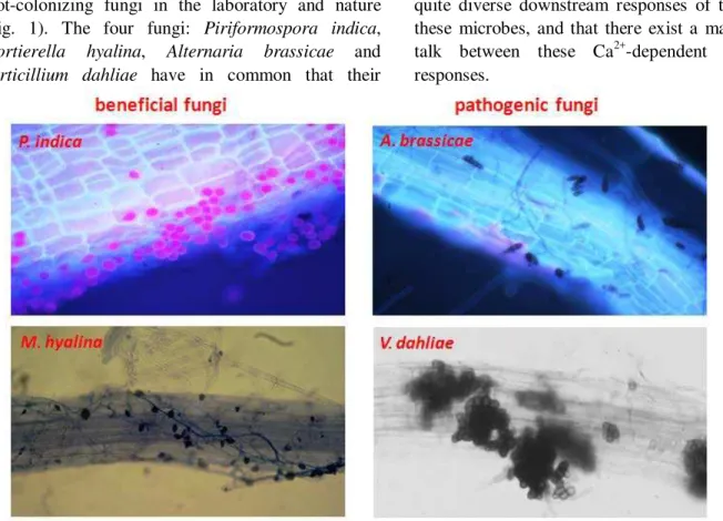
We study the response of the roots of the model plant Arabidopsis thaliana to biomolecules from four root-colonizing fungi in the laboratory and nature (Fig. 1). The four fungi: Piriformospora indica,
Mortierella hyalina, Alternaria brassicae and
Verticillium dahliae have in common that their
exudates contain one or more compounds which induce rapid (within 90 sec) [Ca2+]cyt elevation in the
roots. Genetic studies in Arabidopsis have demonstrated that [Ca2+]cyt elevation is crucial for the
quite diverse downstream responses of the plants to these microbes, and that there exist a massive
cross-talk between these Ca2+-dependent downstream
responses.
Figure 1. Microscopical view of Arabidopsis thaliana roots colonized with different fungi (beneficial and
pathogenic). Piriformospora indica, Mortierella hyalina, Alternaria brassicae, Verticillium dahliae.
2. Beneficial and pathogenic fungi
The endophytic fungus P. indica colonizes the
roots of many plant species. Similar to arbuscular
mycorrhizal fungi, P. indica promotes plant growth,
biomass and seed production and confers resistance to biotic and abiotic stress [3, 21, 22, 24, 25]. P. indica is
a member of Sebacinales, grows inter- and intracellularly and forms pear shaped spores, which accumulate within the roots and on the root surface (Fig. 1). After the establishment of a beneficial interaction, barely any defense or stress genes are activated and no reactive-oxygen species (ROS) are produced by the host against P. indica [3, 8, 16, 24,
25]. The endophyte releases a trihexose that induces root-specific [Ca2+]
cyt elevation in Arabidopsis and N. tabacum, which is followed by a nuclear Ca2+
response in N. tabacum [24, 25].
Similar benefits for the performance of Arabidopsis, N. tabacum and other plant species are
mediated by the root-colonizing fungus M. hyaline
[23]. This fungus (Fig. 1) was originally discovered in
association with the roots of grass plants in golf courts. The fungal exudate contains two biomolecules which strongly induce [Ca2+]cyt elevation in roots and
we are actually working to purify one of these compounds (Ludwig et al. manuscript in preparation).
The necrotrophic fungus A. brassicae (Fig. 1) is a seed-, air- and soil-borne fungus that penetrates through all plant parts and causes lesions on leaves, stems, siliques and roots [2]. It causes black leaf spot disease in crucifers including A. thaliana. The disease progression ultimately results in plant death, mostly caused by host-specific toxins [2, 14, 15, 18, 26] and unknown low molecular weight biomolecules of different chemical classes which induce [Ca2+]cyt
therein]. The fungus forms seed-like long-lived survival structures called microsclerotia, which germinate in response to stimuli from root exudates. The hyphae grow inter- and intracellularly through the root cortex toward the central cylinder of the root (Fig. 1). They enter the xylem cells, proliferate in the vascular system of the root, and at later stages colonize the xylem of the hypocotyl, stem and leaf tissue, where they cause the characteristic symptoms of wilting, by disrupting water transport and ultimately death of aerial tissues [10]. Verticillium species are considered as hemibiotroph with a biotrophic life phase within the nutrient-poor environment of the root xylem and a necrotrophic phase in the aerial plant tissues. V. dahliae releases components, peptides and proteins, probably a cocktail of them, into the medium/environment, which are sufficient to induce disease symptom development in various species including Arabidopsis [9, and references therein]. This exudate preparation contains low molecular weight compounds which rapidly induce [Ca2+]cyt elevations in Arabidopsis root cells.
However, which of these compounds is required for a particular disease phenotype is unknown. We concentrate on two of these biomolecules which induces the strongest Ca2+ response in the roots. Finally, we address questions which need to be answered in order to understand how fungal signals can reprogram the root to either establish a mutualistic interaction with beneficial fungi or to activate the plant immune system against pathogen invaders.
3. Fungal exudates: Induction of cytosolic Ca2+
The exudate preparations from germinating fungal spores in distilled water are used to purify the biomolecules which induce [Ca2+]cyt elevations in the
roots (Fig. 2). The exudate compounds are separated by chromatographic steps (as shown for the P. indica
component in Fig. 3) and fractions with the active compound are identified by the Ca2+ assay.
We use transgenic Arabidopsis lines with the aequorin system [11] to measure [Ca2+]cyt levels in
roots [24, 25]. The Arabidopsis aequorin line was used for ethyl methane sulfonatemutagenesis (EMS). For isolating mutants defective in [Ca2+]cyt elevation to
the different fungal cell wall extracts (CWE) we performed a Ca2+-based screen of approximately 150.000 individual plants (with roots from individual 18-day-old M2 seedlings;for details see [7]). We
identified 12 mutants which completely failed to induce [Ca2+]cyt elevation in response to the CWE
from A. brassicae. Later on, we found out that some of these mutants also failed to respond to CWE preparations for othermicrobes. Under our growth conditions the mutants did not show any visible phenotype compared to wild type (WT) plants [7].
These mutants were named cytoplasmic Ca2+
elevation mutants (cycam) [7]. Genetic analyses of crosses uncovered that four cycam mutants were allelic and two of them, cycam1-1 and cycam1-2, were randomly chosen for further analyses [7]. The
cycam1-1 and cycam1-2 roots did not respond to the Ca2+-inducing extract preparations from mycelia and extract preparation from spores of A. brassicae (data not shown) [7]. Since cycam1 was isolated by a screen in which [Ca2+]cyt elevation was impaired in
Arabidopsis roots, we further tested exudate preparations from other microbes which interact with roots. This list included Rhizoctonia solani, a necrotrophic fungus, Phytophthora parasitica var.
nicotianae, a hemibiotrophicoomycete, and
Agrobacterium tumefaciens, a tumor-inducing bacterium.
Sherameti et al
Figure 3. Purification protocol of the P. indica compound that induces [Ca2+]cyt elevation.
Interestingly, cycam1 did not respond to the exudate preparations from these fungi as well, and less to a CWE from A. tumefaciens, even though these
preparations induced [Ca2+]
cyt elevation in WT. A
CWE preparation from the root-colonizing fungus
Mortierella hyalina [23] induced [Ca2+]cyt elevation in
the roots of the WT and cycam1 mutant. Therefore,
CYCAM1 is involved in [Ca2+]
cyt elevations in
response to different, but not all microbes. Studies are in progress to decipher whether the [Ca2+]
cyt elevation
responses differ between pathogenic and beneficial root-colonizing microbes.
4. Conclusions and perspectives
Our future goal is to compare the responses of the roots of the model plant Arabidopsis thaliana to biomolecules from root-colonizing beneficial and pathogenic fungi. How does a plant distinguish between a beneficial and a pathogenic fungus and how can it mount an appropriate response to signals from
the individual microbes? To address these questions, the bioactive compounds, which induce [Ca2+]
cyt
elevations, must be isolated and characterized in details. The receptors in the roots which become activated after binding of the fungal compound have to be identified. The available mutants which do not show [Ca2+]
cyt elevation in response to the fungal
stimuli are the best tools to identify the responsible genes/protein by map-based cloning strategies and probably Illumina sequencing (next generation sequencing). In this context, it is important to see whether the receptors can be classified into categories which (a) direct beneficial interactions with beneficial microbes and those which (b) activate the plant immune system. The symbiotic signals of mycorrhizal fungi, the Myc factors, and those from rhizobial bacteria, Nod factors, are lipo-chitooligosaccharides. They are perceived by lysin-motif (LysM) receptors which induce a signaling pathway leading to either mycorrhiza or nodule formation. Myc factors from
induce root branching and mycorrhization in
Medicago truncatula [20, and ref. therein]. Interestingly, LysM receptors are also involved in the perception of chitooligosaccharides, fungal cell wall compounds that induce defense responses and resistance to pathogens. This raises the question again of how plants discriminate between beneficial and pathogenic microorganisms and their exudated chemical mediators [cf. 4], and further highlights the importance of this question, for instance for agri- and horticulture. It is reasonable to assume that the perception of fungal signals by root receptors is evolutionarily connected. It is also possible that the exudate compounds from beneficial fungi induce plant immune responses (as signals from pathogens do), which are later on balance by the release of elicitors which reduce or restrict the plant defense response against the beneficial fungi. Such mechanisms have often been described to establish a stable mutualistic interaction, for instance by balancing the growth of the microbes in the roots with the demands of the host.
Another important question is the origin of Ca2+. It is generally believed that it is of apoplastid origin, but a large body of studies also indicates that internal stores, such as the endoplasmatic reticulum, the vacuole and organelles such as nuclei, plastids and
mitochondria store Ca2+ that can be released upon stimulating signals. Most importantly, the Ca2+ channels have to be identified which transport the Ca2+ into the cytoplasm. Since the [Ca2+]cyt elevation
is only transient, there must also be efficient mechanisms which transfer the Ca2+ ions back to its original place. This is most likely mediated by Ca2+ exporters in the plasma membrane or the respective membranes that separate the Ca2+ stores from the cytoplasm.
The Ca2+signature is decoded by Ca2+- binding proteins, and in Arabidopsis, more than 100 different proteins belonging to different protein classes have been identified. Again, the multitude of proteins involved in transducing the Ca2+information to downstream targets maybe important for establishing specificity. Early signaling events also involve reversible phosphorylation events, which can be as fast as seconds after receptor activation. A number of modern mass-spectrometric-based techniques are available now to identify the phosphorylated/dephosphorylated proteins after receptor activation. A comparative analysis of these compounds along the signal transduction pathway may help to decipher events leading to beneficial and pathogenic interactions.
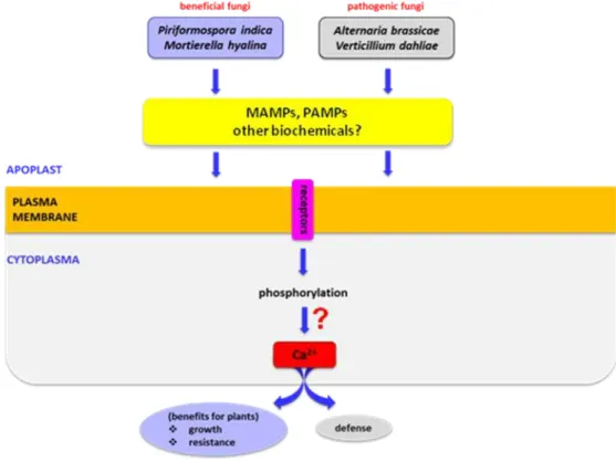
Figure 4. A model describing the apoplastic and cytoplasmic events leading from biomolecules in fungal
Sherameti et al
Finally reprogramming the plant genome requires the activation of specific subsets of genes by transcription factors. These transcription factors need to be identified. However, identification of a specific function associated with a particular transcription factor is often difficult, because they are members of large gene/protein families and thus, individual members of these families often have oberlapping functions. Secondly, the vast majorities of transcription factors do not bind DNA as monomers, but form homo- and particularly heterodimers to activate gene expression. Since one transcription factor can be part of different complexes with quite different functions, it is difficult to pinpoint down a particular function for a transcription factor within a given scenario. The story becomes even more complicated since a particular transcription factor can be part of a complex that functions as a transcriptional activator and of another complex that functions as a transcriptional repressor.
Based on our data and those from the literature we propose a model describing the apoplastic and cytoplasmic events leading from biomolecules in fungal exudates to plant responses (Fig. 4).
We are interested to study early phosphorylation events prior to [Ca2+]cyt elevations, to check whether
differences in the phosphorylation pattern can be detected after application of the biomolecules to the roots. For this we initially useda cerk1 cerk4 knock-out mutant which completely haslost the ability to respond to chitin oligosaccharides [13]. Chitin, a polymer of β-1, 4-linked N-acetyl-glucosamine, is the main component of the fungal cell wall and chitinoligosaccharides which are released from the cell walls of pathogenic fungi function as fungal MAMPs in plant immunity. In A. thaliana, the Chitin Receptor like Kinase CERK1 with three extracellular Lys motifs is responsible for chitinoligosaccharide recognition [7, 19]. CERK1 binds chitin oligomers in vitro, for which all three Lys motifs are required [19] and activates defense genes and the production of ROS. Chitohexaose application to roots induce a rapid increase in [Ca2+]cyt elevations, and this response
depends mainly on CERK1, but also CERK4, one of the four receptors expressed in roots (Mrozinska and Oelmüller, unpublished). It is unclear at present why chitinoligosaccharides from fungal pathogens, but not beneficial fungi, function as PAMPs in plant immunity. Chitin signaling is an excellent example, because the signaling events lead to defense gene activation and many steps in chitin-mediated innate immunity have been elucidate in the past. Therefore,
we will use this information and compare it with the information (and signaling molecules) which become activated after the application of a chemical compound from a beneficial fungus. As mentioned above, also their cell walls contain chitin, and thus, also beneficial fungi can release cell wall compounds which activate immune responses in roots. How can an interaction with a beneficial fungus then lead to the repression of the plant defense system again pathogen attack? By comparing early signaling events in roots after stimulation with either a MAMP (from a beneficial fungus) or a PAMP (from a pathogen), we should be able to answer the question whether chemical mediators in exudate preparations from beneficial and pathogenic fungi trigger the same or different signaling events in Arabidopsis roots.
5. References
1. Aslam SN, Newman MA, Erbs G, Morrissey KL,
Chinchilla D, Boller T, Jensen TT, De Castro C, Ierano T, Molinaro A, Jackson RW, Knight MR,
Cooper RM: Bacterial polysaccharides
suppress induced innate immunity by calcium chelation. Curr. Biol. 2008, 18: 1078-1083. 2. Bains PS, Tewari JP: Purification, chemical
characterization and host-specificity of the toxin produced by Alternaria brassicae.
Physiol. Mol. Plant Pathol. 1987, 30: 259-271. 3. Camehl I, Drzewiecki C, Vadassery J, Shahollari
B, Sherameti I, et al.: The OXI1 kinase
pathway mediates Piriformospora indica -induced growth promotion in Arabidopsis.
PLoS Pathog. 2011, 7: e1002051.
4. Gough C, Cullimore J:
Lipo-chitooligosaccharide signaling in endosymbiotic plant-microbe interactions.
Mol. Plant-Microbe Interact. 2011, 24: 867-878. 5. Harrison MJ: Cellular programs for arbuscular
mycorrhizal symbiosis. Curr. Opin. Plant Biol.
2012, 15: 691-698.
6. Harrison MJ: Signaling in the arbuscular
mycorrhizal symbiosis. Ann. Rev. Microbiol.
2005, 59: 19-42.
7. Johnson JM, Reichelt M, Vadassery J,
Gershenzon J, Oelmüller R: An Arabidopsis
mutant impared in intracellular calcium elevation is sensitive to biotic and abiotic stress. BMC Plant Biol. In Press.
Arabidopsis/Piriformospora indica symbiosis?
In: Molecular Microbial Ecology of the Rhizosphere: FJ de Bruijn, 2013: 833-850.
9. Kanamori N, Madsen LH, Radutoiu S, Frantescu M, Quistgaard EM, Miwa H, Downie JA, James EK, Felle HH, Haaning LL, Jensen TH, Sato S, Nakamura Y, Tabata S, Sandal N, Stougaard J: A nucleoporin is required for induction of Ca2+ spiking in legume nodule development and essential for rhizobial and fungal symbiosis.
Proc. Natl. Acad. Sci. USA 2006, 103: 359-364.
10. Klosterman SJ, Atallah ZK, Vallad GE, Subbarao KV: Diversity, pathogenicity, and management of Verticillium species. Ann. Rev. Phytopathol. 2009, 47: 39-62.
11. Knight MR, Campbell AK, Smith SM, Trewavas
AJ: Transgenic plant aequorin reports the effects of touch and cold-shock and elicitors on cytoplasmic calcium. Nature 1991, 352:
524-526.
12. Lévy J, Bres C, Geurts R, Chalhoub B, Kulikova O, Duc G, Journet EP, Ané JM, Lauber E, Bisseling T, Denarie J, Rosenberg C, Debelle F:
A putative Ca2+ and calmodulin-dependent protein kinase required for bacterial and fungal symbioses. Science 2004, 303: 1361-1364.
13. Miya A, Albert P, Shinya T, Desaki Y, Ichimura K, Shirasu K, Narusaka Y, Kawakami N, Kaku
H, Shibuya N: CERK1, a LysM receptor
kinase, is essential for chitin elicitor signaling in Arabidopsis. Proc. Natl. Acad. Sci. USA
2007, 104: 19613-19618.
14. Moebius N, Hertweck C: Fungal phytotoxins as mediators of virulence. Curr. Opin. Plant Biol.
2009, 12: 390-398.
15. Nishimura S, Kohmoto K: Host-specific toxins and chemical structures from Alternaria species. Annu. Rev. Phytopathol. 1983, 21:
87-116.
16. Nongbri PL, Vahabi K, Mrozinska A, Seebald E, Sun C, et al.: Balancing defense and growth - Analyses of the beneficial symbiosis between
Piriformospora indica and Arabidopsis
thaliana. Symbiosis 2012, 58: 17-28.
17. Oldroyd GE: Speak, friend, and enter:
signalling systems that promote beneficial symbiotic associations in plants. Nat. Rev. Microbiol. 2013, 11: 252-263.
18. Pedras MSC, Khallaf I: Molecular interactions of the phytotoxins destruxin B and sirodesmin PL with crucifers and cereals: Metabolism and elicitation of plant defenses.
Phytochemistry 2012, 77: 129-139.
19. Petutschnig EK, Jones AM, Serazetdinova L, Lipka U, Lipka V: The lysin motif receptor-like kinase (LysM-RLK) CERK1 is a major chitin-binding protein in Arabidopsis thaliana and subject to chitin-induced phosphorylation. J. Biol. Chem. 2010, 285: 28902-28911.
20. Satoh M, Tokaji Y, Nagano AJ, Hara-Nishimura I, Hayashi M, et al.: Arabidopsis mutants affecting oxylipin signaling in photo-oxidative stress responses. Plant Physiol. Biochem. 2013, pii: S0981-9428(13)00418-X.
21. Shahollari B, Vadassery J, Varma A, Oelmüller R: A leucine-rich repeat protein is required for growth promotion and enhanced seed production mediated by the endophytic fungus
Piriformospora indica in Arabidopsis thaliana.
Plant J. 2007, 50: 1-13.
22. Sherameti I, Venus Y, Drzewiecki C, Tripathi S, Dan VM, et al.: PYK10, a beta-glucosidase located in the endoplasmatic reticulum, is crucial for the beneficial interaction between
Arabidopsis thaliana and the endophytic
fungus Piriformospora indica. Plant J. 2008, 54: 428-439.
23. Shinmen Y, Shimizu S, Akimoto K, Kawashima H, Yamada H: Production of arachidonic acid
by Mortierella fungi: selection of a potent
producer and optimization of culture for large-scale production. Appl. Microbiol. Biotechnol. 1989, 31: 11-16.
24. Vadassery J, Ranf S, Drzewiecki C, Mithöfer A, Mazars C, et al.: A cell wall extract from the endophytic fungus Piriformospora indica
promotes growth of Arabidopsis seedlings and induces intracellular calcium elevation in roots. Plant J. 2009, 59: 193-206.
25. Vadassery J, Tripathi S, Prasad R, Varma A,
Oelmüller R: Monodehydroascorbate
reductase 2 and dehydroascorbate reductase 5 are crucial for a mutualistic interaction between Piriformospora indica and Arabidopsis. J. Plant Physiol. 2009, 166:
1263-1274.