J of Evolution of Med and Dent Sci/ eISSN- 2278-4802, pISSN- 2278-4748/ Vol. 4/ Issue 75/ Sept 17, 2015 Page 12985
STUDY OF CRP AND CREATININE HEIGHT INDEX IN PATIENTS OF COPD
Sanskriti Mishra1HOW TO CITE THIS ARTICLE:
Sanskriti Mishra. Study of CRP and Creatinine Height Index in Patients of COPD . Journal of Evolution of Medical and Dental Sciences 2015; Vol. 4, Issue 75, September 17; Page: 12985-12997, DOI: 10.14260/jemds/2015/1872
ABSTRACT: BACKGROUND: This study was conducted in GMCH, Nagpur to measure C-reactive protein and Creatinine Height Index in patients of COPD and to correlate CRP with Creatinine Height Index and Lung Function Test (FEV1%) in these patients. Patients of stable COPD have an on-going systemic inflammation which can be assessed by measuring serum levels of C-Reactive Protein. These patients also suffer from malnutrition causing skeletal muscle wasting which can be assessed by estimation of Creatinine-height index. METHODOLOGY: This is cross sectional observational study carried out in 96 patients with stable COPD attending medicine OPD of Government Medical College, Nagpur over a period of 2 months after obtaining approval from the institute of ethics committee. Informed consent was obtained from each patient. Demographic and clinical data was collected with the help of a questionnaire along with the results of investigations done. Data was analyzed using statistical tool STATA version 10. RESULT: CRP levels were significantly elevated in the study subjects (mean±SD=16.27±8.65). CHI also showed a strong positive correlation with FEV1% the p value being 0.0000 and correlation coefficient=0.7116 suggesting that malnutrition by causing skeletal muscle weakness results in more advanced disease. CONCLUSION: We can conclude that both systemic inflammation and malnutrition are present in patients with stable COPD and working together they cause faster progression of the disease resulting in more advanced stage. Hence these parameters (CRP and CHI) can be applied for follow-up of patients to evaluate treatment methods and their efficacy.
KEYWORDS: COPD, Creatinine height index, CRP.
INTRODUCTION: COPD is characterized by generally progressive airflow limitation in the lungs. Chronic bronchitis defined as presence of cough and sputum for at least 3 months in each of the 2 consecutive years. Emphysema, defined as destruction and enlargement of lung spaces distal to terminal bronchioles, is a pathological term used clinically and describes one of the several structural abnormalities present in COPD.
Chronic obstructive pulmonary disease (COPD) is characterized by chronic airflow limitation and a range of pathological changes in the lung. The chronic airflow limitation characteristic of COPD is caused by a mixture of small airway disease, obstructive bronchiolitis and parenchymal destruction (emphysema). The airflow limitation is usually progressive and associated with an abnormal inflammatory response of the lung to noxious particles or gases. Worldwide, cigarette smoking is the most commonly encountered risk factor for COPD, although in many countries, air pollution resulting from the burning of wood and other biomass fuels has also been identified as a COPD risk factor.1
J of Evolution of Med and Dent Sci/ eISSN- 2278-4802, pISSN- 2278-4748/ Vol. 4/ Issue 75/ Sept 17, 2015 Page 12986 According to crude estimates, 30 million people suffer with COPD in India, and these numbers are only going to increase in the forthcoming years. COPD kills half a million people in India every year, more than those who die due to tuberculosis, malaria or HIV-AIDS.2,3
There is now a large body of data that shows that systemic inflammation exists in stable COPD and that the intensity of the inflammatory process relates to the severity of the underlying disease. CRP levels are raised in COPD patients without clinically relevant IHD and independent of cigarette smoking. CRP may be a systemic marker of the inflammatory process that occurs in patients with COPD.4,5
Many patients with stable COPD suffer malnutrition. Nutritional status is worse with more severe COPD. Depletion involves both fat stores and muscle and visceral protein stores, but the greatest effect is seen in muscle wasting. Skeletal muscle weakness is one of the main systemic effects of COPD and is often accompanied by loss of fat free mass. Muscle wasting has profound effects on morbidity in severe COPD as it results in increased risk of hospitalizations and an increased need for mechanical ventilatory support. Muscle wasting has been identified as significant determinant of mortality in COPD. Assessment of skeletal muscle mass is typically determined by simple measurements and calculations including mid arm circumference (MAC), mid arm muscle circumference (MAMC), and creatinine height ratio (CHI).6,7
We carried out this study to estimate the prevalence of malnutrition and presence of systemic inflammation in stable patients of COPD as both malnutrition and systemic inflammation adversely affect the morbidity and mortality in COPD. There is very limited published literature on this in Indian patients hence this study is important as it would provide some data on prevalence of malnutrition and presence of systemic inflammation in our own patients of COPD.
MATERIAL AND METHODS: Study Design: This was a hospital based cross sectional observational study carried out in 96 patients of stable COPD attending Medicine OPD of Government Medical College Nagpur between June 2013 and July 2013 under the STS program of ICMR with primary objective of measuring CRP and Creatinine height index in patients of stable COPD and secondary objective of correlating CRP with CHI and with lung function test FEV1 in these patients.
Exclusion Criteria: Patients with Acute exacerbation of COPD in last 8 weeks were excluded. Patients with Pulmonary Tuberculosis, Interstitial Lung disease were excluded. Patients with diseases such as malignancies, hepatic, gastrointestinal or kidney abnormalities, metabolic or endocrine diseases, and inflammatory diseases were excluded. Patients with Systemic hypertension, Ischaemic Heart Disease, Cardiomyopathy, Rheumatic Heart Disease, Congenital Heart Disease were excluded.
The study was carried out after obtaining approval of the institutional ethics committee and informed consent of all the study subjects.
J of Evolution of Med and Dent Sci/ eISSN- 2278-4802, pISSN- 2278-4748/ Vol. 4/ Issue 75/ Sept 17, 2015 Page 12987 Expected value Reference Range:
CHI >80% = normal.
CHI =60-80% = mild protein depletion. CHI =40-60% = moderate depletion. CHI <40% = severe depletion.
The CRP levels were correlated with the CHI and the FEV1%. The data was analyzed using statistical software STATA version 10.
OBSERVATIONS AND RESULTS:
Sl.
No. VARIABLE
SUB- VARIABLE
TOTAL (n=96)
1 Gender Male 76(79%)
Female 20(21%) 2 Age(yrs) 41 to 50 20(21%) Mean 59.97 + 9.68 51 to 60 22(23%) 61 to 70 45(46%) 71 to 80 9(10%)
3 Habitat Urban 56(58%)
Rural 40(42%)
4 Smoking Status Smoker 60(62%) Non-smoker 36(38%)
5 Occupation Self employed 28%(27) Farmer 23%(22) Housewife 18%(17) Factory worker 17%(16) Office worker 15%(14)
6 Exposure No exposure 68%(65) Wood smoke 18%(17) Cotton dust 7%(7)
Coal dust 7%(7)
J of Evolution of Med and Dent Sci/ eISSN- 2278-4802, pISSN- 2278-4748/ Vol. 4/ Issue 75/ Sept 17, 2015 Page 12988
Variable Mean±SD
Temp*F 98.20±0.2414
Pulse/min 83±7.352
Resp. Rate/min 16.68±2.340 BP (systolic) mmHg 121.19±10.528 BP(Diastolic) mmHg 78.17±6.555
WBC /uL 8298±2637
Lymphocyte% 30.64±8.90
Monocyte% 3.58±1.96
Granulocyte% 65.53±9.13
RBC /uL 4624063±655197
Hemoglobin gm/dl 12.13±1.73
HCT % 38.19±5.50
RDW 14.94±2.12
Platelets /uL 311270±350253 ESR at 1 hr (Westergren) 31.5±11.2
Total Proteins (gm%) 6.4±0.604 Total Bilirubin (mg%) 0.81±0.331 Alk. Phosphatase (IU) 123.8±25.738
AST/uL 27.45±8.001
ALT/uL 26.79±8.44
Cholesterol (mg%) 162.46±18.57
Urea (mg%) 27.71±9.884
Creatinine (mg%) 1.03±0.257 Blood Glucose Random (mg%) 96.08±14.394
Table 2: Mean and standard deviation of vital parameters, Haemogram and Biochemical
parameters of study subjects. (n=96)
J of Evolution of Med and Dent Sci/ eISSN- 2278-4802, pISSN- 2278-4748/ Vol. 4/ Issue 75/ Sept 17, 2015 Page 12989 The subjects were divided in to 3 subgroups based on FEV1. 32 (33%) were in group 1 with FEV1 between 51%-80%. 47 (49%) were in group 2 with FEV1 between 30%-50%. 17 (18%) were in group 3 with FEV1 <30%.
Chart 2: Column Chart showing duration of symptoms (Yrs) in study subject.
All the study subjects had cough with scanty mucoid expectoration and exertional dyspnoea as the main symptoms. In the present study, the mean duration of cough productive of mucoid sputum in Group 1 (FEV1% 50 to 80, n=32) was 3.28±2.78yrs, in Group 2 (FEV1% 30 to 50, n=47) was 7.10±4.04yrs and in Group 3 (FEV1% <30, n=17) was 7.11±3.95 yrs. The mean duration of exertional breathlessness in Group 1 was 3.78±2.31yrs, in Group 2 was 4.95 ± 2.99yrs and in Group 3 was 8.05±3.92 yrs.
Chart 3: Column Chart showing smoking status (in pack years) in subgroups.
J of Evolution of Med and Dent Sci/ eISSN- 2278-4802, pISSN- 2278-4748/ Vol. 4/ Issue 75/ Sept 17, 2015 Page 12990 VARIABLES GROUP 1
(Mean±SD)
GROUP 2 (Mean±SD)
GROUP 3 (Mean±SD) Temperature(F) 98.1±0.2 98.2±0.2 98.2±0.3
Pulse(/min) 83±7 84±7.2 86±6.3
Respiratory
Rate(/min) 14±2 16±1.9 19±2
BP(mm Hg) 121/78±11/6 119/78±10.1/7.1 124/78±9.8/5.9
TLC 8293±2091 8200±2362 7172±1893
RBC count (/ul) 4687812±641514 4639347±636655 4602777±629059 Haemoglobin (gm%) 12.1±1.6 12.2±1.75 12±1.4
Platelets (/ul) 302593±351263 290173±350345 325277±349253
ESR at 1 hour 21±4.5 32±8.7 32±6.6
Total Proteins (gm%) 6.7±0.3 6.2±0.47 5.7±0.48 Total Bilirubin (mg%) 0.8±0.3 0.8±0.3 0.7±0.3
Alk PO4 (IU) 120±20.1 129±21.8 112±19.2
AST (U/L) 27±8.5 27±7.4 26.7±5.9
ALT (U/L) 28.4±8.3 29±6.9 28.2±11.5
Cholesterol (mg%) 172±18.7 168±19.1 164±19.9
Urea (mg%) 24±6.5 27±9.9 22.8±7
Creatinine (mg%) 1±0.2 1±0.2 0.8±0.1
RBS (mg%) 95±12.8 98±14.8 98±17.7
Table 3: Showing mean and standard deviation of vital parameters, haematological parameters and biochemical parameters of the study subjects in the three
subgroups 1, 2, and 3
The table shows that amongst vital parameters mean respiratory rate in Group 1 is 14±2, in Group 2 is 16±1.9 and in Group 3 is 19±2. Rest of the vital parameters did not show any significant difference across the groups. The mean of all the hematological parameters and biochemical parameters in all the groups was in the normal range and there was no significant difference across the groups.
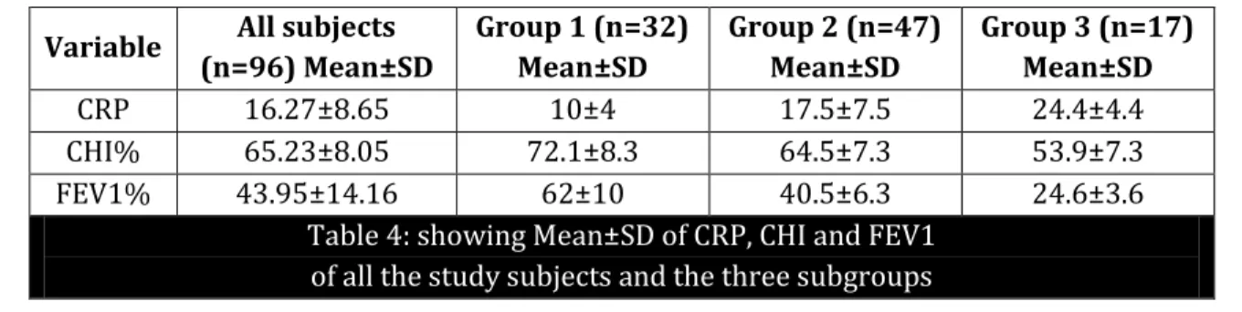
Variable All subjects (n=96) Mean±SD
Group 1 (n=32) Mean±SD
Group 2 (n=47) Mean±SD
Group 3 (n=17) Mean±SD
CRP 16.27±8.65 10±4 17.5±7.5 24.4±4.4
CHI% 65.23±8.05 72.1±8.3 64.5±7.3 53.9±7.3
FEV1% 43.95±14.16 62±10 40.5±6.3 24.6±3.6
J of Evolution of Med and Dent Sci/ eISSN- 2278-4802, pISSN- 2278-4748/ Vol. 4/ Issue 75/ Sept 17, 2015 Page 12991 Chart 4: Column Chart showing mean CRP (mg/L) in different sub groups.
The mean CRP in Group 1 was 10±4, in Group 2 is17.5±7.5 and in Group 3 is 24.4±4.4. The difference in CRP Values of group 1 and 2 is statistically significant with a p value of 0.000, the difference in CRP Values of group 1 and 3 is statistically significant with a p value of 0.000, the difference in CRP Values of group 2 and 3 is statistically significant with a p value of 0.002. This shows that CRP levels were directly proportional to the severity of the disease, higher CRP were associated with more severe disease and lower FEV1, and lower CRP was associated with higher FEV1 and less severe disease.
Chart 5: Column Chart showing CHI (%) in the sub groups.
J of Evolution of Med and Dent Sci/ eISSN- 2278-4802, pISSN- 2278-4748/ Vol. 4/ Issue 75/ Sept 17, 2015 Page 12992 difference in CHI values of Group2 and Group3 is statistically significant with a p value of 0.000, showing as the disease increased in intensity CHI decreased in value showing a negative correlation.
Chart 6: Scatter Diagram showing correlation between CRP and FEV1 of the study subjects.
The scatter diagram has FEV1% of the study subjects plotted along X axis and CRP along Y axis. The line of correlation shows a negative correlation between CRP and FEV1%. The p value is 0.0000 and correlation coefficient= -0.6176.
Chart 7: Scatter diagram showing correlation between FEV1 and CHI.
J of Evolution of Med and Dent Sci/ eISSN- 2278-4802, pISSN- 2278-4748/ Vol. 4/ Issue 75/ Sept 17, 2015 Page 12993 Chart 8: Scatter Diagram showing correlation between CRP and CHI.
The scatter diagram has CRP of the study subjects plotted along X axis and CHI along Y axis. The line of correlation shows negative correlation between CRP and CHI .The P value is 0.0000 and correlation coefficient is -0.6413.
DISCUSSION: This was a hospital based cross sectional observational study which was conducted in 96 patients of stable COPD attending medicine outpatient department. The study was carried out in 2 months between June 2013 and July 2013. The observations were systematically analyzed and compared with available literature:
The mean age of the subjects in our study was 59.97±9.68 yrs. Most subjects 45(46%) were in the age group 61 to 70 yrs followed by 22(23%) in age group 51 to 60 yrs, 20(21%) in the age group 41 to 50 yrs and 9(10%) in the age group 71 to 80 yrs. The prevalence of COPD is known to increase with age. COPD is a disease most commonly observed in middle aged and elderly and is a progressive disease which increases in severity with time.
In our study total of 96 subjects with stable COPD were studied of which 76(79%) were male and 20 (21%) were female. The male to female ratio was 3.8:1. This could be due to smoking which is more common in males. Exposure to other environmental pollutants like smoke, dust and industrial pollutants are also important predisposing factors especially exposure to wood smoke which may be an important risk factor for COPD in women.
The age and sex distribution found in our study is in agreement with Chronic Obstructive Pulmonary Disease fact sheet published in August 2013 by American Lung Association in www.lung.org wherein it is reported that 64% patients of Chronic Bronchitis were over 65 yrs of age and 92% patients of Emphysema were over 45 yrs of age.8
J of Evolution of Med and Dent Sci/ eISSN- 2278-4802, pISSN- 2278-4748/ Vol. 4/ Issue 75/ Sept 17, 2015 Page 12994 In our study of 96 patients of COPD 60(62%) were smoker while 36(38%) were non-smoker.
The ratio of smoker to non-smoker was 1.6:1. Smoking is the most important risk factor associated with COPD as reported by Chronic Obstructive Pulmonary Disease fact sheet published in August 2013 by American Lung Association in www.lung.org.8
Exposure to wood smoke was the most common exposure leading to COPD in the present study followed by exposure to coal dust and cotton dust however majority of patients did not have any occupational exposure to noxious agents.
As shown in Table No. 1, the mean of all the vital parameters of all the study subjects were in the normal range as all the study subjects were in stable status with no co-morbidities.
As shown in Table No. 1 the mean of all haematological variables measured in all the study subjects were in normal range. Stable patients of COPD with no co-morbid condition maintain normal range haematological variables.
As shown in table No. 1 the mean of various biochemical variables measured in all the study subjects were in the normal range for the laboratory. There were no co-morbid conditions in the study subjects.
Depending on the degree of airflow obstruction based on FEV1% of the predicted value the study subjects were divided in to three groups for subgroup analysis.
Group 1 (n=32) 33% 5 % ≤ FEV % < 8 % Group 2 (n=47) 49% 3 % ≤ FEV % <5 % Group 3 (n=17) 18% FEV1% <30%
In the present study, the mean duration of cough productive of mucoid sputum in group 1 was 3.28±2.78 yrs, in Group 2 was 7.10±4.04 yrs and in Group 3 was 7.11±3.95 yrs. The mean duration of exertional breathlessness in Group 1 was 3.78±2.31 yrs, in Group 2 was 4.95±2.99 yrs and in Group 3 was 8.05±3.92 yrs. Thus we found that patients who have longer duration of symptoms of cough and breathlessness have more severe disease. This probably is due to progressive nature of COPD.
The mean duration of smoking in pack years was 15±3.4 pack years in Group 1, 21±4.35 pack years in Group 2 and 32±6.55 pack years in Group 3. Smoking is the major risk factor for COPD and in our study we found that severity of the disease was positively related to the smoking status (In pack years) of the patients. This is possibly due to the cumulative damaging effect of cigarette smoking which makes heavier smokers suffer from more severe disease.
J of Evolution of Med and Dent Sci/ eISSN- 2278-4802, pISSN- 2278-4748/ Vol. 4/ Issue 75/ Sept 17, 2015 Page 12995 CHI and COPD: COPD is often associated with significant nutritional abnormalities, the so called systemic effects of COPD which results in the alterations like weight loss, depletion of fat free mass and to a lesser extent loss of fat mass. Skeletal muscle weakness is one of the main systemic effects of COPD and is often accompanied by loss of fat free mass. CHI which is a sensitive marker of fat free muscle mass is reported to be lower compared to normal in patients of COPD. In the present study we found CHI to be significantly lower compared to normal in the study subjects. Mean CHI% was 65.23±8.05 with normal range being <80%. Also the decrease in CHI was positively related to severity of the disease and subjects with lower CHI had more severe disease. This probably is due to Skeletal muscle wasting and weakness resulting in reduced exercise capacity and faster progression of the disease.
CRP and FEV1: In our study we found that CRP level of the study subjects was negatively associated with the FEV1%. Subjects with higher CRP had more advanced disease with lower FEV1, and those with lower CRP had less advanced disease with higher FEV1. This was found to be statistically significant with a p value of 0.0000.
CRP and CHI: In our study we found a strong negative correlation between CRP and CHI. Those subjects with higher CRP were associated with lower CHI and those with lower CRP were associated with higher CHI. Thus presence of systemic inflammation in patients of COPD and presence of muscle wasting with lower fat free mass go hand in hand and result in more rapid progression of disease.
CHI and FEV1: In our study we found a strong positive correlation between CHI and FEV1. Lower CHI values were associated with lower FEV1 and higher CHI values were associated with Higher FEV1 values. Subjects who suffered lesser degree of muscle wasting due to malnutrition and lesser systemic inflammation suffered from milder disease and those who developed greater muscle wasting due to malnutrition had more advanced disease:
Thus in the present study carried out in 96 patients of stable COPD we found that these patients have presence of systemic inflammation as shown by elevated levels of CRP. Progressively increasing levels of CRP were found with progressively increasing severity of disease. We also found presence of malnutrition in these patients as shown by lower values of CHI compared to normal range. Severity of malnutrition also increased directly with the severity of disease. We can thus conclude that even patients of stable COPD have presence of systemic inflammation and malnutrition and together these factors lead to a more rapid progression of disease process.
Muscle weakness resulting from systemic inflammation and malnutrition can be improved by respiratory muscle exercises and special diet high in calories and rich in proteins, minerals and vitamins. Nutritional supplementation therapy implemented in a pulmonary rehabilitation program is found to be effective in depleted patients with COPD.9 consumption of fresh fruits
and vegetables is positively associated with improved pulmonary function, fewer symptoms, and possibly reduced oxidative stress.10
J of Evolution of Med and Dent Sci/ eISSN- 2278-4802, pISSN- 2278-4748/ Vol. 4/ Issue 75/ Sept 17, 2015 Page 12996 criteria was positively related to the duration of symptoms of cough and breathlessness and also to history of smoking (Pack years).
CRP a marker of systemic inflammation was significantly elevated in the study subjects and it also showed a positive correlation with severity of disease (FEV1%). This confirms the presence of systemic inflammation in patients with stable COPD and it also results in more severe disease.
CHI which is a marker of fat free body mass was significantly lower compared to normal in the study subjects with stable COPD. This confirms presence of malnutrition in these patients. CHI also showed a strong negative correlation with severity disease (FEV1%) suggesting that malnutrition by causing skeletal muscle weakness results in more advanced disease. Thus we can conclude that both systemic inflammation and malnutrition are present in patients with stable COPD and working together they cause faster progression of the disease resulting in more advanced stage.
The present study being cross sectional observational by design had all the shortcomings inherent to it. Larger study especially case control or randomized study is needed to verify the significance of the observations and conclusions of the present study. This can have a therapeutic implication in the management of patients of COPD. Specific nutritional therapy to improve nutritional status can alter the progression of disease. Similarly therapy to reverse systemic inflammation present in these patients can also alter the natural history of the disease process. We have not been able to compare our observations as we could not find any published study in Indian patients.
REFERENCES:
1. Global strategy for the Diagnosis, Management and Prevention of Chronic Obstructive Pulmonary Disease. (GOLD) Updated 2013.
2. Siddharth Shah. COPD. Editorial. JAPI.org/February 2012/COPD/01.
3. S. K. Jindal. COPD: The Unrecognized Epidemic in India. JAPI. org / February 2012 COPD.
4. C- reactive protein in patients with COPD, control smokers and non-smokers. Pinto-Plata V M, Mullerova H, Toso J F, Feudgo-Tepie M, Soriano J B, Vessey R S, Celli B R. Thorax 2006 Jan-61(1):23-8.
5. Wouters E F, Creutzberg E C. Systemic Effects in COPD. Chest. 2002 May; 121 (Suppl):127S-130S.
6. A M W J Schols, P B Soeters, R Mostert, W H M Saris, E F M Wouters Energy Balance in Chronic Obstructive Pulmonary Disease. American Review of Respiratory Disease.1991; 143:1248-1252.
7. Lisa Ezell, G L Jensen. Malnutrition in Chronic Obstructive Pulmonary Disease. American Journal of Clinical Nutrition.org/content/72/6/1415.
8. Chronic Obstructive Pulmonary Disease (COPD) Fact Sheet August 2013. American Lung Association.
9. Creutzberg EC, Wouters EF, Mostert R, Weling-Scheepers CA, Schols AM. Efficacy of nutritional supplementation therapy in depleted patients with COPD. Nutrition.2003 Feb: 19(2):120-7. 10.Llaria St. Florian, Nutrition and COPD- Dietary considerations for a better breathing. Today’s
J of Evolution of Med and Dent Sci/ eISSN- 2278-4802, pISSN- 2278-4748/ Vol. 4/ Issue 75/ Sept 17, 2015 Page 12997
AUTHORS:
1. Sanskriti Mishra
PARTICULARS OF CONTRIBUTORS:
1. Final Year Medical Student, Department of Medicine, Government Medical College, Nagpur.
FINANCIAL OR OTHER
COMPETING INTERESTS: None
NAME ADDRESS EMAIL ID OF THE CORRESPONDING AUTHOR:
Dr. Sanskriti Mishra, #24/A, Shiv Ganga Hospital, Lokmat Square,
Dhantoli-440012, Nagpur.
E-mail: [email protected]