Stress hormones increase cell proliferation and regulates interleukin-6 secretion
in human oral squamous cell carcinoma cells
Daniel G. Bernabé
a,b,⇑, Adriano C. Tamae
b, Éder R. Biasoli
a, Sandra H.P. Oliveira
baOral Oncology Center and Department of Pathology and Clinical Propedeutics, School of Dentistry of Araçatuba, UNESP – Univ. Estadual Paulista, Araçatuba, São Paulo, Brazil bLaboratory of Pharmacology, Department of Basic Sciences, School of Dentistry of Araçatuba, UNESP – Univ. Estadual Paulista, Araçatuba, São Paulo, Brazil
a r t i c l e
i n f o
Article history:
Received 6 July 2010
Received in revised form 16 December 2010 Accepted 18 December 2010
Available online 25 December 2010
Keywords:
Psychological stress Squamous cell carcinoma Oral cancer
Interleukin-6
Beta-adrenergic receptor Norepinephrine Cortisol
a b s t r a c t
Patients with oral cancer can have high psychological distress levels, but the effects of stress-related hor-mones on oral cancer cells and possible mechanisms underlying these relationships are unknown. In this study, we have investigated the effects of stress-related hormones on interleukin-6 (IL-6) secretion and proliferation of oral squamous cell carcinoma (OSCC) cells. The effects of norepinephrine (NE), and cor-tisol were studied in SCC9, SCC15, and SCC25 cells and effects of isoproterenol in SCC9 and SCC25 cells. Real-time PCR studies revealed constitutiveb1- andb2-adrenergic receptors (b-ARs) expression in the SCC9, SCC15, and SCC25 cells. The results showed that NE and isoproterenol significantly enhanced IL-6 mRNA expression and protein production in supernatants of SCC9 and SCC25 cells. Physiological stress levels of NE and isoproterenol (10lM) at 1 h elicited the most robust IL-6 increase. Regarding IL-6 secretion, 10lM NE induced a 5-fold increase at 1 h, 3.7-fold increase at 6 h, and 3.2-fold at 24 h in SCC9 cells. These effects were blocked by theb-adrenergic antagonist propranolol, supporting a role forb-ARs in IL-6 secretion. The effects of cortisol varied according to the hormone concentration. Phar-macological concentrations of cortisol (1000 nM) inhibited IL-6 production by SCC9 and SCC25 cells. Cor-tisol dose that simulates stress conditions (10 nM) tended to increase IL-6 expression in SCC9 cells. Hormonal doses that simulate stress conditions (10lM NE, at 6 h in SCC9 and SCC15 cells and 10 nM cor-tisol, at 48 h in SCC15 cells) stimulated increased cell proliferation. Treatment of SCC9 cells with IL-6 neu-tralizing ab (10lg/mL) partially inhibited NE-induced proliferation. Finally, 20 OSCC biopsies were shown to expressb1- andb2-ARs. These findings suggest that stress hormones can affect oral cancer cells behavior.
Ó2010 Elsevier Inc. All rights reserved.
1. Introduction
A growing number of studies have shown that hormonal and immune alterations resulting from chronic stress and other behav-ioral conditions may influence cancer development and progres-sion (Reiche et al., 2004; Thaker et al., 2007; Antoni et al., 2006; Lillberg et al., 2003). Chronic stress is associated with dysregula-tion of the hypothalamic–pituitary–adrenal (HPA) axis, with con-sequent increase in the production of the hormone cortisol, and elevated levels of norepinephrine (NE) and epinephrine (E), which are catecholamines released from the adrenal medulla and the neurons of the sympathetic nervous system (SNS) (Thaker et al., 2007; Glaser and Kiecolt-Glaser, 2005).
Neurohormonal products derived from chronic stress may re-duce the natural killer cell cytotoxicity by inhibiting the response
of cells to certain cytokines such as interferon-gamma (IFN-
c
) and interleukin-2 (IL-2) (Kiecolt-Glaser and Glaser, 1999; Esterling et al., 1996). Stress hormones also have the ability to act directly on tumor cells and to deregulate the production of cytokines, chemo-kines, and growth factors that are related to cancer development and progression (Reiche et al., 2004; Antoni et al., 2006; Ardestani et al., 1999). For example, studies on ovarian cancer have shown that catecholamines enhance the expression of substances such as vascular endothelial growth factor (VEGF) and matrix metallo-proteinases (MMPs), which are known to influence tumor progres-sion (Lutgendorf et al., 2003; Sood et al., 2006; Yang et al., 2008). Other investigations have demonstrated that the neurohormonal products derived from chronic stress influence skin (Saul et al., 2005), breast (Ben-Eliyahu et al., 1991), lung (Melamed et al., 2005), and colon (Lointier et al., 1992) cancer progression.Interleukin-6 (IL-6) is a cytokine that plays an important role in angiogenesis and tumoral progression (Heikkilä et al., 2008). The head and neck squamous cell carcinoma (HNSCC) cell line secrete IL-6 (Chakravarti et al., 2006), and a high level of this cytokine has been detected in the saliva and blood of patients with HNSCC
0889-1591/$ - see front matterÓ2010 Elsevier Inc. All rights reserved.
doi:10.1016/j.bbi.2010.12.012
⇑Corresponding author. Address: Oral Oncology Center, School of Dentistry of Araçatuba, São Paulo State University – UNESP, José Bonifácio St. 1193, CEP 16015-050, Araçatuba, São Paulo, Brazil. Fax: +55 18 3636 3233.
E-mail address:[email protected](D.G. Bernabé).
Contents lists available atScienceDirect
Brain, Behavior, and Immunity
(Rhodus et al., 2005; Duffy et al., 2008). In-vitro studies have de-scribed that IL-6 stimulates cell proliferation (Chakravarti et al., 2006) and bone invasion of OSCC cells (Okamoto et al., 2000). High IL-6 levels in OSCC tissue and plasma have also been associated with recurrence, lymph node involvement, and a poor prognostic survival (Duffy et al., 2008; Nagata et al., 2003). IL-6 has been directly related to chronic stress. Individuals who experience emotional stress dis-play high IL-6 circulating levels (Kiecolt-Glaser et al., 2003). Recently, it has been shown that NE can increase IL-6 expression in malignant melanocytes (Yang et al., 2009) and human ovarian carcinoma cells (Nilsson et al., 2007), thereby contributing to the development of these tumors.
There are few studies investigating the influence of stress hor-mones on HNSCC. Recently, the presence ofb-adrenergic receptors
(b-AR) for NE and E has been identified in oral (Shang et al., 2009)
and esophagus cancer (Liu et al., 2008) cell lines. These investiga-tions also showed that the proliferation of these cell lines is stim-ulated by NE and E, respectively. Nevertheless, there is no evidence that IL-6 expression in oral cancer can be influenced by stress hor-mones. In this study, we have evaluated the effects of stress-related hormones on IL-6 expression and proliferation of OSCC cells, and evidence that OSCC biopsies expressb-ARs is provided.
2. Materials and methods
2.1. Cell culture and hormone treatment
The OSCC-derived cell lines SCC9, SCC15, and SCC25 were used in the evaluation of the effects of stress hormones. The cell lines were kindly provided by Dr. Ricardo Della Coletta (School of Den-tistry, State University of Campinas, Piracicaba, São Paulo, Brazil). These cells were maintained and propagated in a 1:1 mixture of Dulbecco’s modified Eagle’s medium (DMEM) and Ham’s F12 med-ium (DMEM/F12; Invitrogen, Carlsbad, CA, USA) supplemented with 10% fetal bovine serum (FBS), 100
l
g/mL penicillin, 100l
g/ mL streptomycin, and 0.1% gentamicin, at 37°C, in 5% CO2humidified atmosphere. Experiments were carried out with 80% confluent cultures. SCC9, SCC15, and SCC25 cells were seeded in 24-well plates (1.0105 cells per well) and cultured for 24 h in
serum-reduced medium (0.1% FBS). The following hormones were tested: NE (Calbiochemical Co, La Jolla, CA), cortisol (Sigma– Aldrich, St. Louis, MO) and isoproterenol (Sigma–Aldrich, St. Louis, MO), ab-adrenergic agonist. The cells SCC9 and SCC25 were then
treated with NE or isoproterenol at 0, 0.1, 1, and 10
l
M, or cortisol at 0, 1, 10, 100, and 1000 nM. These concentrations were used in the subsequent experiments. The cells SCC15 were treated with NE and cortisol. For blocking experiments, 1l
M propranolol was added to the cell cultures 1 h before addition of 10l
M NE. Cell-free supernatants and cells were collected at 1, 6, and 24 h, and kept at 80°C until the assays were performed. The hormoneconcentra-tions employed were defined by taking the physiological levels that usually reaching in the tumor microenvironment. NE basal cir-culating levels range between 10 pM and 1 nM (Sood et al., 2006), and studies have suggested that stress increases these levels to approximately 100 nM, and they may reach 10
l
M in the microen-vironment of some types of tumors (Antoni et al., 2006; Sood et al., 2006). The concentrations of 10 and 100 nM cortisol reflect similar levels to those found in stress conditions, and higher concentra-tions (1000 nM) simulate pharmacological doses of glucocorticoids (Miller and O’Callaghan, 2002).2.2. Real-time PCR assessment of IL-6 gene expression
Quantitative real-time reverse transcription-PCR (RT-PCR) was used to assess the IL-6 gene expression in SCC9, SCC15, and
SCC25 cells treated with stress hormones. Total RNA from cell lines was isolated with TRIzol, following the manufacturer’s instructions (Invitrogen Life Technologies, Carlsbad, CA). The RNA concentra-tion was measured by spectrophotometry. First strand cDNAs were synthesized using 1
l
g of total RNA and Superscript II RNase H reverse transcriptase (Invitrogen Life Technologies). IL-6 mRNA levels were measured by means of the SYBR Green system and amplified in the Stepone Real-Time PCR system (Applied Biosys-tems). The housekeeping gene b-actin was employed as internalpositive control. The primers were as follows: b-actin (forward,
50-TGGATCAGCAAGCAGGAGTATG-30; reverse, 50
GCATTTGCGGTG-GACGAT-30) and IL-6 (forward, 50-AGGGCTCTTCGGCAAATGTA-30;
reverse, 50-GAAGGAATGCCCATTAACAACAA-30). Primers were
drawn using the Primer Express software (Applied Biosystems). Reactions were carried out using a volume of 20
l
L, and each sam-ple was run in duplicate. The PCR thermal cycle conditions used in the experiments were those recommended by the manufacturer. The IL-6 mRNA expression levels in each sample were normalized to the b-actin mRNA level. The results were analyzed using thecomparative threshold cycle (CT) method. Results were presented on fold increase of the IL-6 mRNA expression in cells treated with hormones as compared to untreated cells.
2.3. Determination of IL-6 protein expression
The total IL-6 protein concentrations in the supernatants of the SCC9 and SCC25 cells treated with stress hormones were deter-mined. Serum-reduced conditioned medium from cultures of oral cancer cells was collected at 1, 6, and 24 h following exposure to NE, isoproterenol, or cortisol. Quantification of serum IL-6 levels was accomplished by the quantitative sandwich enzyme immuno-assay technique (ELISA) (R&D Systems, Minneapolis, MN) following the manufacture’s protocol. The resulting color was read in a spec-trophotometer set to the wavelength of 450 nm.
2.4.b1- andb2-AR expression in OSCC cell lines
To assess whether the SCC9, SCC15, and SCC25 cell lines express mRNA forb1- andb2-AR, real-time PCR assay was performed as
described previously. The utilized primers were b1 (forward,
50-GCGTGTGATGCATCTTTAGATTTT-30; reverse, 50- CCTAACCCACC
CATCTTCCA-30) andb2 (forward, 50
-TTGAAGGCCTATGGGAATGG-30; reverse, 50-TCCACTCTGCTCCCCTGTGT-30). Primers were drawn
using the Primer Express software. Theb-actin gene was used as
endogenous control.
2.5. MTT cell proliferation assay
OSCC cells SCC9 and SCC15 were seeded in 96-well plates (1.0103per well) and grown in 100
l
L 10% FBS-supplementedDMEM/F12 medium. After 20% confluence had been reached, cells were cultured for 24 h in serum-reduced medium (0.1% FBS). Cells were treated with NE or cortisol. Blocking experiments were per-formed with propranolol (1
l
M added 1 h before addition of 10l
M NE). The MTT solution was carried out by dissolving 5 mg of MTT [3-(4,5-dimethyl-2-thiazolyl)-2,5-diphenyl-2H-tetrazolium bromide] (Sigma) in 1 mL of PBS, followed by filtration and steril-ization in Millipore filter 0.22l
m. The MTT solution (100l
L) diluted 10in serum-reduced medium (0.1%) was added to eachwell after 6, 24, and 48 h of hormone treatment. The cells remained for 4 h at 37°C and 5% CO2humidified atmosphere.The MTT
set to the wavelength of 570 nm. The experiments were carried out in six replicates and were repeated three times.
2.6. Effects of IL-6 neutralizing ab on NE-induced proliferation
To evaluate whether NE-induced OSCC proliferation is mediated by IL-6, anti-IL-6 ab (R&D Systems, Minneapolis, MN) was employed to neutralize the action of IL-6. Briefly, after SCC9 cells had reached 20% confluence, cells were cultured for 24 h in ser-um-reduced medium (0.1% FBS). Then, the SCC9 cells were pre-treated with IL-6 neutralizing ab (1 and 10
l
g/mL) for 30 min prior to the addition of NE (10l
M). Cells were further incubated for 6 h, and proliferation was evaluated by MTT assay.2.7.b1- andb2-AR expression in OSCC biopsies, oral leukoplakia biopsies, and normal oral mucosa
To assess whether OSCC cells expressb1- andb2-AR, 20 tumor
specimens were collected from patients with OSCC who had not re-ceived any treatment yet. All the OSCC cases had the diagnosis con-firmed histologically. Once removed from the surgery site, the specimens were washed in saline solution, placed in a tube con-taining TRIzol reagent (Invitrogen Life Technologies, Carlsbad, CA), and immediately stored in liquid nitrogen. For comparative analysis, 17 specimens of oral leukoplakia (considered a precursor lesion of OSCC) and 15 samples of normal oral mucosa were col-lected and stored following the same protocol. The samples were then thawed and ground in TRIzol with an electric homogenizer.
The total RNA was then extracted, cDNA was synthesized, and real-time PCR assay was performed as previously described.
2.8. Statistical analysis
Data were checked for normality, and statistical analysis was performed by one-way analysis of variance (ANOVA) followed by the Bonferroni’s multiple-comparison test. P values <0.05 were considered significant.
3. Results
3.1. Adrenergic stimulation upregulates IL-6 gene expression in OSCC cells
In all the evaluated times (1, 6, and 24 h), treatment of SCC9, SCC15, and SCC25 cells with physiological stress levels of NE (10
l
M) elevated IL-6 mRNA expression. Maximum IL-6 expression peaked 1 h after stimulation with 10l
M NE, leading to an increase of 501.5 ± 34.8%, 317.1 ± 32.65%, and 237.7 ± 37.6% in IL-6 mRNA expression in SCC9 (p< 0.001), SCC15 (p< 0.05), and SCC25 cells (p< 0.05), respectively (Fig. 1A–C). A smaller but significant enhancement in IL-6 mRNA levels in the SCC9 and SCC25 cell lines was also observed after 6 h of stimulation with NE, which did not continue after 24 h (Fig. 1A and B). The synthetic b-adrenergicreceptor agonist isoproterenol also induced a significant rise in IL-6 mRNA expression in SCC9 and SCC25 cells (SCC15 cells were not tested for isoproterenol). Specifically, after 1 h of treatment of SCC9 cells with 1 and 10
l
M isoproterenol, IL-6 RNAm levelsincreased 269.7 ± 16.4% (p< 0.001) and 395.6 ± 4.4% (p< 0.001), respectively (Fig. 1D). As in the case of SCC9 cells, after 1 h, 10
l
M isoproterenol induced a significant increase in IL-6 mRNA production by SCC25 cells (267.2 ± 43.5%;p< 0.05). However, after longer periods, higher IL-6 mRNA levels were observed with 1l
M isoproterenol, where only the increase after 6 h was significant (194.1 ± 5.8%;p< 0.05) (Fig. 1E).3.2. Adrenergic stimulation increases IL-6 protein levels in culture supernatants
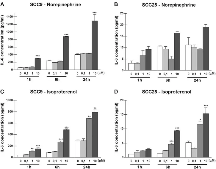
IL-6 protein levels were measured in supernatants of the SCC9 and SCC25 cells. Production of IL-6 protein by SCC9 cells at the three tested times was enhanced compared to the production by SCC25 cells. For example, the mean basal levels of IL-6 production by SCC9 and SCC25 cells at 1 h with no stimulation were 58.63 ± 3.42 pg/mL and 3.11 ± 1.06 pg/mL, respectively. The basal level of IL-6 production by SCC9 and SCC25 cells with no stimula-tion were detectable at 1 h and increased over the time period examined (Figs. 2 and 3). For both cell lines, physiological stress levels of NE (10
l
M) elicited the most robust IL-6 increase. Maxi-mum elevations in IL-6 occurred at 1 h of incubation. As depicted inFig. 2A, stimulation of SCC9 cells with 10l
M NE for 1 h pro-duced 301.3 ± 3.45 pg/mL of IL-6 protein, resulting in an approxi-mately 5-fold increase (p< 0.001) compared to the control. After 6 h, 10l
M NE induced a 3.7-fold increase, whereas after 24 h a3.2-fold enhancement in IL-6 production (p< 0.001) was detected. As for SCC25 cells, treatment with 1
l
M NE for 1 h produced a 2.1-fold increase in IL-6 production, and 10l
M NE induced an eleva-tion of approximately 3-fold (Fig. 2B). For both SCC9 and SCC25 cells, a maximum IL-6 rise was observed after 6 h in the presence of 10l
M isoproterenol. The mean basal level of IL-6 secretion by SCC9 cells after 6 h was 83.18 ± 3.23 pg/mL. The IL-6 levels in-creased to 272.3 ± 12.42 pg/mL after treatment with 1l
M isopro-terenol (p< 0.001), and to 487.1 ± 15.27 pg/mL after treatment with 10l
M isoproterenol (p< 0.001) (Fig. 2C). The patterns of the IL-6 increase in SCC25 cells after isoproterenol stimulation were similar to those found in SCC9 cells, except for the stimulus with 0.1l
M isoproterenol after 24 h, which reduced IL-6 levels (but this result was not significant) (Fig. 2D).3.3. The effects of cortisol on IL-6 expression in OSCC cells
The pattern of IL-6 mRNA expression after treatment with cor-tisol was distinct from that found for NE and isoproterenol. The effects of cortisol varied according to the hormone concentration. In SCC9 cells, in general, higher concentrations of cortisol (100 and 1000 nM) determined lower IL-6 mRNA and protein produc-tion. For 1000 nM cortisol, a dose that is approximately equiva-lent to pharmacological levels of glucocorticoid, there was a significant decrease in IL-6 mRNA expression at all the tested periods. A larger suppression in IL-6 mRNA expression and IL-6
protein levels was observed after treatment with 1000 nM cortisol at 24 h. This treatment reduced IL-6 mRNA expression by 298 ± 1.9% compared to the control (p< 0.01) (Fig. 3A) and induced a 2.7-fold decrease in IL-6 protein levels (p< 0.001) (Fig. 3B). In contrast, after 1 h, 10 nM cortisol (simulating physi-ological stress levels) promoted increase IL-6 mRNA expression (129% compared to control) (Fig. 3A) and protein levels (Fig. 3B) in SCC9 cells, but these changes did not reach signifi-cance. These cortisol effects were blocked by glucocorticoid inhibitor Mefipristone (data not shown). SCC25 cells did not ex-hibit a significant response to cortisol treatment. Specifically, SCC25 cells treated with 1000 nM cortisol at 6 h produced 292.2 ± 17.40 pg/mL of IL-6, resulting in a 1.25-fold decrease compared to the control (p< 0.05) (Fig. 3D). In these same cells, lower IL-6 mRNA levels were detected at 1 h with 100 nM corti-sol (131.1 ± 0.03% compared to the control) and 1000 nM corticorti-sol (152.1 ± 2.7%), while an increase in IL-6 mRNA levels took place at 24 h using 10 nM cortisol (138 ± 12.96%) and 100 nM cortisol (147 ± 28.75%), but these results were not significant (Fig. 3C).
Similar results were found in SCC15 cells, in which lower cortisol concentrations (1 and 10 nM) did not determine large variations in IL-6 mRNA levels, whereas high concentrations simulating pharma-cological concentrations (e.g., 1000 nM) decreased IL-6 expression (but these results were not significant) (Fig. 3E).
3.4. Stress hormones increase OSCC cell proliferation
To examine the effects of stress hormones on OSCC cell prolifer-ation, SCC9 and SCC15 cells were treated with different doses of NE and cortisol, and cell proliferation was assayed by MTT at 6, 24, and
48 h. The SCC25 cell line was not assayed by MTT because it did not respond well (absence of cell growth) to culture in serum-reduced medium (0.1% FBS). Stimulation of SCC9 and SCC15 cells with physiological NE stress levels (10
l
M) induced an enhancement of 170 ± 17.7% (p< 0.05) and 124 ± 13.7% (p< 0.05) in cell prolifer-ation at 6 h compared with non-treated cells, respectively (Fig. 4A). These NE-induced effects of SCC9 and SCC15 cells were not signif-icant at subsequent times (24 and 48 h) (data not shown). In SCC9 cells, treatment with pharmacological levels of cortisol (1000 nM) produced at later time point (48 h) a rise of approximately 200 ± 36.1% in cell proliferation (p< 0.05) (Fig. 4B). Cortisol doses that simulate stress conditions (10 nM) induced at 48 h an increase in cell proliferation in SCC9 (non-significant) (Fig. 4B) and in SCC15 cells (135 ± 17.5%;p< 0.05) (Fig. 4B). There was no significant in-crease in the cell proliferation index after 6 and 24 h of stimulus with cortisol (data not shown).3.5. Regulation of IL-6 expression and OSCC cell proliferation by stress hormones requiresb-ARs activation
Real-time PCR assays confirmed that SCC9, SCC15, and SCC25 cells express mRNA for b1- and b2-AR (Fig. 5A). To determine
whether the increase in IL-6 expression was mediated through
b-adrenergic receptors, the cell lines were pre-treated with a
non-specificbantagonist (propranolol), at the time point of maximum
mRNA IL-6 expression (10
l
M NE at 1 h). Propranolol pretreatment inhibited NE-induced IL-6 mRNA expression in the three cell lines investigated (Fig. 5B). Next, whether the increase in cell prolifera-tion induced by NE was also mediated byb-ARs was assessed. SCC9cells were treated with propranolol before stimulation with 10
l
MNE at 6 h, and cell proliferation was assayed by MTT. Inhibition of
b-ARs produced significant decrease in NE-induced cell
prolifera-tion, showing that this event is b-AR-dependent (Fig. 5C). This
decreasing in NE-induced cell proliferation afterb-ARs inhibition
also was found in the SCC15 cells (results not shown).
3.6. Effects of IL-6 neutralizing ab on NE-induced proliferation
Since NE may stimulate IL-6 production by OSCC, whether NE-induced OSCC proliferation is mediated by IL-6 was subsequently tested. To this end, anti-IL-6 ab was used to neutralize the action of IL-6 in SCC9 cells. As illustrated inFig. 5C, treatment of SCC9 cells with 10
l
g/mL of anti-IL-6 induced significant inhibition of NE-induced proliferation (p< 0.05). Anti-IL-6 in lower concentra-tion (1l
g/mL) was not able to inhibit NE-induced proliferation (Fig. 5C). Recombinant IL-6 increased SCC9 cell proliferation (data not shown).3.7. OSCC biopsies expressb1- andb2-ARs
To determine the clinical relevance of our results, expression of
b1- andb2-ARs mRNAs were examined in 20 tumor specimens of
OSCC and compared with the expression in 17 specimens of oral leukoplakia and 15 specimens of normal oral mucosa. Clinical char-acteristics of patients from whom samples were obtained are sum-marized inTable 1.b1- andb2-AR mRNAs were expressed in all 20
cases of OSCC. Of the 17 cases of leukoplakia, five were negative for
b1-AR and one was negative forb2-AR. Of the 15 specimens of
nor-mal mucosa, three did not expressb1-AR and one was negative for Fig. 4.Stress hormones increase OSCC cell proliferation. The human OSCC cell lines
SCC9 and SCC15 were stimulated with 0.1, 1, and 10lM norepinephrine or with 1, 10, 100, and 1000 nM cortisol, and MTT cell proliferation assays were determined at 6, 24, and 48 h. The non-stimulated cells (0lM or 0 nM) were set as 100%, and the treated cells were determined relatively to non-stimulated cells. Significant results were found with NE at 6 h (A) and cortisol at 48 h (B). Bars represent the mean ± SEM of six replicates, and the assays were performed in triplicate.⁄P60.05.
b2-AR. Quantitatively, the mean expression of the b1-AR mRNA
levels in OSCC specimens was 2.7-fold higher compared to normal mucosa (p< 0.05), while in specimens of leukoplakia the expres-sion was 1.6-fold higher (p> 0.05) (Fig. 6A). In contrast, b2-AR
mRNA mean expression was lower in leukoplakia compared to normal mucosa and OSCC, but these results were not significant (Fig. 6A). Theb-AR expression for each studied case can be better
seen inFig. 6B and C.
4. Discussion
This study provides strong evidence that OSCC cells are influ-enced by neurohormonal mediators. The results demonstrated that stress-related mediators (NE and isoproterenol) can enhance the production of the pro-angiogenic cytokine IL-6 in human OSCC cell lines. IL-6, originally identified as a B-cell growth factor, is pro-duced by many cell types, including T-cells, macrophages, and stromal cells. As seen in this study, OSCC cells are also capable of producing IL-6, and basal levels are already detectable at 1 h. Se-creted cytokine products, including IL-6, are available to interact with cellular receptors; thus, they are able to exert paracrine or autocrine effects. The concentrations of IL-6 secreted by OSCC cells in this study, even by non-stimulated cells, are clearly within the range expected to have biological activity. This activity is related to autocrine growth stimulation of cancer cells (Hodge et al., 2005) and could explain the non-stimulated increase in IL-6 ob-served over the time period. Previous studies on melanoma (Yang et al., 2009) and ovarian cancer cells (Nilsson et al., 2007) have shown that IL-6 expression is upregulated via adrenergic stimula-tion. Enhanced IL-6 production after NE treatment has also been reported in myocytes (Briest et al., 2003) and human pancreatic duct epithelial cells (Chan et al., 2008).
The NE and isoproterenol concentrations that determined max-imum increase in IL-6 expression were within the levels that would be produced from stress-related catecholamine secretion (10
l
M). Maximum elevations in IL-6 occurred at an early time(1 h), giving evidence of fast metabolism of adrenergic mediators by OSCC cells.Nilsson et al. (2007)found that maximum increases in IL-6 expression in ovarian carcinoma cells occurred only after 6 h of incubation with NE. Nilsson’s results after 3 h of treatment of these same cells with NE showed just a minimum rise in IL-6 production. These data indicate that distinct tumors may have var-iable sensitivity to catecholamines. The responses to NE were med-iated by b-adrenergic receptors, whereas the b1- and b2-ARs
antagonist propranolol inhibited the NE-dependent upregulation of IL-6 expression and protein release. This inhibition reached con-trol levels in SCC15 and SCC25 cells and was partial in SCC9 cells, indicating that other receptors can be involved in the SCC9 cell activation during the NE-induced IL-6 production.
To our knowledge, this is the first study showing that IL-6 expression and production in OSCC cells can be upregulated by NE. The activation of the IL-6 complex is related to growth stimu-lation of OSCC cells (Chakravarti et al., 2006). Moreover, high IL-6 production in tumor cells and plasma of patients with OSCC has been associated with recurrence, regional metastasis, and poor sur-vival (Duffy et al., 2008; Nagata et al., 2003). As a result, upregu-lated IL-6 production in response to NE found in this study can be a way for stress-related OSCC progression. It has also been found that NE treatment increase the expression of other sub-stances that contribute to angiogenesis (such as VEGF) in nasopha-ryngeal carcinoma tumor cells, an EBV-associated malignant tumor (Yang et al., 2006), and multiple myeloma-derived cells (Yang et al., 2008).
Similarly to what happens in terms of IL-6 expression, treat-ment with NE at physiological stress levels (10
l
M) induced SCC9 and SCC15 cell proliferation. Furthermore, IL-6 neutralizing ab partially inhibited the NE-induced proliferation in SCC9 cells, indicating a possible pathway among NE/IL-6/cell growth in OSCC cells. The NE-induced SCC9 and SCC15 cell proliferation was med-iated byb-adrenergic receptors and was significant at 6 h,com-pared to 24 and 48 h. The effects of b-adrenergic agonists on
malignant cell proliferation has been reported for several tumors, including lung (Schuller et al., 1999), pancreas (Askari et al., 2005), breast (Cakir et al., 2002), and gastric (Shin et al., 2007) can-cers, all of which are adenocarcinomas. Studies investigating the influence of catecholamines on human HNSCC cell proliferation, as in our case, are still scarce.Liu et al. (2008)have demonstrated that epinephrine stimulates esophageal squamous cell carcinoma cell proliferation. This effect occurred viab-AR-dependent
transac-tivation of the extracellular signal-regulated kinase/cyclooxygen-ase-2 pathway. Recently,Shang et al. (2009)have reported that the OSCC cell line TCa8113 expresses b2-AR and presents
NE-induced proliferation, an effect that was also inhibited by propran-olol. However, the authors presented no data concerning the expression of theb1-receptor subtype. Here, constitutive
expres-sion of bothb1- andb2-ARs in the three studied OSCC cell lines
has been demonstrated. Collectively, the results obtained by us and byShang et al. (2009)provide evidence that catecholamines such as NE may play an important role in the progression of oral cancer.
Effects of cortisol on IL-6 expression differ according to the hor-mone dose. At different times, cortisol at a concentration compat-ible with physiological stress levels in humans (10 nM) enhanced IL-6 expression in SCC9, SCC15, and SCC25 cells, but these results were not significant. In contrast, cortisol concentrations closer to pharmacological levels (1000 nM) promoted reduction in IL-6 expression at all analyzed time points in SCC9 and SCC15 cells. These data suggest the possibility of cortisol have a dual role on IL-6 expression in OSCC cell, in which doses that simulate physio-logical stress levels (e.g., 10 nM) could have a proinflammatory effect, while pharmacological doses inhibit the proinflammatory cytokine IL-6. Inhibitory effects of glucocorticoids on the
Table 1
Clinical characteristics of patients from whom samples were collected for evaluation of mRNA expression forb1- andb2-AR by real-time PCR.
Clinical Characteristics
Normal mucosa (n= 15)
Leukoplakia (n= 17)
OSCC (n= 20) Mean age (years) 62.2 (±8.36) 56.5 (±8.31) 60.25 (±11.22)
Range 40–79 42–82 43–80
Gender: n (%)
Male:n(%) 7 (47) 8 (47) 15 (75)
Female:n(%) 8 (53) 9 (53) 5 (15)
Sample site: n (%)
Floor of mouth 2 (13.3) 2 (11.8) 4 (20)
Gum 1 (6.7) 1 (5.9) 0 (0)
Tongue 2 (13.3) 3 (17.6) 4 (20)
Buccal mucosa 3 (20) 4 (23.5) 2 (10)
Hard palate 1 (6.7) 3 (17.6) 2 (10)
Alveolar ridge 4 (26.7) 2 (11.8) 4 (20)
Retromolar area 2 (13.3) 2 (11.8) 4 (20)
Tobacco use: n (%)
No 2 (13) 1 (5.9) 3 (15)
Yes 13 (87) 16 (94.1) 17 (85)
Alcohol consumption: n (%)
No 13 (86.7) 11 (64.7) 5 (8.8)
Yes 2 (13.3) 6 (35.3) 15 (35.4)
Clinical stage: n (%)
I – – 2 (10)
II 4 (20)
III 4 (20)
expression of cytokines such as IL-6 and IL-8 have been reported previously (Hasan et al., 2003; Yano et al., 2006). Nevertheless, in these studies the cortisol was generally tested at pharmacological concentrations (1000 nM or more).Lutgendorf et al. (2003) also found different effects of cortisol on VEGF in ovarian carcinoma cells, depending on the hormone dose. In line with our results on IL-6, pharmacological doses of cortisol inhibited VEGF secretion, while cortisol simulating physiological stress levels (10 nM) in-duced significant increase in VEGF. Although some types of non-steroidal anti-inflammatory drugs (NSAIDs) cause antiproliferative effects and induce apoptosis in HNSCC cell lines (Thurnher et al., 2001; Pelzmann et al., 2004), it seems that the effects of glucocor-ticoids on the growth of these cells are not as clear. For example, previous experiments with a high dose of hydrocortisone
(3000 nM) did not reveal relevant effects on the HNSCC cell prolif-eration rate (Thurnher et al., 2001). Our findings showed that 10 nM cortisol increased the number of SCC15 cells after 48 h of treatment, while doses of 1000 nM increase the number of SCC9 cells after the same time period. As observed in the case of cyto-kines expression regulation, this result may suggest that the corti-sol effect on the cell cycle proteins may be dependent on the hormone levels. Further studies are necessary to evaluate which underlying mechanisms are activated in OSCC cells after variations of the systemic and tissue levels of cortisol in response to chronic and acute stress conditions.
In addition to confirming that OSCC cell lines expressb1- and b2-AR, we have also demonstrated that these receptors are
expressed in specimens of OSCC, oral leukoplakia, and normal oral
mucosa. The b-adrenergic receptors are members of the large
family of G protein-coupled receptors (GPCR), and their activation involves protein-tyrosine-kinase-activated pathways, as well as cyclic-adenosine-monophosphate (cAMC)-linked pathways. It has been shown that several types of cancer express b-AR, which
may affect proliferation and migration as well as induce metastasis (Askari et al., 2005; Cakir et al., 2002; Shin et al., 2007). b1-AR
expression in OSCC and oral leukoplakia specimens has not yet been reported. Quantitatively, the mean b1-AR expression level
in OSCC was approximately 3- and 2-fold those encountered in the normal mucosa and leukoplakia, respectively. These findings suggest that the changes in epithelial and mesenchymal cells dur-ing oral carcinogenesis can be accompanied by modifications in
b1-AR expression. Moreover,b1-adrenergic receptor agonists, such
as NE, could determine more pronounced effects in neoplastic tissues compared to normal tissues. b2-AR expression in OSCC
biopsies has been previously analyzed by Shang et al. (2009). Immunohistochemistry analysis showed that 67.7% of OSCC cases were positive forb2-AR protein expression, while only 20% of
adja-cent normal mucosa specimens were positive forb2-AR staining
(Shang et al., 2009). However,b1-AR expression was not evaluated.
In our cases, only one specimen of normal mucosa was negative for
b2-AR, and there was no expressive difference in its expression
when tumor and normal mucosa specimens were compared. This distinct result in terms ofb2-AR expression obtained by us and
Shang et al. may be due to the use of different methods. In real-time PCR assay other cells of the tumor microenvironment that also expressb-ARs in addition to epithelial cells are also included
in the analysis.
Previous studies have shown that patients with oral cancer can have high psychological distress levels (Kugaya et al., 2000; Chen et al., 2009). The effects of stress-related hormones on oral cancer cells are still poorly understood. Although this study has limita-tions because it is composed mainly of in-vitro assays, the results reveal that stress-related mediators, mainly NE at concentration compatible with physiological stress levels in humans, can upregulate IL-6 expression and induce OSCC cell proliferation. These findings provide one of the first evidences that stress hormones may act directly on OSCC cells and possibly affect tumor progression.
Conflict of interest statement
No potential conflicts of interest are disclosed.
Acknowledgments
This work was supported by FAPESP (Fundação de Amparo à Pesquisa do Estado de São Paulo). D.G.B was supported by a fellow-ship from FAPESP (2006/59835-0). The authors thank Dr. Ricardo Della Coletta (State University of Campinas – UNICAMP, Piracicaba, SP, Brazil) for providing the cell lines used in this study.
References
Antoni, M.H., Lutgendorf, S.K., Cole, S.W., Dhabhar, F.S., Sephton, S.E., McDonald, P.G., Stefanek, M., Sood, A.K., 2006. The influence of bio-behavioural factors on tumour biology: pathways and mechanisms. Nat. Rev. Cancer 6, 240– 248.
Ardestani, S.K., Inserra, P., Solkoff, D., Watson, R.R., 1999. The role of cytokines and chemokines on tumor progression: a review. Cancer Detect. Prev. 23, 215–225.
Askari, M.D., Tsao, M.S., Schuller, H.M., 2005. The tobacco-specific carcinogen, 4-(methylnitrosamino)-1-(3-pyridyl)-1-butanone stimulates proliferation of immortalized human pancreatic duct epithelia through beta-adrenergic transactivation of EGF receptors. J. Cancer Res. Clin. Oncol. 131, 639–648. Ben-Eliyahu, S., Yirmiya, R., Liebeskind, J.C., Taylor, A.N., Gale, R.P., 1991. Stress
increases metastatic spread of a mammary tumor in rats: evidence for mediation by the immune system. Brain Behav. Immun. 5, 193–205.
Briest, W., Rassler, B., Deten, A., Leicht, M., Morwinski, R., Neichel, D., Wallukat, G., Ziegelhöffer, T., Zimmer, H.G., 2003. Norepinephrine-induced interleukin-6 increase in rat hearts: differential signal transduction in myocytes and non-myocytes. Pflugers Arch. 446, 437–446.
Cakir, Y., Plummer 3rd, H.K., Tithof, F.P.K., Schuller, H.M., 2002. Beta-adrenergic and arachidonic acid-mediated growth regulation of human breast cancer cell lines. Int. J. Oncol. 21, 153–157.
Chakravarti, N., Myers, J.N., Aggarwal, B.B., 2006. Targeting constitutive and interleukin-6-inducible signal transducers and activators of transcription 3 pathway in head and neck squamous cell carcinoma cells by curcumin (diferuloylmethane). Int. J. Cancer 119, 1268–1275.
Chan, C., Lin, H.J., Lin, J., 2008. Stress-associated hormone, norepinephrine, increases proliferation and IL-6 levels of human pancreatic duct epithelial cells and can be inhibited by the dietary agent, sulforaphane. Int. J. Oncol. 33, 415–419. Chen, S.C., Liao, C.T., Lin, C.C., Chang, J.T., Lai, Y.H., 2009. Distress and care needs in
newly diagnosed oral cavity cancer patients receiving surgery. Oral Oncol. 45, 815–820.
Duffy, S.A., Taylor, J.M., Terrell, J.E., Islam, M., Li, Y., Fowler, K.E., Wolf, G.T., Teknos, T.N., 2008. Interleukin-6 predicts recurrence and survival among head and neck cancer patients. Cancer 113, 750–757.
Esterling, B.A., Kiecolt-Glaser, J.K., Glaser, R., 1996. Psychosocial modulation of cytokine-induced natural killer cell activity in older adults. Psychosom. Med. 58, 264–272.
Glaser, R., Kiecolt-Glaser, J.K., 2005. Stress-induced immune dysfunction: implications for health. Nat. Rev. Immunol. 5, 243–251.
Hasan, Q., Tan, S.T., Xu, B., Davis, P.F., 2003. Effects of five commonly used glucocorticoids on haemangioma in vitro. Clin. Exp. Pharmacol. Physiol. 30, 140–144.
Heikkilä, K., Ebrahim, S., Lawlor, D.A., 2008. Systematic review of the association between circulating interleukin-6 (IL-6) and cancer. Eur. J. Cancer 44, 937–945. Hodge, D.R., Hurt, E.M., Farrar, W.L., 2005. The role of IL-6 and STAT3 in
inflammation and cancer. Eur. J. Cancer 41, 2502–2512.
Kiecolt-Glaser, J.K., Glaser, R., 1999. Psychoneuroimmunology and cancer: fact or fiction? Eur. J. Cancer 35, 1603–1607.
Kiecolt-Glaser, J.K., Preacher, K.J., MacCallum, R.C., Atkinson, C., Malarkey, W.B., Glaser, R., 2003. Chronic stress and age-related increases in the proinflammatory cytokine IL-6. Proc. Natl. Acad. Sci. USA 100, 9090–9095. Kugaya, A., Akechi, T., Okuyama, T., Nakano, T., Mikami, I., Okamura, H., Uchitomi, Y.,
2000. Prevalence, predictive factors, and screening for psychologic distress in patients with newly diagnosed head and neck cancer. Cancer 88, 2817–2823. Lillberg, K., Verkasalo, P.K., Kaprio, J., Teppo, L., Helenius, H., Koskenvuo, M., 2003.
Stressful life events and risk of breast cancer in 10,808 women: a cohort study. Am. J. Epidemiol. 157, 415–423.
Liu, X., Wu, W.K., Yu, L., Sung, J.J., Srivastava, G., Zhang, S.T., Cho, C.H., 2008. Epinephrine stimulates esophageal squamous-cell carcinoma cell proliferation via beta-adrenoceptor-dependent transactivation of extracellular signal-regulated kinase/cyclooxygenase-2 pathway. J. Cell. Biochem. 105, 53–60. Lointier, P., Wildrick, D.M., Boman, B.M., 1992. The effects of steroid hormones on a
human colon cancer cell line in vitro. Anticancer Res. 12, 1327–1330. Lutgendorf, S.K., Cole, S., Costanzo, E., Bradley, S., Coffin, J., Jabbari, S., Rainwater, K.,
Ritchie, J.M., Yang, M., Sood, A.K., 2003. Stress-related mediators stimulate vascular endothelial growth factor secretion by two ovarian cancer cell lines. Clin. Cancer Res. 9, 4514–4521.
Melamed, R., Rosenne, E., Shakhar, K., Schwartz, Y., Abudarham, N., Ben-Eliyahu, S., 2005. Marginating pulmonary-NK activity and resistance to experimental tumor metastasis: suppression by surgery and the prophylactic use of a beta-adrenergic antagonist and a prostaglandin synthesis inhibitor. Brain Behav. Immun. 19, 114–126.
Miller, D.B., O’Callaghan, J.P., 2002. Neuroendocrine aspects of the response to stress. Metabolism 51, 5–10.
Nagata, M., Fujita, H., Ida, H., Hoshina, H., Inoue, T., Seki, Y., Ohnishi, M., Ohyama, T., Shingaki, S., Kaji, M., et al., 2003. Identification of potential biomarkers of lymph node metastasis in oral squamous cell carcinoma by cDNA microarray analysis. Int. J. Cancer 106, 683–689.
Nilsson, M.B., Armaiz-Pena, G., Takahashi, R., Lin, Y.G., Trevino, J., Li, Y., Jennings, N., Arevalo, J., Lutgendorf, S.K., Gallick, G.E., et al., 2007. Stress hormones regulate interleukin-6 expression by human ovarian carcinoma cells through a Src-dependent mechanism. J. Biol. Chem. 282, 29919–29926.
Okamoto, M., Hiura, K., Ohe, G., Ohba, Y., Terai, K., Oshikawa, T., Furuichi, S., Nishikawa, H., Moriyama, K., Yoshida, H., et al., 2000. Mechanism for bone invasion of oral cancer cells mediated by interleukin-6 in vitro and in vivo. Cancer 89, 1966–1975.
Pelzmann, M., Thurnher, D., Gedlicka, C., Martinek, H., Knerer, B., 2004. Nimesulide and indomethacin induce apoptosis in head and neck cancer cells. J. Oral Pathol. Med. 33, 607–613.
Reiche, E.M., Nunes, S.O., Morimoto, H.K., 2004. Stress, depression, the immune system, and cancer. Lancet Oncol. 5, 617–625.
Rhodus, N.L., Ho, V., Miller, C.S., Myers, S., Ondrey, F., 2005. NF-kappaB dependent cytokine levels in saliva of patients with oral preneoplastic lesions and oral squamous cell carcinoma. Cancer Detect. Prev. 29, 42–45.
Saul, A.N., Oberyszyn, T.M., Daugherty, C., Kusewitt, D., Jones, S., Jewell, S., Malarkey, W.B., Lehman, A., Lemeshow, S., Dhabhar, F.S., 2005. Chronic stress and susceptibility to skin cancer. J. Natl. Cancer Inst. 97, 1760–1767.
beta-adrenergic receptor-mediated release of arachidonic acid. Cancer Res. 59, 4510–4515.
Shang, Z.J., Liu, K., Liang, D.F., 2009. Expression of beta(2)-adrenergic receptor in oral squamous cell carcinoma. J. Oral Pathol. Med. 38, 371–376.
Shin, V.Y., Wu, W.K., Chu, K.M., Koo, M.W., Wong, H.P., Lam, E.K., Tai, E.K., Cho, C.H., 2007. Functional role of beta-adrenergic receptors in the mitogenic action of nicotine on gastric cancer cells. Toxicol. Sci. 96, 21–29.
Sood, A.K., Bhatty, R., Kamat, A.A., Landen, C.N., Han, L., Thaker, P.H., Li, Y., Gershenson, D.M., Lutgendorf, S., Cole, S.W., 2006. Stress hormone-mediated invasion of ovarian cancer cells. Clin. Cancer Res. 12, 369–375.
Thaker, P.H., Lutgendorf, S.K., Sood, A.K., 2007. The neuroendocrine impact of chronic stress on cancer. Cell Cycle 6, 430–433.
Thurnher, D., Bakroeva, M., Formanek, M., Knerer, B., Kornfehl, J., 2001. Non-steroidal anti-inflammatory drugs inhibit telomerase activity in head and neck squamous carcinoma cell lines. Head Neck 23, 1049–1055.
Yang, E.V., Sood, A.K., Chen, M., Li, Y., Eubank, T.D., Marsh, C.B., Jewell, S., Flavahan, N.A., Morrison, C., Yeh, P.E., et al., 2006. Norepinephrine up-regulates the expression of vascular endothelial growth factor, matrix metalloproteinase (MMP)-2, and MMP-9 in nasopharyngeal carcinoma tumor cells. Cancer Res. 66, 10357–10364.
Yang, E.V., Donovan, E.L., Benson, D.M., Glaser, R., 2008. VEGF is differentially regulated in multiple myeloma-derived cell lines by norepinephrine. Brain Behav. Immun. 22, 318–323.
Yang, E.V., Kim, S.J., Donovan, E.L., Chen, M., Gross, A.C., Webster Marketon, J.I., Barsky, S.H., Glaser, R., 2009. Norepinephrine upregulates VEGF, IL-8, and IL-6 expression in human melanoma tumor cell lines: implications for stress-related enhancement of tumor progression. Brain Behav. Immun. 23, 267–275. Yano, A., Fujii, Y., Iwai, A., Kageyama, Y., Kihara, K., 2006. Glucocorticoids suppress