CLINICAL SCIENCE
Angiogenesis in the progression of cutaneous
squamous cell carcinoma: an immunohistochemical
study of endothelial markers
Michelle Etienne Baptistella Florence,IJuliana Yumi Massuda,IEva-Bettina Bro¨cker,IIKonradin Metze,III Maria Letı´cia Cintra,IIIElemir Macedo de SouzaI
IDepartment of Internal Medicine, Medical School University of Campinas, Sa˜o Paulo, Brazil.IIDepartment of Pathology, Medical School University of
Campinas, Sa˜o Paulo, Brazil.IIIDepartment of Dermatology, University of Wuerzburg, Germany.
OBJECTIVE:To demonstrate the role of angiogenesis in the progression of cutaneous squamous cell carcinoma.
INTRODUCTION: Angiogenesis is a pivotal phenomenon in carcinogenesis. Its time course in cutaneous squamous cell carcinoma has not yet been fully established.
METHODS:We studied the vascular bed in 29 solar keratoses, 30 superficially invasive squamous cell carcinomas and 30 invasive squamous cell carcinomas. The Chalkley method was used to quantify the microvascular area by comparing panendothelial (CD34) with neoangiogenesis (CD105) immunohistochemical markers. The vascular bed from non-neoplastic adjacent skin was evaluated in 8 solar keratoses, 10 superficially invasive squamous cell carcinomas and 10 invasive squamous cell carcinomas.
RESULTS: The microvascular area in CD105-stained specimens significantly increased in parallel with cutaneous squamous cell carcinoma progression. However, no differences between groups were found in CD34 sections. Solar keratosis, superficially invasive squamous cell carcinoma and invasive squamous cell carcinoma samples showed significant increases in microvascular area for both CD34- and CD105-stained specimens compared with the respective adjacent skin.
DISCUSSION:The angiogenic switch occurs early in the development of cutaneous squamous cell carcinoma, and the rate of neovascularization is parallel to tumor progression. In contrast to panendothelial markers, CD105 use allows a dynamic evaluation of tumor angiogenesis.
CONCLUSION:This study demonstrated the dependence of skin carcinogenesis on angiogenesis.
KEYWORDS: Pathologic neovascularization; CD105 antigen, human; CD34 antigen; skin neoplasms; keratosis, actinic.
Florence MEB, Massuda JY, Bro¨cker EB, Metze K, Cintra ML, Souza EM. Angiogenesis in the progression of cutaneous squamous cell carcinoma: an immunohistochemical study of endothelial markers. Clinics. 2011;66(3):465-468.
Received for publication onNovember 1, 2010;Review completed onDecember 7, 2010;Accepted for publication onDecember 7, 2010 E-mail: etienneflorence@yahoo.com.br
Tel.: 55 19 3521 7169
INTRODUCTION
Non-melanoma skin cancer is the most common cancer in humans and is more prevalent among fair-skinned indivi-duals.1 However, if the in situ component of cutaneous
squamous cell carcinoma (CSCC), that is, solar keratosis (SK), were accounted for in epidemiology, CSCC would be the most frequent human malignancy.2
Although the complex events leading to cancer progres-sion are not yet entirely clear, they certainly involve close interactions between neoplastic cells and the microenviron-ment.3,4 The recruitment of new capillary blood vessels (angiogenesis) is a prerequisite for clonal expansion, tumor growth, invasion and metastasis in most cancers.2,5 The dependence on neovascularization for CSCC invasion remains controversial.
A tumor vascular bed may be quantified by different means, including the microvascular density (MVD) and Chalkley microvascular area methods.6 The evaluation of MVD using panendothelial markers, for example, CD34, may assess the vascular status of a tumor bed but not its angiogenic activity.7
CD105, also known as endoglin, is a cell membrane glycoprotein responsible for the modulation of endothelial
Copyrightß2011CLINICS– This is an Open Access article distributed under the terms of the Creative Commons Attribution Non-Commercial License (http:// creativecommons.org/licenses/by-nc/3.0/) which permits unrestricted non-commercial use, distribution, and reproduction in any medium, provided the original work is properly cited.
CLINICS 2011;66(3):465-468 DOI:10.1590/S1807-59322011000300018
responses to transforming growth factorb(TGFb).8Endoglin
is associated with proliferation and may be induced by hypoxia, thus playing an important role in vascular devel-opment and remodelling.6,8 The expression of CD105 has
been repeatedly demonstrated to be strongly positive in tumor vessels when compared with normal tissue.9,10
The aim of this study was to quantify the vasculature at the different stages of carcinogenesis of CSCC by comparing panendothelial to neoangiogenesis markers.
MATERIALS AND METHODS
CSCC specimens from dorsal hands or forearms had been routinely formalin fixed and paraffin embedded, and we randomly retrieved those dated between 2001 and 2009 from the files of the Department of Pathology, Faculty of Medical Sciences, University of Campinas (UNICAMP). A total of 89 hematoxylin-eosin-stained slides were reviewed and divided into 3 groups: 1) thein situ component, solar
keratosis (SK, N = 29); 2) superficially invasive squamous cell carcinoma (siSCC, N = 30); and 3) invasive squamous cell carcinoma (iSCC, N = 30). Because the borderline between SK and early CSCC is not clear cut and SK satisfies all of the histopathological criteria for CSCC, SK is considered to be a kind ofin situCSCC.2
Tumors from patients with xeroderma pigmentosum, albinism, arsenicism, any kind of imunosuppression, or a history of previous radiotherapy were not included.
This study was approved by the Research Ethics Committee of the State University of Campinas, Brazil.
Immunohistochemical staining
The following primary antibodies were used: CD34 (QBEnd-10; DAKO, Glostrup, Denmark) at a dilution of 1:150 and CD105 (SN6h; DAKO, Carpinteria, USA) at a dilution of 1:10. All slides were incubated for 25 minutes and 12 hours at room temperature with CD34 and CD105, respectively. For CD34, antigen retrieval was effected using 0.25% proteinase K (DAKO, Carpinteria, USA) at 37
˚
C for 10 min. For CD105, antigen retrieval was effected using 0.4% pepsin at 37˚
C for 30 min. CD105 sections were also incubated with serum-free protein block (DAKO, Carpinteria, USA) at 37˚
C for 30 min. The EnVision Plus polymer (DAKO, Carpinteria, USA) was used as a reaction amplifier. Appropriate control (granulation tissue) was included in each test. Negative control was obtained by omitting the primary antibody. Both CD34 and CD105 are cell membrane proteins; therefore, immunoreactivity was considered positive when a membrane staining pattern was found in endothelial cells.Microvessel quantification
The microvessel area was estimated using the Chalkley point counting system,11 a reproducible method for the immunohistochemical evaluation of angiogenesis.12,13
Brie-fly, a 25-point Chalkley eyepiece graticule was applied to the areas of the tumor with the highest microvessel profiles, called hot spots.6The intersecting points were recorded at a
specific magnification, as was the corresponding defined Chalkley grid area. The Chalkley count was calculated as the mean value of three or four graticule counts.
Histological sections were blindly scanned at low magnification (1006) by two observers to identify the hot
spots in tumor fronts, i.e., in the boundary stroma from
typical areas of the three groups. A 25-point Chalkley eyepiece graticule was then applied to each hot spot, using a magnification of 4006(Chalkley grid area 0.041 mm2). The
Chalkley count (CC) was obtained by finding the mean value of three graticule counts. Specimens with adjacent non-neoplastic skin that exhibited sufficient stroma for the evaluation of vascular hot spots were also studied. Therefore, we analyzed 28 slides: eight from skin adjacent to SK, ten from siSCC, and ten from iSCC.
Statistical analysis
The data were tested using Winstat 3.1 software (Kalmia Company Inc, Cambridge, USA). T-test dependent and analysis of variance (ANOVA) were used to evaluate differences between groups.
RESULTS
Whereas the CD105 Chalkley count significantly increased with CSCC progression from SK to iSCC (ANOVA, P = 0.006) (Figures 1 and 2), the CD34 Chalkley count was not correlated with tumor stage (ANOVA, P = 0.55). In addition, the CD34 and CD105 Chalkley counts of the advancing tumor fronts were significantly higher than those of the respective adjacent non-neoplastic tissue (P values in Table 1). When the Chalkley counts of adjacent non-neoplastic tissue were compared between the groups, no significant differences were found for either the CD34 (P = 0.44) or CD105 (P = 0.89) immunomarkers.
Endothelial cell immunoreactivity for CD105 was positive in 28.6% (eight) of 28 adjacent non-neoplastic tissue samples and 78.7% (70) of 89 tumor lesions. For CD34, these rates were 96.4% and 100%, respectively.
DISCUSSION
There is an ongoing debate over the malignant nature of SK.2,14-16It has been estimated that 10% of SKs invade the
dermis.14 The tendency to spread, the lethal potential of untreated lesions and the full CSCC histopathological criteria found in SK make considering SK a type ofin situ
CSCC reasonable.2 Understanding the pathophysiology of
Figure 1 -CD105 mean Chalkley counts for solar keratosis (SK),
superficially invasive squamous cell carcinoma (siSCC) and invasive squamous cell carcinoma (iSCC).
Skin Cancer Angiogenesis, Florenceet al.
Florence MEB et al. CLINICS 2011;66(3):465-468
carcinogenesis is crucial for developing therapies for and preventing complications of this common neoplasm.
After centuries of observation and research, the vascular bed of tumors is still an area of interest due to its role in malignancy progression. Several mechanisms are responsi-ble for tumor blood supply, including angiogenesis. The angiogenic theory supports the possibility that tumor cells can elicit growth of new capillary endothelium. A positive balance between angiogenesis inducers and countervailing inhibitors activates the ‘‘angiogenic switch,’’ the transfor-mation of a quiescent vascular bed into a proliferating one.5 The quantification of vasculature in human tumors began in 1991, when vessels were immunohistochemically
high-lighted with antibodies to factor VIII-related antigen.11Since
then, several immunohistochemical markers have been used to stain and study vessels.6,14 However, panendothelial markers, such as CD34, CD31, factor VIII and von Willebrand factor, do not distinguish between small and large vessels.17One alternative to panendothelial markers is CD105, which is over-expressed in proliferating endothelial cells and is strongly up-regulated in the endothelium of various neoplastic tissues compared with normal ones.8-10,18 Our results reinforced the neoangiogenic character of this marker because CD105 expression was less evident in vessels from chronic sun-damaged adjacent non-neoplastic tissue (28.6%) than in those from the tumor front (78.7%) (Figure 2), in contrast to the indiscriminate immunoreactiv-ity found in CD34 samples.
Among the different techniques used for the immunohis-tochemical study of angiogenesis, we chose the Chalkley method because it is considered to be the most accurate.6,12 Specifically, the Chalkley method abolishes the observer-dependent judgment in quantifying adjoining immunos-tained structures, such as deciding whether a vessel is vessel single or two distinct blood vessels.13Whereas early CSCC cells distribute parallel to the epidermis, the developed CSCC cells spread out though the dermis, and vessels accompany the tumor architecture. Because the Chalkley method considers intersecting points within the circumfer-ential area of the ocular, the distribution of the structures influences the count. Of the magnifications normally used (200, 250 and 4006),6,18we considered it more reliable to use
the highest (4006) so that vessels from different groups
could be distributed in the circumference.
In this study, vessels were assessed in the stroma of the tumor front, regardless of whether they were intra- or peri-tumoral, because islands of desmoplasia and angiogenesis are created at sites where multiple cutaneous carcinoma protrusions invade the dermis.
Angiogenesis can be activated at different stages of tumor progression.4For example, Smith-McCuneet al. studied the
multi-stage progression of invasive CSCC in transgenic mice and concluded that the angiogenic switch occurs during the premalignant stages of tumorigenesis.19 In addition, Coussenet al. proposed that the disruption of the stromal
architecture in the dermis at the dysplastic stage could be related to the release of matrix-bound heparin-binding growth factors and the consequent activation of angiogen-esis.20
The data regarding human CSCC are controversial in part because researchers evaluate angiogenesis in different ways. For example, Striethet al.analyzed mean vascular density in
CD31-immunostained vessels from normal skin, SK, hyper-trophic SK, and early- and late-stage CSCC samples via computer-assisted imagery.21Only late stage CSCCs
exhib-ited a significant increase in vascularization using this method. In contrast, Nijstenet al.compared maximum and mean CD34
Chalkley counts in vascular hot spots of normal skin, SK and iSCC and found significantly higher counts in both SK and iSCC than in normal skin.22This group also studied the active angiogenesis rate through the endothelial cell proliferation (ECP) fraction using double labeling of Ki-67/CD34 endothe-lial cells. The ECP was found to be significantly increased and was parallel to CSCC progression.22
The present study showed that the angiogenic switch occurs early in the development of CSCC, which was demonstrated using both panendothelial and neoangiogenesis markers.
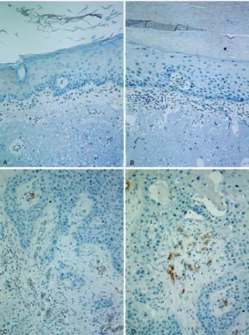
Figure 2 - Vascular hot spots in CD105-stained adjacent
non-neoplastic skin (A), SK (B), siSCC (C), and iSCC (D).
Table 1 -Mean CD34 and CD105 Chalkley counts in stroma from tumor fronts and adjacent non-neoplastic skin.
CD34 CC CD105 CC
Adjacent skin
Tumor
front P
Adjacent skin
Tumor
front P
SK 1.91 3.25 0.049 0.08 1.66 0.046
siSCC 2.06 3.50 0.016 0.36 2.46 0.0008
iSCC 2.16 3.96 0.028 0.46 3.73 0.001
CC: mean Chalkley count, SK: solar keratosis, siSCC: superficially invasive squamous cell carcinoma, iSCC: invasive squamous cell carcinoma. Differences between tumor front and adjacent skin were evaluated by T-test dependent.
CLINICS 2011;66(3):465-468 Skin Cancer Angiogenesis, Florenceet al.
Florence MEB et al.
Using the panendothelial CD34 stain, no differences were found in the microvascular area between the different stages of CSCC. However, the CD105 stain revealed an induction of newly formed vessels that accompany the progression of CSCC. Therefore, Nijsten’s data22were corroborated by our findings, which emphasizes the necessity of assessing tumor angiogenesis through active angiogenesis markers (ECP and CD105).
It has been proposed that angiogenesis onset occurs not only for metabolic supply but also for the establishment of invasion pathways by producing proteases and/or for guiding tumor cells.4,21 Because SKs are limited lesions, angiogenesis induction in this stage supports their invasive carcinomatous behavior.
A lower MVD has been found in neoplastic tissue in glioblastomas and renal, colon and mammary carcinomas compared with their respective normal tissues.5As a result, Eberhard et al. and Hlatky et al. have suggested that
techniques for assessing microvessel density using pan-endothelial markers estimate the vascular bed amount of a tissue but not the angiogenic status, that is, the rate of evolving neovasculature.5,23Because neo-angiogenesis mar-kers allow the dynamic evaluation of tumor-dependent angiogenesis, they should be considered for assessing angiogenic status in the future.
CONCLUSION
The dependence of skin carcinogenesis on angiogenesis demonstrated in this study is useful for understanding CSCC evolution and metastatic dissemination and for the development of new therapeutic strategies, such as anti-angiogenic therapies, that specifically target CD105.
ACKNOWLEDGMENTS
This study was supported by FAPESP (Sao Paulo Research Foundation), CNPq (National Council of Technological and Scientific Development) and the Department of Dermatology at the University of Wuerzburg, Germany. We are grateful to Adilson Abilio Piaza, Ana Claudia Piaza, and Lı´dia Marı´lia Frey for technical support. Konradin Metze is a researcher of CNPq.
REFERENCES
1. Diepgen TL, Mahler V. The Epidemiology of Skin Cancer. Br J Dermatol. 2002;146(suppl 61):1-6, doi: 10.1046/j.1365-2133.146.s61.2.x.
2. Ackerman AB, Mones JM. Solar (actinic) Keratosis is Squamous Cell Carcinoma. Br J Dermatol. 2006:155;9-22, doi: 10.1111/j.1365-2133.2005. 07121.x.
3. Hanahan D, Weinberg A. The Hallmarks of Cancer. Cell 2000;100:57-70, doi: 10.1016/S0092-8674(00)81683-9.
4. Bergers G, Benjamin LE. Tumorigenesis and the Angiogenic Switch. Nat Rev Cancer. 2003;3:401-10, doi: 10.1038/nrc1093.
5. Hanahan D., Folkman J. Patterns and Emerging Mechanisms of the Angiogenic Switch during Tumorigenesis. Cell. 1996;86:353–64, doi: 10. 1016/S0092-8674(00)80108-7.
6. Sharma S, Sharma MC, Sarkar C. Morphology of Angiogenesis in Human Cancer: A Conceptual Overview, Histoprognostic Perspective and Significance of Neoangiogenesis. Histopathology. 2005;46:481-9, doi: 10.1111/j.1365-2559.2005.02142.x.
7. Eberhard A, Kahlert S, Goede V, Hemmerlein B, Plate KH, Augustin HG. Heterogeneity of Angiogenesis and Blood Vessel Maturation in Human Tumors: Implications for Antiangiogenic Tumor Therapies. Cancer Res. 2000;60:1388-93.
8. Fonsatti E, Altomonte M, Nicotra MR, Natali PG, Maio M. Endoglin (CD105): a powerful therapeutic target on tumor-associated angiogenetic blood vessels. Oncogene 2003;22:6557-63, doi: 10.1038/sj.onc.1206813. 9. Fonsatti E, Del Vecchio L, Altomonte M, Sigalotti L, Nicotra MR, Coral S,
et al. Endoglin: An Accessory Component of the TGF-b-Binding Receptor-Complex With Diagnostic, Prognostic, and Bioimmunotherapeutic Potential in Human Malignancies J Cell Physiol. 2001;188:1–7.
10. Minhajat R, Mori D, Yamasaki F, Sugita Y, Satoh T, Tokunaga O. Organ-specific endoglin (CD105) expression in the angiogenesis of human cancers. Pathol Int. 2006;56:717–23, doi: 10.1111/j.1440-1827.2006.02037.x. 11. Fox SB and Harris AL. Histological Quantitation of Tumour Angiogenesis. APMIS. 2004;112:413–30, doi: 10.1111/j.1600-0463.2004. apm11207-0803.x.
12. Vermeulen PB, Gasparini G, Fox SB, Colpaert C, Marson LP, Gion M. Second International Consensus on the Methodology and Criteria of Evaluation of Angiogenesis Quantification in Solid Human Tumours. Eur J Cancer. 2002;38:1564–79.
13. Hansen S, Sorensen FB, Vach W, Grabau DA, Bak M, Rose C. Microvessel Density Compared with the Chalkley Count in a Prognostic Study of Angiogenesis in Breast Cancer Patients. Histopathology. 2004;44:428–36, doi: 10.1111/j.1365-2559.2004.01848.x.
14. Glogau RG. The risk of progression to invasive disease. J Am Acad Dermatol. 2000;42:S23–4, doi: 10.1067/mjd.2000.103339.
15. Lee AD, Jorizzo JL. Optimizing Management of Actinic Keratosis and Photodamaged Skin: Utilizing a Stepwise Approach. Cutis. 2009;84:169-75.
16. Fuchs A, Marmur E. The Kinetics of Skin Cancer: Progression of Actinic Keratosis to Squamous Cell Carcinoma. Dermatol Surg. 2007;33:1099– 101, doi: 10.1111/j.1524-4725.2007.33224.x.
17. Hasan J, Byers R, Jayson GC. Intra-tumoural Microvessel Density in Human Solid Tumours. Br J Cancer. 2002;86:1566–77.
18. Bluff JE, Menakuru SR, Cross SS, Higham SE, Balasubramanian SP, Brown NJ, et al. Angiogenesis is Associated With the Onset of Hyperplasia in Human Ductal Breast Disease. Br J Cancer. 2009; 101:666–72.
19. Smith-McCune K, Zhu Y-H, Hanahan D, Arbeit J. Cross-species Comparison of Angiogenesis during the Premalignant Stages of Squamous Carcinogenesis in the Human Cervix and K14-HPV16 Transgenic Mice. Cancer Res. 1997;57:1294–300.
20. Coussens LM, Hanahan D, Arbeit JM. Genetic Predisposition and Parameters of Malignant Progression in K14-HPV 16 Transgenic Mice. Am J Pathol. 1996;149:1899–1917.
21. Strieth S, Hartschuh W, Pilz L, Fusenig NE. Angiogenic Switch Occurs Late in Squamous Cell Carcinomas of Human Skin. Br J Cancer. 2000;82:591-600.
22. Nijsten T, Colpaert CG, Vermeulen PB, Harris AL, Van Marck E, Lambert J. Cyclooxygenase-2 Expression and Angiogenesis in Squamous Cell Carcinoma of the Skin and its Precursors: A Paired Immunohistochemical Study of 35 Cases. Br J Dermatol. 2004;151:837– 45, doi: 10.1111/j.1365-2133.2004.06214.x.
23. Hlatky L, Hahnfeldt P, Folkman J. Clinical Application of Antiangiogenic Therapy: Microvessel Density, What it Does and Doesn’t Tell Us. J Natl Cancer Inst. 2002;94;883-93.
Skin Cancer Angiogenesis, Florenceet al.
Florence MEB et al. CLINICS 2011;66(3):465-468