Association between human papillomavirus (HPV) and
the oral squamous cell carcinoma: a systematic review
Associação entre o papilomavírus humano (HPV) e o carcinoma
de células escamosas oral: uma revisão sistemática
Marcos Antonio Pereira de Lima1; Cláudio Gleidiston Lima da Silva2; Silvia Helena Barem Rabenhorst3
First submission on 11/06/13; last submission on 13/11/13; accepted for publication on 19/11/13; published on 20/02/14
1. Doctorate in Biotechnology in Health from Rede Nordeste de Biotecnologia (Renobio)/Universidade Federal do Ceará (UFC); master’s degree in Medical Microbiology from UFC; professor of Medical Microbiology at the Medical School of UFC.
2. Doctorate in Pharmacology from UFC; master’s degree in Pathology from UFC; assistant professor of Pathology and Bioethics at the Medicine School of UFC.
3. Post-doctorate in Molecular Genetics from the University of Colorado Health Sciences Center – Medical Oncology; doctorate in Biological Sciences from Universidade Estadual Paulista Júlio de Mesquita Filho (UNESP); master’s degree in Biological Sciences from UNESP; associate professor IV at UFC.
ABSTRACT
The human papillomavirus (HPV) is an epitheliotropic agent whose high-risk genotypes have a well-established link with the development of cervical cancer. Although the relation of HPV to the oral squamous cell carcinoma (OSCC) has been studied since the beginning of the 1980s, its role in the oral carcinogenesis and the probable underlying molecular mechanisms are still not fully elucidated. We performed a systematic review of the worldwide scientiic literature, published until the preparation of the present paper, concerning the association of HPV with OSCC, scrutinizing the samples, prevalence levels, the techniques utilized and relevant indings of the studies. The results showed that HPV is associated with approximately one quarter of OSCCs. Another interesting feature is the distinct pattern of infection in these oral tumors, including the participation of genotypes that are uncommon in cervical malignant lesions, such as HPV-38, 44, 53 and 70. Equally interesting is the possibility of carcinogenic action without the occurrence of viral integration, veriied by the high expression of messenger ribonucleic acid (mRNA) of E6 and E7 from high-risk genotypes in cases whose virus remain in the episomal form. These
indings support the assumptionof HPV involvement in the genesis of OSCC, whereas warn about the possibility of unexpected viral
behaviors that sometimes are not perceived or understood due to the technological limitations of the time and to the shortage of studies with the adequate approaches.
Key words: HPV; oral cancer; squamous cell carcinoma.
INTRODUCTION
Oral cancer is a serious worldwide public health problem, with high incidence and mortality rates. According to data from the International Agency for Research on Cancer (IARC), approximately 263,900 new cases and 128,000 deaths by cancer of the oral cavity are estimated to have occurred in the world in
2008(22). Among the malignant tumors of this anatomic site, more
than 90% are oral squamous cell carcinomas (OSCC). Several risk factors are related to oral cancer, with the main being: tobacco use, alcohol consumption, and infection by high-risk genotypes of
human papillomavirus (HPV)(41).
HPV is an epitheliotropic virus implicated in the development
of skin warts and papillomatous lesions in mucosae(46, 55).
Nowadays, more than 100 HPV genotypes are known, and based on their potential for induction of malignant transformation, the several genotypes are classiied as “low risk” and “high risk” for
the development of genital malignancy(24). Structurally, this virus
is characterized by a non-enveloped icosahedral capsid, with circular double-stranded deoxyribonucleic acid (DNA) genome,
approximately 8,000 base pairs (pb) long(33, 40). Its genome may
and pRb, respectively, an event considered crucial for the neoplastic transformation of infected cells(26, 35, 36, 39).
The mechanism that makes a high-risk HPV induce the malignant progression of previously benign lesions is, primarily, this genotype capacity to integrate its genome to that of the host (viral integration). This provokes the break of a viral DNA segment that contains E2 gene, which, among other functions, inhibits the expression of E6 and E7 genes, culminating in the overexpression of E6 and E7(18, 40). This cascade of events leads to excessive and unregulated cell
proliferation, with involvement of repair mechanisms, which favors the accumulation of mutations and the occurrence of chromosomal aberrations, as well as apoptosis inhibition(12, 26).
The relationship between HPV and OSCC was irst suggested
in 1983 by Syrjänen et al.(49), when they discovered koilocytotic
atypias in malignant oral lesions by optical microscopy. But the presence of viral DNA was only conirmed two years later, by
means of in situ hybridization (ISH)(29). HPV infection in the
oral cavity is associated with risky sexual behaviors, mainly to orogenital sex. However, mouth-to-mouth contact, vertical birth-transmission and autoinoculation resulting from chewing warts
are also transmission modes of this virus to the oral mucosa(10).
The viruses isolated in OSCC are low-risk genotypes, including HPV-6, 11, 16, 18, 22, 31, 33, 35, 38, 58, 68, and 70(51). Nevertheless,
in around 80% of the cases of infected oral squamous carcinomas, the identiied genotypes were HPV-16/18. Co-infections with two HPV genotypes have been reported; the indings reveal that in these cases, neoplasms tend to occur, on average, a decade earlier than in individuals infected by a single genotype, or not infected(32).
In spite of the methodological advances and the innumerable works developed since then, the role of HPV in oral carcinogenesis has not been fully elucidated. The several indings that reinforce the hypothesis of this virus involvement in oral neoplasia were followed by conlicting results. In this article, we reviewed the international scientiic studies published until the preparation of this manuscript, concerning the association between HPV and OSCC, scrutinizing the sample characteristics, prevalence rates, the used techniques and the relevant study indings.
MATERIALS AND METHODS
The articles used for the conduction of this systematic review were selected on Pubmed (http://www.ncbi.nlm.nih.gov/pubmed). The full versions of almost all the articles were retrieved either from that search engine or at the periodical portal of Coordenação de Aperfeiçoamento de Pessoal de Nível Superior (CAPES) (http:// www.periodicos.capes.gov.br/). The descriptors used in the search
were: human papillomavirus, HPV, oral carcinoma, oral squamous cell carcinoma, and OSCC.
In situ approach studies were given preference in this review, having in mind the existence of a smaller number of works using
in situ detection in oral carcinomas, when compared with those
using ampliication of genetic material, as well as the importance of these methods for assessing HPV role in tumors, because they allow for the precise localization of the virus in tissue.
The inclusion criteria for the original articles were: 1. discuss
the detection of HPV in samples of oral carcinoma, involving in
situ techniques; or 2. combine HPV detection, regardless of the used technique, with the expression of tumor markers strictly related to HPV tumorigenic pathways; or 3. compare HPV detection with that of another virus with recognized carcinogenic potential; or 4. point out aspects that may contribute to comprehension of viral involvement in oral carcinoma, such as case-control studies, viral expression assays and those of malignant transformation in oral epithelial cells. Exclusion criteria: 1. not investigate HPV presence; 2. discuss exclusively other neck and head carcinomas; 3. use only ampliication of nucleic acid for HPV detection, with
neither in situ approach, nor assessment of tumor markers or the
presence of another virus.
Based on the established criteria, we retrieved 41 articles, including the irst reports, so that the current review comprises works published from 1983 to June 2013, in English and Spanish. In the development of this paper we also considered meta-analyses, which encompass information about virus detection through genetic ampliication techniques, and review articles to deepen the discussion.
RESULTS AND DISCUSSION
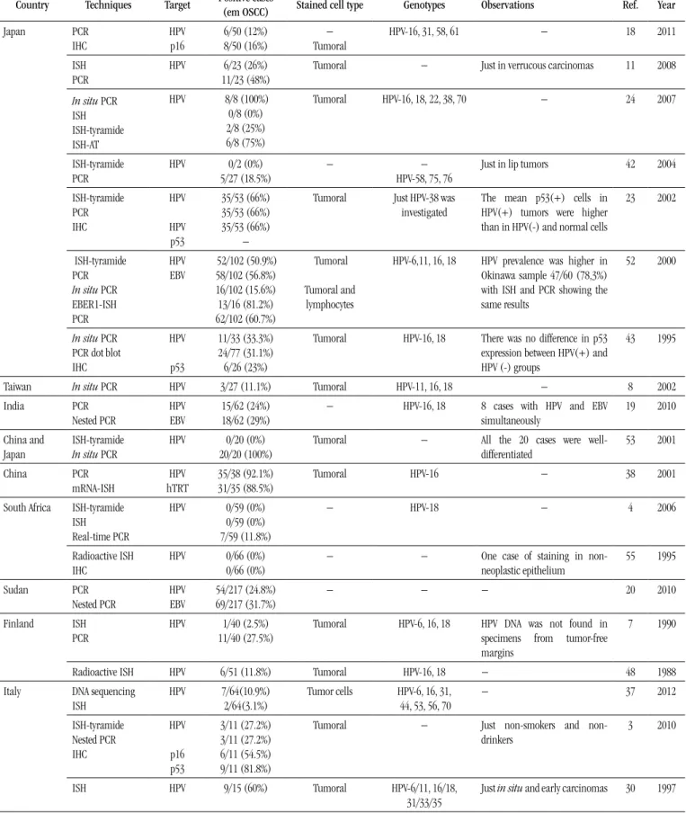
The Table provides a list of studies carried out in the last three decades around the world and their results as to HPV detection in
oral carcinomas, highlighting those that used in situ methods. The
average prevalence observed in those studies was approximately 25%, ranging from 0% to 100%. The works that displayed higher prevalence rates, in general, used the ISH technique with a
biotinyl-tyramide-based detection system, or the in situ polymerase chain
TABLE – Summary of studies, grouped by world region, assessing HPV
association with oral carcinomas, highlighting those using in situ approaches
Country Techniques Target Positive cases (em OSCC) Stained cell type Genotypes Observations Ref. Year
Japan PCR IHC HPV p16 6/50 (12%) 8/50 (16%) − Tumoral
HPV-16, 31, 58, 61 − 18 2011
ISH PCR
HPV 6/23 (26%)
11/23 (48%)
Tumoral − Just in verrucous carcinomas 11 2008
In situ PCR ISH ISH-tyramide ISH-AT
HPV 8/8 (100%)
0/8 (0%) 2/8 (25%) 6/8 (75%)
Tumoral HPV-16, 18, 22, 38, 70 − 24 2007
ISH-tyramide PCR
HPV 0/2 (0%)
5/27 (18.5%)
− −
HPV-58, 75, 76
Just in lip tumors 42 2004
ISH-tyramide PCR IHC HPV HPV p53 35/53 (66%) 35/53 (66%) 35/53 (66%) −
Tumoral Just HPV-38 was investigated
The mean p53(+) cells in HPV(+) tumors were higher than in HPV(-) and normal cells
23 2002
ISH-tyramide PCR In situ PCR EBER1-ISH PCR HPV EBV 52/102 (50.9%) 58/102 (56.8%) 16/102 (15.6%) 13/16 (81.2%) 62/102 (60.7%) Tumoral Tumoral and lymphocytes
HPV-6,11, 16, 18 HPV prevalence was higher in Okinawa sample 47/60 (78,3%) with ISH and PCR showing the same results
52 2000
In situ PCR PCR dot blot IHC HPV p53 11/33 (33.3%) 24/77 (31.1%) 6/26 (23%)
Tumoral HPV-16, 18 There was no difference in p53 expression between HPV(+) and HPV (-) groups
43 1995
Taiwan In situ PCR HPV 3/27 (11.1%) Tumoral HPV-11, 16, 18 − 8 2002
India PCR Nested PCR HPV EBV 15/62 (24%) 18/62 (29%)
− HPV-16, 18 8 cases with HPV and EBV
simultaneously
19 2010
China and Japan
ISH-tyramide In situ PCR
HPV 0/20 (0%)
20/20 (100%)
Tumoral − All the 20 cases were
well-differentiated 53 2001 China PCR mRNA-ISH HPV hTRT 35/38 (92.1%) 31/35 (88.5%)
Tumoral HPV-16 − 38 2001
South Africa
ISH-tyramide ISH Real-time PCR
HPV 0/59 (0%)
0/59 (0%) 7/59 (11.8%)
− HPV-18 − 4 2006
Radioactive ISH IHC
HPV 0/66 (0%)
0/66 (0%)
− − One case of staining in
non-neoplastic epithelium 55 1995 Sudan PCR Nested PCR HPV EBV 54/217 (24.8%) 69/217 (31.7%)
− − − 20 2010
Finland ISH
PCR
HPV 1/40 (2.5%)
11/40 (27.5%)
Tumoral HPV-6, 16, 18 HPV DNA was not found in specimens from tumor-free margins
7 1990
Radioactive ISH HPV 6/51 (11.8%) Tumoral HPV-16, 18 − 48 1988
Italy DNA sequencing ISH HPV 7/64(10.9%) 2/64(3.1%)
Tumor cells HPV-6, 16, 31, 44, 53, 56, 70
− 37 2012
ISH-tyramide Nested PCR IHC HPV p16 p53 3/11 (27.2%) 3/11 (27.2%) 6/11 (54.5%) 9/11 (81.8%)
Tumoral − Just smokers and
non-drinkers
3 2010
ISH HPV 9/15 (60%) Tumoral HPV-6/11, 16/18,
31/33/35
Country Techniques Target Positive cases (em OSCC) Stained cell type Genotypes Observations Ref. Year
Spain ISH HPV 10/27 (37%) Tumoral − HPV more frequent in
well-differentiated tumors
13 1994
Germany ISH HPV 7/12 (58.3%) Tumoral HPV-6, 11, 16, 18 Just smoking and
non-drinking patients with T2N0M0 tongue tumors
17 1992
Hungary PCR
IHC
HPV p16 p53 pRb
33/79 (41.7%) 13/79 (16.4%) 45/79 (56.9%) 63/79 (79.7%)
Tumoral HPV-16 − 33 2006
Czech Republic
ISH-tyramide PCR IHC
HPV
p16
6/24 (25%) 3/24 (13%) 7/24 (29%)
Tumoral HPV-16 Just smokers and non-drinkers
Lip tumors were not included
25 2011
Netherlands ISH-tyramide PCR
HPV 0/7 (0%)
0/7 (0%)
− − Just smokers and
non-drinkers with tongue tumors
45 2008
Serbia PCR
dPCR SSCP
HPV c-myc
p53
6/60 (10%) 21/60 (35%) 36/60 (60%)
− HPV16 − 39 2010
USA
PCR IHC
HPV p16 p53
4/26 (15.3%) 15/29 (51.7%) 27/29 (93.1%)
Tumoral − − 14 2008
ISH HPV 2/36 (5.5%) Non-neoplastic
epithelium
HPV6/11, 16/18 Smoking and drinking patients 1 1991
ISH PCR
HPV 1/10 (10%)
1/10 (10%)
Tumoral HPV-16/18 − 44 1991
ISH HPV 0/17 (0%) − − − 57 1991
ISH HPV 0/20 (0%) − − Five in situ carcinomas were
included
58 1991
ISH HPV 3/50 (6%) Tumoral HPV-16, 18, 33 − 15 1990
USA Venezuela ISH In situ PCR
HPV 0/30 (0%)
20/30 (66.7%)
Tumoral HPV-16, 18 − 31 1994
Brazil PCR dot blot IHC
HPV p53 Bcl-2
26/88 (29.5%) 26/43 (60.4%) 17/43 (39.5%)
− HPV-16, 18 − 35 2009
ISH-tyramide HPV 3/10 (30%) Tumoral HPV-16/18, 31/33 − 2 2008
PCR dot blot
IHC
HPV pRb p21
11/33 (33.3%) 24/33 (72.7) 12/33 (36.3%)
− HPV-16, 18 − 47 2008
ISH-tyramide HPV 2/12 (16.6%) Tumoral HPV-16/18 HPV-positive cases compatible
with in situ carcinoma
46 2002
Argentina ISH HPV 8/33 (24.2%) Tumoral − Five HPV(+) cases were
verrucous carcinomas
5 1999
dPCR: differential PCR; EBER1-ISH: ISH using probes complementary to EBV-encoded RNA 1 (EBER1); EBV: Epstein-Barr virus; HPV: human papillomavirus; hTRT: human telomerase reverse transcriptase; IHC: immunohistochemistry; in situ PCR: PCR followed by ISH, both on slide; ISH: conventional in situ hybridization; ISH-AT: ISH-AT Tailing; ISH-tyramide: ISH with a biotinyl-tyramide-based signal ampliication system; mRNA-ISH: ISH with probes directed towards a target messenger ribonucleic acid; nested PCR: two sets of primers used in two successive PCR runs, aiming at reducing non-speciic ampliication; PCR dot blot: PCR ampliication followed by dot blot hybridization for virus typing; PCR: polymerase chain reaction; qPCR: quantitative PCR; radioactive ISH: ISH using radioactive labelled probes; Ref.: reference; SSCP: single strand conformation polymorphism.
Considerations about viral prevalence
According to a meta-analysis carried out by Miller and
White(32), in which 58 studies were reviewed, accounting for 1,051
FIGURE – HPV prevalence rates found in studies carried out in different parts of the world using in situ hybridization techniques
was 38.1%(51). In both studies the prevalence rates ranged from
0% to 100%. Such a variation depends on the sensitivity of the employed technique, the sample size, the state of conservation of the clinical specimens, and epidemiological factors of the studied
population(6). The irst meta-analysis found out that the studies in
which carcinoma specimens were collected fresh and kept frozen, and then submitted to PCR technique, presented a prevalence rate of 51.6% (115 out of 223). On the other hand, the researches that used ISH in parafin-embedded specimens demonstrated a prevalence of 21.7% (136 out of 628)(32). In the study by Termine et
al.(51), the average prevalence obtained by ISH was 29.8%, whereas
by PCR was 39.9%. The Figure presents the attained prevalence
rates, by ISH methods, in several researches carried out around the world.
Considerations about oral and oropharyngeal
topography
Another factor that inluences knowledge about the actual HPV prevalence in oral lesions includes the precise deinition of oral and oropharyngeal tumors. From the anatomical point of view, the border between oral cavity and oropharynx is the posterior third of the tongue. However, it is not always so simple to clinically deine it, and the result is that some tumors of a site
may be included and analyzed in the other(16). Some authors, like
Tsuhako et al.(52), have indistinctly considered the cases of oral
and oropharyngeal squamous cell carcinomas in their casuistics. This may be the cause of the elevated determined prevalence rates, considering that oropharyngeal carcinomas demonstrate higher prevalence, and the relationship between HPV and carcinogenesis in this anatomic site is well accepted(37, 50).
Considerations about viral detection
The screening of a limited number of genotypes is another important factor that may lead to false negative results, and consequently, to an underestimate of HPV role in OSCC. A remarkable example is the study by Kojima et al.(23), in which
ISH-tyramide exposed an elevated prevalence of HPV-38 (35/53 [66%]), a genotype rarely investigated in other studies.
Meanwhile, Pannone et al.(37) detected genotypes 44, 53, and 70,
by DNA sequencing following PCR, but were not successful with the ISH method, as the available commercial kits do not include the corresponding probes: they are designed to detect high-risk genotypes associated with anogenital lesions.
The amount of viral copies may vary depending on the differentiation stage of the host cell, reaching the highest levels in differentiated keratinocytes of the most supericial layers, where viruses are in their vegetative phase. About the sensitivity of the employed techniques, ISH is only able to detect HPV when the rate
USA 0-10%
Brazil 16.6-30%
Argentina 24.2%
Spain 37%
Netherlands 0%
China 0%
Japan 0-75% Italy
3.1-60%
South Africa
0%
Finland 2.5-11.8% Germany
58.3% Czech Republic
is higher than 10 copies of viral DNA per cell, being considered of low sensitivity. The methods of moderate sensitivity, like Southern blot, dot blot, and reverse blot hybridization, can detect it from a rate of one viral copy per cell. PCR demonstrates an expressive ratio, able to detect HPV at a rate lower than a copy of viral DNA per cell, being considered of high sensitivity. Immunoluorescence
and immunoperoxidase assays are considered of low sensitivity(32).
Some authors afirm that ISH sensitivity could be improved by means of a biotinyl-tyramide-based detection system, amplifying the signal and, thus, allowing the detection even of a single viral copy per infected cell(9, 28). According to Syrjänen et al.(50), ISH is
perhaps more sensitive than PCR, especially when few cells in the specimen contain viral copies that may not be detected by PCR, depending on the tissue portion used for DNA obtainment, what
demonstrates the importance of studies using in situ approaches.
It is interesting to note that technical speciicity may also inluence HPV detection, and consequently, prevalence rates. Still according to the meta-analysis by Miller and White(32), studies
involving PCR using primers to detect early HPV genes yielded an average prevalence of 42.7% (88 out of 206), whereas those using primers designed for HPV late regions presented prevalence of 22.5% (48 out of 213). The discrepancy is enlarged as one considers only studies that detected HPV-16/18, with prevalence rates of 46.7% (84 out of 180) for the early region, and 15.9% (33 out of 208) for the late region. Probably, such differences are due to the disruption of the viral genome, most frequently in sites located in the late region, during integration to the host genome. As a result, false negative results may occur when primers are used in the late regions. However, disruption may eventually occur in early regions, with a possible underestimate of prevalence when using primers for this region of viral genome(16).
Although the Southern blot requires a signiicant amount of DNA for its conduction, it allows deining whether HPV is in the episomal or integrated form. Moreover, it is not prone to contamination errors, an event that may occur with PCR and is dificult to control, because PCR is very sensitive, besides offering simply a binary inding. Thus, techniques as the Southern blot and quantitative PCR (qPCR) provide means for quantiication to differentiate low-level positivity from contamination(16).
Controversy about the role of HPV in the oral
carcinoma
Although the presence of HPV in a percentage of oral carcinomas has been conirmed, as previously exposed, viral detection only does not prove the causal relationship between this agent and the aforementioned neoplasia. These results, from
studies of diverse designs, allow inferring the possibility of the cited virus participation in oral carcinogenesis, but direct experimental evidence establishing cause and effect is required for conirmation.
For Feller et al.(10), it is necessary that a signiicant number of
individuals harboring HPV in the oral mucosa develop carcinoma in a certain period, as can be seen in cases of squamous carcinoma
of the uterine cervix. The presence of high-risk genotypes with conirmed expression of E6 and E7 oncoproteins in the malignant cells, both in the primary site and in metastases, and viral monoclonality are also decisive.
The existence of a substantial number of viral DNA copies (viral load) in the neoplastic tissue also provokes an involvement of the microbial agent. However, evidence warns that this inding is not a reliable prediction of HPV-induced neoplastic progression, as the determination of viral load does not distinguish between an infection of many cells containing few copies of viral DNA per cell and an infection of few cells with a large number of viral copies per cell, or even between long-standing infection and recent infection(10).
Although the mucosae of the oral cavity and uterinecervix
share histologic similarities, signiicant differences are observed as to neoplastic development. The oral mucosa, for example, is more exposed to a series of carcinogens such as alcohol, tobacco and betel, which play an important role in the etiology of oral neoplasms. In
the uterine cervix, HPV seems to be the main triggering factor, with more than 90% of association. Regarding HPV in squamous oral carcinomas, other differences have been observed, among which, the infrequent viral integration and isolation of genotypes 31, 33, and 35, commonly observed in cervical carcinomas, as well as the relatively lower overall prevalence in oral carcinomas. These examples illustrate the differences in mechanisms of oncogenesis between both sites; it is possible, then, that HPV presents a distinct behavior in the oral mucosa, and still plays a role in the neoplastic process(10, 32).
Concerning viral integration into the host genome, although it is a strong indicator of the viral oncogenic role, the presence of high viral load followed by the high-risk E6 and E7 mRNA active expression may occur, for instance, in malignant cells of
squamous carcinoma of the oropharynx in the episomal form(56).
Other aspects that hinder deine the role of HPV in oral carcinogenesis include its identiication in samples of normal mucosa, benign leukoplakia and intraepithelial neoplasia, with signiicant prevalence rates of 13.5%, 14.8%, and 27%, respectively,
according to a meta-analysis carried out by Miller and White(32).
for which HPV is the only known risk factor, that is, without including tobacco use and/or alcohol consumption, what would provide a more accurate analysis of the isolated viral action. In general, this group of patients, when observed, represents a small
part, around 7.3% of the studied samples(32). The limited number
of studies assessing the expression of viral oncoproteins and their effects on oral lesions also makes it dificult to understand viral
behavior in situ.
Castro and Bussoloti Filho(6) highlight also the fact that
the virus is detected just in a subset of tumor cells when cell localization techniques are used, usually with low viral load. Both indings, added to the presence of HPV in samples of normal oral mucosa and pre-neoplastic lesions, may suggest that this virus is merely a passenger in the oral cavity. On the other hand, we cannot exclude the possibility of a progressive loss of viral genome copies after the establishment of neoplasia, a mechanism named “hit and run”. Thus, HPV would play an important role in tumor development that could be underestimated due to the loss of viral copies during the process, with the resulting failure of the detection techniques(10, 32).
Another intriguing inding is the relatively favorable prognosis of HPV-associated oral carcinomas. In this context, the
meta-analysis conducted by O’Rorke et al.(36) concluded that not only
OSCC cases, but head and neck carcinomas in general exhibited better survival in relation to HPV-negative cases. It is admitted that
HPV-positive tumors commonly present the wild-type TP53 gene,
whereas cases not related to HPV harbor mutations in the referred gene which, in their turn, are associated with worse prognosis. That would help explain the prognostic proiles veriied among
HPV-positive and HPV-negative cases(26).
Evidence confirming the role of HPV
The indings that conirm HPV participation in the development of oral squamous carcinomas include the predominant detection of high-risk genotypes (HPV-16/18), the increased prevalence of HPV in dysplasia and squamous carcinomas compared with the normal mucosa, mainly as to high-risk genotypes, indicating HPV as an independent risk factor for the mentioned neoplastic type(6, 21, 50). Correspondence around 76% has been found between
genotypes identiied in tumor sites and metastatic lymph nodes(32).
Lind et al.(27) reported that seven of 13 cases of HPV-positive
leukoplakias progressed to oral carcinoma within a 10-year period, but genotypes were not reported. Nonetheless, studies show that oral human keratinocytes expressing E6 and E7 genes from HPV-16 become immortal, as previously demonstrated in other keratinocyte cell lines(34).
We must remember there is no certainty as to the necessity of viral integration, in the oral cavity, for the activation of viral oncogenesis(16). HPV isolation, however, especially high-risk, in
non-tumoral oral mucosa may be related to the latency period of the virus in the tissue. Subclinical infections of the oral and oropharyngeal mucosae by HPV are common. It is possible that these sites function as reservoirs where viruses remain quiescent, and later, when activated, may play a role in the development of local carcinomas(10). Besides, viral presence does not necessarily
indicate active viral expression, as demonstrated in studies with cases of HPV-positive head and neck squamous carcinoma, in which less than half of the cases (9 out of 20) showed E6 mRNA expression(54).
Miller and White(32), based on the body of evidence observed
until then, ascertain that HPV involvement in the neoplastic process is clear, and warn that possibly some genotypes still have not been identiied in oral lesions, what would explain the relatively low prevalence. If HPV were transient, at least one of the following characteristics should be veriied: 1. similar prevalence of HPV among samples of tumor, normal mucosa and pre-neoplastic lesions, including non-tumoral specimens obtained from sites far from the lesions in patients affected by squamous carcinoma; 2. viral prevalence differences in biopsy samples and in oral squamous carcinoma cell lines, due to the potential selectivity of HPV-negative cells during culture; 3. indifferent prevalence of high- and low-risk genotypes in oral cancer samples. Such characteristics were not veriied in retrospective analyses, though.
As stated by Syrjänen et al.(50) when considering the modiied
Koch’s postulates, at least three conditions necessary to formally conirm the role of HPV as an etiological agent of OSCC have already been satisied: 1. presence of viral genome in tumor lesions or tumor cells; 2. ability of the virus or viral protein to transform cells in vitro; 3. ability of the virus or viral protein to
promote tumor formation in animals. As to the other criteria: 4. viral infection precedes cancer development – in spite of the few prospective studies, we must not forget the work by Lind et al.(27), in
which some cases of infected leukoplakia progressed to carcinoma within a 10-year period; 5. epidemiologic association between presence of the virus and development of cancer – although the association has not been proved, several works demonstrate that the prevalence of HPV is higher in pre-neoplastic lesions and
carcinomas when compared with the normal mucosa(21, 50); and
CONCLUSIONS
Although there still is a certain skepticism about the association of HPV and OSCC, it is important to remember examples like that of Helicobacter pylori and HPV itself: time, technological advances and successive studies have been able to conirm their participations in the development of, respectively, gastric and cervical carcinomas, many years after the irst suggestion. The relationship between HPV and oral carcinomas have been investigated for three decades, and as previously stated, in spite of the conlicting indings, there is a mass of evidence conirming the involvement of this virus in a percentage OSCC, which may reach a little more than a quarter of OSCC. Another interesting aspect is the distinct pattern of infections in these oral tumors, including the participation of uncommon genotypes in
cervical malignant lesions, such as HPV-38, 44, 53, and 70, which may be underestimated, as the available kits for the detection of HPV are based on genotypes considered of high risk for anogenital lesions. Equally interesting is the possibility of carcinogenic action without the occurrence of viral integration, veriied by the high E6 and E7 mRNA expression, of high-risk genotypes, in whose cases the virus was in its episomal form. All these pieces of evidence reinforce the thesis of HPV participation in the genesis of oral carcinomas, while warn about the possible unexpected viral behaviors that, sometimes, are not noticed or understood due to the technological limitation of the time and the shortage of studies with the adequate approach. From now on, it is fundamental to develop appropriate research designs, with emphasis on case-control studies that may bring more consistent information about the cause-effect relationship between HPV and OSCC.
RESUMO
O papilomavírus humano (HPV) é um agente epiteliotrópico cujos genótipos de alto risco têm uma ligação já bem estabelecida com o desenvolvimento de cânceres cervicais. Embora a relação do HPV com o carcinoma de células escamosas oral (CCEO) venha sendo estudada desde o início da década de 1980, seu papel na carcinogênese oral e os prováveis mecanismos moleculares subjacentes ainda não estão completamente elucidados. Realizou-se uma revisão sistemática dos trabalhos existentes na literatura cientíica internacional até o momento da elaboração deste manuscrito concernente à associação do HPV com o CCEO, esquadrinhando as características das amostras, das prevalências veriicadas, das técnicas utilizadas e os achados relevantes dos estudos. Os resultados demonstram que o HPV está associado a cerca de um quarto dos CCEO. Outro aspecto interessante refere-se ao padrão distinto das infecções nesses tumores orais, incluindo a participação de genótipos incomuns em lesões malignas cervicais, tais como HPV-38, 44, 53 e 70. Igualmente interessante é a possibilidade de atuação carcinogênica sem a ocorrência de integração viral, constatada pela elevada expressão de ácido ribonucleico mensageiro (RNAm) de E6 e E7, de genótipos de alto risco, em casos cujo vírus encontrava-se em estado epissomal. Essas evidências reforçam a tese do envolvimento do HPV na gênese dos CCEO, ao mesmo tempo em que alertam para a possibilidade de comportamentos virais inesperados que, por vezes, não são percebidos ou compreendidos devido à limitação tecnológica da época e à carência de estudos com abordagem apropriada.
Unitermos: HPV; câncer oral; carcinoma de células escamosas.
REFERENCES
1. ABDELSAYED, R. Study of human papillomavirus in oral epithelial dysplasia and epidermoid carcinoma in the absence of tobacco and alcohol use. Oral Surg Oral MedOral Pathol, v. 71, n. 6, p. 730-2, 1991.
2. ACAY, R.; et al. Human papillomavirus as a risk factor in oral carcinogenesis: a study using in situ hybridization with signal ampliication. Oral Microbiol Immunol, v. 23, n. 4, p. 271-4, 2008. 3. ANGIERO, F. et al. Frequency and role of HPV in the progression of epithelial dysplasia to oral cancer. Anticancer Res, v. 30, n. 9, p. 3435-40, 2010.
4. BOY, S.; et al. HPV detection in primary intra-oral squamous cell carcinomas – commensal, aetiological agent or contamination? J Oral Pathol Med, v. 35, n. 2, p. 86-90, 2006.
5. BUSTOS, D. A. et al. Human papillomavirus detection in oral cancer lesions in the city of Córdoba. Rev Fac Cien Med Univ Nac Cordoba, v. 56, n. 1, p. 65-71, 1999.
6. CASTRO, T. P. P.G.; BUSSOLOTI FILHO, I. Prevalência do papilomavírus humano (HPV) na cavidade oral e na orofaringe. Rev Bras Otorrinolaringol, v. 72, n. 2, p. 272-82, 2006.
8. CHEN, P. C. et al. Risk of oral cancer associated with human papillomavirus infection, betel quid chewing, and cigarette smoking in Taiwan – an integrated molecular and epidemiological study of 58 cases. J Oral Pathol Med, v. 31, n. 6, p. 317-22, 2002.
9. EVANS, M. F.; ALIESKY, H. A.; COOPER, K. Optimization of biotinyl-tyramide-based in situ hybridization for sensitive background-free applications on formalin-ixed, parafin-embedded tissue specimens. BMC Clin Pathol, v. 3, n. 2, p. 1-17, 2003.
10. FELLER, L. et al. Human papillomavirus-mediated carcinogenesis and HPV-associated oral and oropharyngeal squamous cell carcinoma. Part 2: Human papillomavirus associated oral and oropharyngeal squamous cell carcinoma. Head Face Med, v. 6, n. 15, p. 1-6, 2010. 11. FUJITA, S.; et al. Human papillomavirus infection in oral verrucous carcinoma: genotyping analysis and inverse correlation with p53 expression. Pathobiology, v. 75, n. 4, p. 257-64, 2008.
12. GARCÍA-TAMAYO, J.; MOLINA, J.; BLASCO-OLAETXEA, E. El virus del papiloma humano y el cáncer cervical. Una revisión de la historia actualizada sobre la investigación del cáncer del cuello uterino en Venezuela. Invest Clin, v. 51, n. 2, p. 193-208, 2010.
13. GONZALEZ-MOLES, M. A. et al. Detection of HPV DNA by in situ hybridization in benign, premalignant and malignant lesions of the oral mucosa. Bull Group Int Rech Sci Stomatol Odontol, v. 37, n. 3-4, p. 79-85, 1994.
14. GREER, R. O. et al. Is p16INK4a protein expression in oral ST lesions a reliable precancerous marker? Int J Oral Maxillofac Surg, v. 37, p. 840-6, 2008. 15. GREER, R. O.; EVERSOLE, L. R.; CROSBY, L. K. Detection of human papillomavirus-genomic DNA in oral epithelial dysplasias, oral smokeless tobacco-associated leukoplakias, and epithelial malignancies. J Oral MaxillofacSurg, v. 48, n. 11, p. 1201-5, 1990.
16. HA, P. K.; CALIFANO, J. A. The role of human papillomavirus in oral carcinogenesis. Crit Rev Oral Biol Med, v. 15, n. 4, p. 188-96, 2004. 17. HÖNIG, J. F. Non radioactive in situ hybridization for detection of human papilloma virus DNA in squamous cell carcinoma of tongue. Bull Group Int Rech Sci Stomatol Odontol, v. 35, n. 3-4, p. 107-15, 1992. 18. ISHIBASHI, M. et al. The prevalence of human papillomavirus in oral premalignant lesions and squamous cell carcinoma in comparison to cervical lesions used as a positive control. Int J Clin Oncol, v. 16, n. 6, p. 646-53, 2011.
19. JALOULI, J. et al. Prevalence of viral (HPV, EBV, HSV) infections in oral submucous ibrosis and oral cancer from India. Acta Otolaryngol, v. 130, n. 11, p. 1306-11, 2010a.
20. JALOULI, J. et al. Presence of human papilloma virus, herpes simplex virus and Epstein-Barr virus DNA in oral biopsies from Sudanese patients with regard to toombak use. J. Oral Pathol Med, v. 39, n. 8, p. 599-604, 2010b.
21. JAYAPRAKASH, V. et al. Human papillomavirus types 16 and 18 in epithelial dysplasia of oral cavity and oropharynx: a meta-analysis, 1985-2010. Oral Oncol, v. 47, n. 11, p. 1048-54, 2011.
22. JEMAL, A. et al. Global cancer statistics. CA Cancer J Clin, v. 61, n. 2, p. 69-90, 2011.
23. KOJIMA, A. et al. Human papillomavirus type 38 infection in oral squamous cell carcinomas. Oral Oncol, v. 38, n. 6, p. 591-6, 2002.
24. KOYAMA, K.; UOBE, K.; TANAKA, A. Highly sensitive detection of HPV-DNA in parafin sections of human oral carcinomas. J. Oral Pathol Med, v. 36, n. 1, p. 18-24, 2007.
25. LACO, J. et al. The role of high-risk human papillomavirus infection in oral and oropharyngeal squamous cell carcinoma in non-smoking and non-drinking patients: a clinicopathological and molecular study of 46 cases. Virchows Arch, v. 458, n. 2, p. 179-87, 2011.
26. LEEMANS, C. R.; BRAAKHUIS, B. J.; BRAKENHOFF, R. H. The molecular biology of head and neck cancer. Nat Rev Cancer, v. 11, n. 1, p. 9-22, 2011. 27. LIND, P. O. et al. Local immunoreactivity and human papillomavirus (HPV) in oral precancer and cancer lesions. Scand J Dent Res, v. 94, n. 5, p. 419-26, 1986.
28. LIZARD, G. et al. In situ hybridization detection of single-copy human papillomavirus on isolated cells, using a catalyzed signal ampliication
system: GenPoint. DiagnCytopathol, v. 24, n. 2, p. 112-6, 2001. 29. LONING, T. et al. Analysis of oral papillomas, leukoplakias, and invasive carcinomas for human papillomavirus type related DNA. J Invest Dermatol, v. 84, n. 5, p. 417-20, 1985.
30. MIGNOGNA, M. D. et al. The presence of human papillomavirus (HPV) in microinvasive in situ spinocellular carcinoma of the oral cavity. Preliminary report. Minerva Stomatol, v. 46, n. 6, p. 287-91, 1997. 31. MILLER, C. S. et al. Detection of HPV DNA in oral carcinoma using polymerase chain reaction together with in situ hybridization. Oral Surg Oral MedOral Pathol, v. 77, n. 5, p. 480-6, 1994.
32. MILLER, C. S.; WHITE, D. K. Human papillomavirus expression in oral mucosa, premalignant conditions, and squamous cell carcinoma: a retrospective review of the literature. Oral Surg Oral Med Oral Pathol Oral Radiol Endod, v. 82, n. 1, p. 57-68, 1996.
33. NEMES, J. A. et al. Expression of p16INK4A, p53, and Rb proteins are independent from the presence of human papillomavirus genes in oral squamous cell carcinoma. Oral Surg Oral Med Oral Pathol Oral Radiol Endod, v. 102, n. 3, p. 344-52, 2006.
34. ODA, D. et al. HPV immortalization of human oral epithelial cells: a model for carcinogenesis. Exp Cell Res, v. 226, n. 1, p. 164-9, 1996. 35. OLIVEIRA, M. C. et al. High-risk human papillomavirus (HPV) is not associated with p53 and bcl-2 expression in oral squamous cell carcinomas. Auris Nasus Larynx, v. 36, p. 450-6, 2009.
36. O’RORKE, M. A. et al. Human papillomavirus related head and neck cancer survival: a systematic review and meta-analysis. Oral Oncol, v. 48, n. 12, p. 1191-201, 2012.
37. PANNONE, G. et al. Evaluation of a combined triple method to detect causative HPV in oral and oropharyngeal squamous cell carcinomas: p16 immunohistochemistry, Consensus PCR HPV-DNA, and in situ
hybridization. Infect Agent Cancer, v. 7. n. 4, 2012.
38. PATIMAN; ZHANG, Z.; CAO, J. Research on expression of human papillomavirus type 16 and telomerase in oral lesions. Zhonghua Kou Qiang Yi Xue Za Zhi, v. 36, n. 2, p. 119-21, 2001.
41. ROSEBUSH, M. S. et al. Oral cancer: enduring characteristics and
emerging trends. J Tenn Dent Assoc, v. 91, n. 2, p.24-7, 2011.
42. SHIMIZU, M. et al. Detection of various types of human papillomavirus DNA, mainly belonging to the cutaneous-group, more frequently in normal tissue than in squamous cell carcinomas of the lip. J Dermatol Sci, v. 36, n. 1, p. 33-9, 2004.
43. SHINDOH, M. et al. Detection of human papillomavirus DNA sequences in oral squamous cell carcinomas and their relation to p53 and proliferating cell nuclear antigen expression. Cancer, v. 76, n. 9, p. 1513-21, 1995.
44. SHROYER, K. R.; GREER, R. O. Detection of human papillomavirus DNA by in situ DNA hybridization and polymerase chain reaction in premalignant and malignant oral lesions. Oral Surg Oral Med Oral Pathol, v. 71, n. 6, p. 708-13, 1991.
45. SIEBERS, T. J. H. et al. No high-risk HPV detected in SCC of the oral tongue in the absolute absence of tobacco and alcohol – a case study of seven patients. Oral Maxillofac Surg, v. 12, n. 4, p. 185-8, 2008. 46. SOARES, C. P. et al. Presença do papilomavírus humano em lesões malignas de mucosa oral. Rev Soc Bras Med Trop, v. 35, n. 5, p. 439-44, 2002.
47. SOARES, R. C. et al. Detection of HPV DNA and immunohistochemical expression of cell cycle proteins in oral carcinoma in a population of Brazilian patients. J Appl Oral Sci, v. 16, n. 5, p. 340-4, 2008.
48. SYRJÄNEN, S. M.; SYRJÄNEN, K. J.; HAPPONEN, R. Human papillomavirus (HPV) DNA sequences in oral precancerous lesions and squamous cell carcinoma demonstrated by in situ hybridization. J Oral Pathol, v. 17, n. 6, p. 273-8, 1988.
49. SYRJÄNEN, K. et al. Morphological and immunohistochemical evidence suggesting human papillomavirus (HPV) involvement in
oral squamous cell carcinogenesis. Int J Oral Surg, v. 12, n. 6, p. 418-24, 1983.
50. SYRJÄNEN, S. et al. Human papillomaviruses in oral carcinoma and oral potentially malignant disorders: a systematic review. Oral Dis, v. 17, suppl. 1, p. 58-72, 2011.
51. TERMINE, N. et al. HPV in oral squamous cell carcinoma vs. head
and neck squamous cell carcinoma biopsies: a meta-analysis (1988-2007). Ann Oncol, v. 19, n. 10, p. 1681-90, 2008.
52. TSUHAKO, K. et al. Comparative study of oral squamous cell carcinoma in Okinawa, Southern Japan and Sapporo in Hokkaido, Northern Japan; with special reference to human papillomavirus and Epstein-Barr virus infection. J Oral Pathol Med, v. 29, n. 2, p. 70-9, 2000.
53. UOBE, K. et al. Detection of HPV in Japanese and Chinese oral carcinomas by in situ PCR. Oral Oncol, v. 37, n. 2, p. 146-52, 2001. 54. VAN HOUTEN, V. M. et al. Biological evidence that human papillomaviruses are etiologically involved in a subgroup of head and neck squamous cell carcinomas. Int J Cancer, v. 93, n. 2, p. 232-5, 2001. 55. VAN RENSBURG, E. J. et al. Detection of EBV DNA in oral squamous cell carcinomas in a black African population sample. In Vivo, v. 9, n. 3, p. 199-202, 1995.
56. WEINBERGER, P. M. et al. Molecular classiication identiies a subset of human papillomavirus-associated oropharyngeal cancers with favourable prognosis. J Clin Oncol, v. 24, n. 5, p. 736-47, 2006. 57. YOUNG, S. K.; MIN, K. W. In situ DNA hybridization analysis of oral papillomas, leukoplakias, and carcinomas for human papillomavirus. Oral Surg Oral Med Oral Pathol, v. 71, n. 6, p. 726-9, 1991.
58. ZEUSS, M. S. et al. In situ hybridization analysis of human papillomavirus DNA in oral mucosal lesions. Oral Surg Oral Med Oral Pathol, v. 71, n. 6, p. 714-20, 1991
MAILING ADDRESS
Marcos Antonio Pereira de Lima