Volume 2013, Article ID 437123,12pages http://dx.doi.org/10.1155/2013/437123
Research Article
Stimulation of MMP-1 and CCL2 by NAMPT in PDL Cells
Marjan Nokhbehsaim,
1,2Sigrun Eick,
3Andressa Vilas Boas Nogueira,
1,4Per Hoffmann,
5,6Stefan Herms,
5,6Holger Fröhlich,
7Søren Jepsen,
2,8Andreas Jäger,
2,9Joni Augusto Cirelli,
4and James Deschner
1,21Experimental Dento-Maxillo-Facial Medicine, Center of Dento-Maxillo-Facial Medicine, University of Bonn,
53111 Bonn, Germany
2Clinical Research Unit 208, Center of Dento-Maxillo-Facial Medicine, University of Bonn, Welschnonnenstraße 17,
53111 Bonn, Germany
3Department of Periodontology, Laboratory of Oral Microbiology, University of Bern, 3010 Bern, Switzerland
4Department of Diagnosis and Surgery, School of Dentistry, SP, UNESP, 14801-903 Araraquara, Brazil
5Institute of Human Genetics, Biomedical Center, University of Bonn, 53127 Bonn, Germany
6Division of Medical Genetics, University Hospital Basel and Department of Biomedicine, University of Basel,
4058 Basel, Switzerland
7Bonn-Aachen International Center for IT, Algorithmic Bioinformatics, University of Bonn, 53113 Bonn, Germany
8Department of Periodontology, Operative and Preventive Dentistry, University of Bonn, 53111 Bonn, Germany
9Department of Orthodontics, University of Bonn, 53111 Bonn, Germany
Correspondence should be addressed to James Deschner; [email protected]
Received 14 May 2013; Accepted 18 July 2013
Academic Editor: Timo Sorsa
Copyright © 2013 Marjan Nokhbehsaim et al. his is an open access article distributed under the Creative Commons Attribution License, which permits unrestricted use, distribution, and reproduction in any medium, provided the original work is properly cited.
Periodontitis is an inlammatory disease caused by pathogenic microorganisms and characterized by the destruction of the periodontium. Obese individuals have an increased risk of periodontitis, and elevated circulating levels of adipokines, such as nicotinamide phosphoribosyltransferase (NAMPT), may be a pathomechanistic link between both diseases. he aim of this in vitro study was to examine the regulation of periodontal ligament (PDL) cells by NAMPT and its production under inlammatory and infectious conditions. NAMPT caused a signiicant upregulation of 9 genes and downregulation of 3 genes, as analyzed by microarray analysis. Eight of these genes could be conirmed by real-time PCR: NAMPT induced a signiicant upregulation of EGR1, MMP-1, SYT7, ITPKA, CCL2, NTM, IGF2BP3, and NRP1. NAMPT also increased signiicantly the MMP-1 and CCL2 protein synthesis. NAMPT was signiicantly induced by interleukin-1�and the periodontal microorganismP. gingivalis. NAMPT may contribute to periodontitis through upregulation of MMP-1 and CCL2 in PDL cells. Increased NAMPT levels, as found in obesity, may therefore represent a mechanism whereby obesity could confer an increased risk of periodontitis. Furthermore, microbial and inlammatory signals may enhance the NAMPT synthesis in PDL cells and thereby contribute to the increased gingival and serum levels of this adipokine, as found in periodontitis.
1. Introduction
Periodontitis is a chronic inlammatory disease, which is characterized by the destruction of the tooth-supporting tissues, such as the periodontal ligament (PDL), and caused by pathogenic microorganisms, such as Porphyromonas
gingivalis, Tannerella forsythia, Treponema denticola, and
Aggregatibacter actinomycetemcomitans. hese
metalloproteinase-1 (MMP-1), by iniltrating and resident cells of the periodontium. As a consequence of the exag-gerated immunoinlammatory and proteolytic processes, the periodontal tissues are subjected to degradation and resorp-tion, which can inally result in tooth loss [1,2]. Data from the National Health and Nutrition Examination Survey III, which analyzed the health and nutritional status in the United States, demonstrate that approximately half of the US population aged≥30 years is alicted with periodontitis [3]. Periodontitis is also associated with systemic diseases and conditions, such as cardiovascular diseases, diabetes mellitus, and obesity, and has a negative inluence on a wide range of physical, psychological, and social aspects of quality of life [4–6].
he exact mechanisms underlying the association between periodontitis and obesity are as yet unknown. However, it has been suggested that increased serum levels of adipokines derived from adipose tissue could make obese individuals more susceptible to periodontitis. he adipose tissue is not only an energy storage tissue but also acts as an endocrine organ, from which adipokines, such as nicoti-namide phosphoribosyltransferase (NAMPT, also known as visfatin or pre-B-cell colony-enhancing factor 1), are secreted. Adipokines not only regulate insulin sensitivity and energy expenditure but also inlammatory and healing processes [7]. NAMPT is mainly produced by macrophages and adipocytes in the adipose tissue and induces production of inlammatory mediators and nuclear factor-kappaB (NF�B) activation [8]. High serum levels of NAMPT are found in obesity, metabolic syndrome, type 2 diabetes, atherosclerosis, and other diseases [9–11]. herefore, increased serum levels of NAMPT could be at least one mechanism, whereby these diseases contribute to periodontitis. Recently, it has been demonstrated that NAMPT is also present in high levels in gingival crevicular luid (GCF), gingival tissues, and serum from periodontally diseased patients, indicating that NAMPT might also be produced locally in the periodontium and play a role in the etiopathogenesis of periodontitis [12, 13]. However, whether NAMPT regulates the gene expression and protein synthesis of periodontal cells has yet to be elucidated. Furthermore, whether NAMPT is induced in these cells by periodontopathogens and IL-1�, which has been shown to be increased in GCF and gingival tissues at inlamed sites, is also largely unknown. his study was therefore established to examine the actions of NAMPT and its regulation by microbial and inlammatory signals in human PDL cells.
2. Materials and Methods
2.1. Culture and Treatment of Cells. PDL cells were harvested
from periodontally healthy teeth that had to be extracted for orthodontic reasons. Written informed parental consent and approval of the Ethics Committee of the University of Bonn were obtained (#043/11). he cells were cultured in Dulbecco’s minimal essential medium (DMEM, Invitrogen, Karlsruhe, Germany) supplemented with 10% fetal bovine serum (FBS, Invitrogen), 100 units penicillin, and 100�g/mL streptomycin (Biochrom, Berlin, Germany) at 37∘C in a humidiied atmosphere of 5% CO2. PDL cells from passages 3
to 5 were seeded (50,000 cells/well) on cell culture plates and grown to 80% conluence. One day prior to the experiment, the FBS concentration was reduced to 1%. Medium was changed every other day.
In order to study the actions of NAMPT on PDL cells, various concentrations of NAMPT (30–300 ng/mL; Biomol, Hamburg, Germany) were added to cells. To mimic an inf-lammatory environment, cells were incubated with IL-1� (0.2–5 ng/mL; Calbiochem, San Diego, CA, USA), as done in our previous studies [14–16]. In order to simulate an infectious environment in vitro, cells were stimulated with the inactivated oral periodontopathogens Porphyromonas
gingivalis ATCC 33277, Tannerella forsythia ATCC 43037,
Treponema denticolaATCC 35405, andAggregatibacter
acti-nomycetemcomitansY4 (optical density: 0.025, 0.05, and 0.1).
Bacteria were suspended in PBS (OD660nm = 1, equivalent
to 1.2 × 10⁹ bacterial cells/mL) and exposed two times to ultrasonication (160 W for 15 min) resulting in a com-plete killing. In the present study, cells were exposed to NAMPT, IL-1�, or periodontopathogens for up to 3 d. In order to unravel the intracellular mechanisms underlying the efects of NAMPT, PDL cells were preincubated with speciic inhibitors against NF�B (pyrrolidine dithiocarba-mate, PDTC; 10�M; Calbiochem), JNK (SP600125; 10�M; Calbiochem), p38 (SB203580; 10�M; Calbiochem), MEK1/2 (U0126; 10�M; Calbiochem), or PI3 K (50�M; Calbiochem) signaling pathways 1 h before experiments.
2.2. Microarray Analysis. he regulatory efects of NAMPT
on the gene expression of PDL cells were analyzed by a genomewide expression proiling using Illumina’s HumanHT-12 v4 Expression BeadArrays (Illumina, San Diego, CA, USA). he RNA was reverse transcribed, ampliied, and subsequently biotinylated using the Illumina TotalPrep-96 RNA Ampliication Kit (Life Technologies, Carlsbad, CA, USA). he resulting cRNA was hybridized to the arrays according to the manufacturers manual using an automated liquid handling pipeline and scanned on an IScan System. he intensity signals were QC checked and exported using Illumina’s GenomeStudio 2011 v1.1 sotware suite.
itting 2-way ANOVA model. Ater model itting, diferential gene expression for all clinically relevant diferences can be extracted as contrasts from the model and corresponding� values can be computed (multiple testing correction) [23]. We refer the reader to the limma user guide for an excellent introduction [24].
2.3. Real-Time PCR. RNA was extracted using an RNA
extraction kit (Qiagen, Hilden, Germany), and a total of 1�g of RNA was reverse transcribed using iScriptTM Select cDNA Synthesis Kit (Bio-Rad Laboratories, Munich, Ger-many) at 42∘C for 90 min followed by 85∘C for 5 min. Expression of early growth response 1 (EGR1), MMP-1, synaptotagmin 7 (SYT7), inositol MMP-1,4,5-trisphosphate 3-kinase a (ITPKA), CCL2, neurotrimin (NTM), insulin-like growth factor 2 mRNA-binding protein 3 (IGF2BP3), neuropilin 1 (NRP1), potassium channel tetramerization domain-containing protein 12 (KCTD12), heat-shock 27 kD protein 3 (HSPB3), transmembrane 4 L six family member 20 (TM4SF20), regulator of G protein signaling 4 (RGS4), NAMPT, COX2, and glyceraldehyde-3-phosphate dehydro-genase (GAPDH) was detected by real-time PCR using the iCycler iQ detection system (Bio-Rad Laboratories), SYBR Green (Bio-Rad Laboratories), and speciic primers (QuantiTect Primer Assay, Qiagen). One�L of cDNA was ampliied as a template in a 25�L reaction mixture containing 12.5�L 2x QuantiFast SYBR Green PCR Master Mix (Qia-gen), 2.5�L of primers, and deionized water. he mixture was heated initially at 95∘C for 5 min and then followed by 40 cycles with denaturation at 95∘C for 10 s and combined annealing/extension at 60∘C for 30 s. GAPDH was used as an endogenous control. he data were analyzed by the comparative threshold cycle (CT) method.
2.4. ELISA. he levels of NAMPT, MMP-1, and CCL2 in the
supernatants of PDL cells were analysed by commercially available enzyme-linked immunoassay (ELISA) kits (Ray-Biotech, Norcross, GA, USA) according to the manufacturer’s instructions. he absorbance was measured with a microplate reader (PowerWave x, BioTek Instruments, Winooski, VT, USA) at 450 nm. he data were normalized by the cell number, which was measured with an automatic cell counter (Moelab, Hilden, Germany).
2.5. Immunocytochemistry. PDL cells were cultured in the
presence and absence of P. gingivalis ATCC 33277 or IL-1�on glass coverslips in 24-well plates. Ater 3 days, cells were ixed in 4% paraformaldehyde (Sigma-Aldrich, Munich, Germany) at pH 7.4 and room temperature (RT) for 15 min and then permeabilized in 0.1% Triton X-100 (Sigma-Aldrich) for 5 min. Non speciic antigens were blocked by incubation with serum block (LSAB System; Santa Cruz Biotechnology, Santa Cruz, CA, USA) for 20 min. Subsequently, cells were incubated at RT for 90 min with rabbit polyclonal antibody to NAMPT (Santa Cruz Biotechnology). Aterwards cells were labeled with goat anti-rabbit IgG-HRP secondary antibody (Cell Signalling Technology, Danvers, MA, USA) for 45 min.
For staining, cells were exposed to DAB chromogen (Dako, Hamburg, Germany) for 3 min at RT. Ater each incubation step, cells were washed twice with PBS (Sigma-Aldrich). Counterstaining was performed with Mayer’s Haematoxylin (Merck Eurolab, Dietikon, Switzerland) for 2 min. Cover-slips were mounted in DePex mounting medium (Serva Electrophoresis, Heidelberg, Germany). Standardized pho-tomicrographs were taken using an Axioplan 2 imaging microscope (Carl Zeiss MicroImaging, Jena, Germany).
2.6. Immunoluorescence. PDL cells were ixed with 4%
paraformaldehyde in PBS pH 7.4 for 10 min at RT, washed with PBS, and treated with 0.1% Triton X-100 for 5 min at RT. hen cells were washed again with PBS and blocked with a blocking bufer (nonfat dry milk; Bio-Rad Laboratories) for 1 h at RT. Ater washing, the cells were incubated with primary rabbit antibody NF�B p65 (1 : 400; Cell Signalling Technology) for 90 min and with secondary antibody CY3 (1 : 2,000; Abcam, Cambridge, MA, USA) for 45 min. Cells were observed under a 20x objective using an Axioplan 2 imaging microscope (Carl Zeiss MicroImaging). he images were captured with a PVCAM camera and the VisiView capturing sotware (Visitron Systems, Puchheim, Germany).
2.7. Statistical Analysis. he IBM SPSS Statistics 20 sotware
was used for analysis. Mean values and standard errors of the mean (SEM) were calculated. All experiments were per-formed in triplicate and repeated at least twice. For statistical analysis, parametric (�test and ANOVA followed by the post-hoc Dunnett’s test) and non parametric tests (Wilcoxon and Mann-Whitney �tests) were applied. Diferences between groups were considered signiicant at� < 0.05. Microarray data were analyzed in the Bonn-Aachen International Center for IT, Algorithmic Bioinformatics, University of Bonn, Germany (see Section2.2).
3. Results
3.1. Regulation of Gene Expression by NAMPT in PDL Cells.
Table 1: Regulation of gene expression by NAMPT, as analyzed by microarray.
Gene LogFC adj.�value
EGR1 2.184 0.008
MMP-1 1.545 0.010
SYT7 1.443 0.010
ITPKA 1.292 0.010
CCL2 1.150 0.028
NTM 1.114 0.028
IGF2BP3 1.040 0.020
NRP1 1.024 0.010
KCTD12 1.019 0.048
HSPB3 −1.035 0.030
TM4SF20 −1.164 0.028
RGS4 −1.397 0.025
Efect of NAMPT on gene expression of PDL cells from 3 donors at 1 d. Only genes with statistically signiicant (FDR<5%) logFC ≥ 1(upregulation by at least 2-fold) or logFC ≤ −1 (downregulation by at least 50%) were considered. Abbreviations: EGR1: early growth response 1; MMP-1: matrix metalloproteinase-1; SYT7: synaptotagmin 7; ITPKA: inositol 1,4,5-trisphosphate 3-kinase a; CCL2: chemokine, cc motif, ligand 2; NTM: neurotrimin; IGF2BP3: insulin-like growth factor 2 mRNA-binding protein 3; NRP1: neuropilin 1; KCTD12: potassium channel tetramerization domain-containing protein 12; HSPB3: heat-shock 27-kD protein 3; TM4SF20: transmembrane 4 L six family member 20; RGS4: regulator of G protein signaling 4.
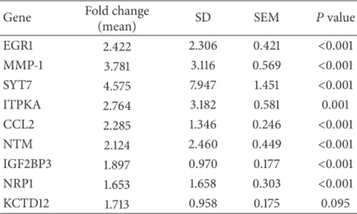
Table 2: Upregulation of gene expression by NAMPT, as analyzed by real-time PCR.
Gene Fold change
(mean) SD SEM �value EGR1 2.422 2.306 0.421 <0.001 MMP-1 3.781 3.116 0.569 <0.001 SYT7 4.575 7.947 1.451 <0.001 ITPKA 2.764 3.182 0.581 0.001 CCL2 2.285 1.346 0.246 <0.001 NTM 2.124 2.460 0.449 <0.001 IGF2BP3 1.897 0.970 0.177 <0.001 NRP1 1.653 1.658 0.303 <0.001 KCTD12 1.713 0.958 0.175 0.095
Efect of NAMPT on gene expression of PDL cells from 10 donors at 1 d. Abbreviations: EGR1: early growth response 1; MMP-1: matrix metalloproteinase-1; SYT7: synaptotagmin 7; ITPKA: inositol 1,4,5-trisphosphate 3-kinase a; CCL2: chemokine, cc motif, ligand 2; NTM: neurotrimin; IGF2BP3: insulin-like growth factor 2 mRNA-binding protein 3; NRP1: neuropilin 1; KCTD12: potassium channel tetramerization domain-containing protein 12.
3.2. Efect of NAMPT on MMP-1 and CCL2. Since MMP-1
and CCL2 are molecules known to be strongly associated with periodontitis, we further studied their regulation by NAMPT. As observed at 1 d, NAMPT increased signiicantly the MMP-1 (MMP-1.4-fold) and CCL2 (MMP-1.8-fold) expressions at 3 d (Fig-ure1(b)). Furthermore, the stimulatory efects of NAMPT on MMP-1 and CCL2 expressions were dose-dependent at both time points, as shown in Figures1(c)–1(f). In addition,
the NAMPT-induced upregulation of the MMP-1 and CCL2 expressions was parallelled by increased MMP-1 and CCL2 protein levels in the supernatants of NAMPT-stimulated cells, as compared to those of the control, at 1 d and 3 d (Figures1(g) and1(h)).
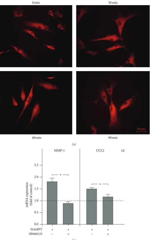
3.3. Exploitation of the JNK Pathway by NAMPT. Next, we
studied how the efects of NAMPT on MMP-1 and CCL2 are mediated intracellularly. As evidenced by immunoluores-cence microscopy, NAMPT stimulated the nuclear translo-cation of NF�B and caused a maximal NF�B accumulation within the nucleus at 60 min (Figure 2(a)). However, pre-incubation of cells with PDTC, a speciic inhibitor of NF�B, had not efect on the NAMPT-stimulated upregulation of MMP-1 and CCL2 (data not shown). Moreover, the upreg-ulation of both molecules by NAMPT was also not afected by inhibitors against the p38, MEK1/2 and PI3K signaling pathways. However, SP600125, an inhibitor of JNK signaling, blocked completely the stimulatory efect of NAMPT on the MMP-1 expression at 1 d (Figure2(b)). In addition, SP600125 reduced signiicantly the NAMPT-stimulated upregulation of CCL2 by 69% at this time point (Figure2(b)).
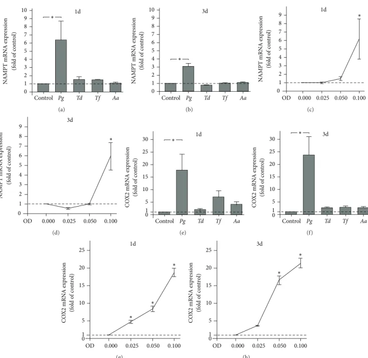
3.4. Stimulation of NAMPT Production by P. gingivalis.
We then wondered whether pathogens that are strongly associated with periodontitis are capable of inducing the production of NAMPT in PDL cells. WhereasT. denticola,
T. forsythiaandA. actinomycetemcomitanshad no signiicant
efect,P. gingivalisincreased signiicantly the NAMPT mRNA expression at 1 d and 3 d (Figures3(a)and3(b)). As shown in Figures3(c)and3(d), the actions ofP. gingivalison NAMPT mRNA expression were dose-dependent. Interestingly, pre-incubation with a speciic inhibitor against NF�B signaling blocked completely theP. gingivalis-induced stimulation of NAMPT at 1 d. Furthermore, inhibitors against JNK and p38 signaling reduced the stimulatory efects of NAMPT by 87% and 37%, respectively, at this time point.
he stimulation of NAMPT expression by P. gingivalis
was also observed at protein level, as analyzed by ELISA: at 1 d, PDL cells in the presence and absence of P. gingivalis
produced 28.83 ± 0.29ng NAMPT protein/104 cells and
17.91 ± 0.58ng NAMPT protein/104 cells, respectively. At
3 d,P. gingivalis-stimulated cells and control cells synthesized
30.97 ± 3.09ng NAMPT protein/104 cells and 21.65 ±
2.94ng NAMPT protein/104 cells, respectively. he difer-ences between both groups at each time point were sig-niicant. he stimulation of NAMPT protein production by
P. gingivalis was also evidenced by immunocytochemistry
(Figure4(c)).
Like NAMPT, COX2 was signiicantly upregulated byP.
gingivalisat 1 d and 3 d (Figures3(e)and3(f)). By contrast, all
other periodontopathogens were not capable of stimulating the COX2 expression at both time points (Figures 3(e) and 3(f)). he P. gingivalis-induced stimulation of COX2 expression was dose-dependent (Figures3(g)and3(h)).
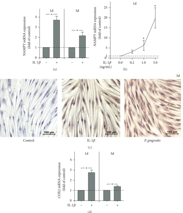
3.5. Stimulation of NAMPT Production by IL-1�. Finally,
1d 5 6 7 8 0 2 1 3 4
EGR1 MMP-1 SYT7 ITPKA CCL2 NTM IGF2BP3 NRP1 KCTD12 ∗ ∗ ∗ ∗ mRN A exp res sio n (f o ld o f co n tr o l) ∗ ∗ ∗ ∗ ∗ (a) ∗ 1.5
2.0 MMP-1 CCL2∗ 3d
0.0 0.5
NAMPT − + − +
1.0 mRN A exp res sio n (f o ld o f co n tr o l) (b) ∗ ∗ ∗ 2 3 1d 4 1 0 30 100 NAMPT (ng/mL) 300 0 MMP -1 mRN A exp res sio n (f o ld o f co n tr o l) (c) ∗ ∗ 2 3 3d 4 1 0 30 100 NAMPT (ng/mL) 300 0 MMP -1 mRN A exp res sio n (f o ld o f co n tr o l) (d) ∗ 2 3 1d 4 ∗ 1 0 30 100 NAMPT (ng/mL) 300 0 C CL2 mRN A exp re ssio n (f o ld o f co n tr o l) (e) ∗ 2 3 3d 4 ∗ ∗ 1 0 30 100 NAMPT (ng/mL) 300 0 C CL2 mRN A exp re ssio n (f o ld o f co n tr o l) (f) 125 75 100 175 150 200 1d ∗ 3d 0 25 50 ∗
NAMPT − + − +
MMP -1 p ro tein (pg/ 10 3 cells) (g) 50 20 30 40 70 60 80 1d ∗ ∗ 3d 0 10
NAMPT − + − +
C CL2 p ro tein (pg/ 10 4 cells) (h)
Figure 1: Stimulation of gene expression and protein synthesis by NAMPT. Upregulation of genes by NAMPT (100 ng/mL) in PDL cells from 10 donors at 1 d (a). Upregulation of MMP-1 and CCL2 expression by NAMPT (100 ng/mL) in PDL cells from 10 donors at 3 d (b). Stimulation of MMP-1 expression by various concentrations of NAMPT in PDL cells from 3 donors at 1 d (c) and 3 d (d). Stimulation of CCL2 expression by various concentrations of NAMPT in PDL cells from 3 donors at 1 d (e) and 3 d (f). Stimulation of MMP-1 protein synthesis by NAMPT (100 ng/mL) in PDL cells from 6 donors at 1 d and 3 d (g). Stimulation of CCL2 protein synthesis by NAMPT (100 ng/mL) in PDL cells from 6 donors at 1 d and 3 d (h). All experiments were performed in triplicate and repeated at least twice. Mean±SEM;∗signiicantly (� < 0.05) diferent from NAMPT-untreated cells (control).
PDL cells. Interestingly, IL-1�upregulated signiicantly the NAMPT expression at 1 d and 3 d (Figure4(a)). Moreover, the actions of IL-1� on NAMPT expression were dose-dependent at 1 d (Figure1(b)) but not at 3 d (data not shown). When cells were preincubated with an inhibitor against the MEK1/2 signaling pathway, the IL-1�-induced stimulation of NAMPT expression was completely suppressed. Moreover, inhibitors against JNK, NF�B, and p38 signaling reduced the
stimulatory efect of IL-1�on NAMPT by 80%, 78%, and 46%, respectively.
50 �m
0 min 30 min
60 min 90 min
(a)
∗
1.0 1.5 2.0 2.5
1d CCL2
∗ MMP-1
0.0 NAMPT SP600125
+ +
+ + +
+ 0.5
− −
mRN
A exp
res
sio
n
(f
o
ld o
f co
n
tr
o
l)
(b)
1d ∗ 0 N AMPT mRN A exp re ssio n (f o ld o f co n tr o l) 2 1 3 4 5 6 7 8 9 10
Control Pg Td Tf Aa
(a) 3d ∗ 0 N AMPT mRN A exp re ssio n (f o ld o f co n tr o l) 2 1 3 4 5 6 7 8 9 10
Control Pg Td Tf Aa
(b) ∗ 8 7 9 1d 0 5 1 2 3 4 6 0.025 0.050
OD 0.000 0.100
N AMPT mRN A exp re ssio n (f o ld o f co n tr o l) (c) ∗ 8 7 9 3d 0 5 1 2 3 4 6 0.025 0.050
OD 0.000 0.100
N AMPT mRN A exp re ssio n (f o ld o f co n tr o l) (d) 1d ∗ 20 30 0 5 1 10 15
Control Pg Td Tf Aa
C O X2 mRN A exp re ssio n (fo ld o f co n tr o l) 25 (e) 3d ∗ 20 25 30 0 5 1 10 15
Control Pg Td Tf Aa
C O X2 mRN A exp re ssio n (fo ld o f co n tr o l) (f) ∗ 15 20 25 1d 0.025 0.050 0 OD 0.100 1 5 10 0.000 ∗ ∗ C O X2 mRN A exp re ssio n (fo ld o f co n tr o l) (g) ∗ 15 20 25 3d ∗ 0 0.025 0.050 OD 0.100 1 5 10 0.000 C O X2 mRN A exp re ssio n (fo ld o f co n tr o l) (h)
Figure 3: Regulation of NAMPT and COX2 expressions by periodontopathogens. NAMPT expression in PDL cells from 3 donors in response to various periodontopathogens (OD: 0.1;P. gingivalis, Pg; T. denticola, Td; T. forsythia, Tf; A. actinomycetemcomitans, Aa)at 1 d (a) and 3 d (b). Stimulation of NAMPT expression in PDL cells from 3 donors by various concentrations ofP. gingivalis(OD: 0.025, 0.050, and 0.100) at 1 d (c) and 3 d (d). COX2 expression in PDL cells from 3 donors in response to various periodontopathogens (OD: 0.1;P. gingivalis, Pg; T.
denticola, Td; T. forsythia, Tf; A. actinomycetemcomitans, Aa) at 1 d (e) and 3 d (f). Stimulation of COX2 expression in PDL cells from 3 donors
by various concentrations ofP. gingivalis(OD: 0.025, 0.050, and 0.100) at 1 d (g) and 3 d (h). All experiments were performed in triplicate and repeated at least twice. Mean±SEM;∗signiicantly (� < 0.05) diferent fromP. gingivalis-untreated cells (control).
control cells produced30.97 ± 3.82ng NAMPT protein/104 cells and20.43 ± 0.76ng NAMPT protein/104 cells, respec-tively. he diferences between groups were signiicant at both time points. he stimulation of NAMPT protein synthesis by IL-1�was also observed by immunocytochemistry (Fig-ure4(c)).
IL-1�also induced a signiicant upregulation of COX2 expression at 1 d and 3 d (Figure4(d)).
4. Discussion
N
AMPT mRN
A exp
re
ssio
n
(fo
ld
o
f co
n
tr
o
l)
∗
1 2 3 4
1d 3d
∗
0
+ +
− −
IL-1𝛽
(a)
∗ 1d
15
5 10 25
N
AMPT mRN
A exp
re
ssio
n
(fo
ld
o
f co
n
tr
o
l) 20
∗
0
0.2 1.0
(ng/mL)
5.0 1
0.0 IL-1𝛽
(b)
Control P. gingivalis
3d
IL-1𝛽
(c)
1d 3d
C
O
X2 mRN
A exp
re
ssio
n
(fo
ld
o
f co
n
tr
o
l)
∗
0 1 2 3 4
∗
+ +
− −
IL-1𝛽
(d)
Figure 4: Regulation of NAMPT and COX2 expressions by IL-1�. NAMPT expression in PDL cells from 6 donors in response to IL-1� (1 ng/mL) at 1 d and 3 d (a). Stimulation of NAMPT expression in PDL cells from 3 donors by various concentrations of IL-1�(0.2, 1.0, and 5.0 ng/mL) at 1 d (b). NAMPT protein synthesis in PDL cells in the presence and absence of IL-1�andP. gingivalisat 3 d. Images from one representative donor are shown (c). COX2 expression in PDL cells from 6 donors in response to IL-1�(1 ng/mL) at 1 d and 3 d (d). All experiments were performed in triplicate and repeated at least twice. Mean±SEM;∗signiicantly (� < 0.05) diferent from IL-1�-untreated cells (control).
and inlammatory signals may use this adipokine for their detrimental efects on the periodontium.
MMP-1 degrades speciically type I collagen and, addi-tionally, types II, III, V, IX, and X collagen, thereby playing a critical role for modeling and remodeling of the extracel-lular matrix [25]. Several studies have revealed that MMP-1 levels are increased in GCF and human gingiva from periodontitis patients [26, 27]. Furthermore, MMP-1 levels
CCL2 in human PDL cells, as evidenced by several assays and in a high number of donors. hese indings are in accordance with previous observations in nonperiodontal cells and underline the proinlammatory and catabolic role of NAMPT in the pathophysiology of periodontitis [32–34]. Since increased NAMPT levels are found in obesity, our data suggest at least one mechanism whereby obesity could confer an increased risk of periodontitis in obese individuals [5,9].
Surprisingly, NAMPT also upregulated a number of genes, most of which have mainly been reported in oncol-ogy and neurosciences. he transcription factor EGR1 is a regulator of multiple tumor suppressors but, paradoxically, can also promote tumor progression [35,36]. EGR1 is altered by hypoxia [37], stimulates angiogenesis [35], and regulates the immune system [38]. Synaptotagmins are transmem-brane proteins with two Ca2+-binding C(2) domains in their cytosolic region. Syt7 regulates the exocytosis of lysosomes and thereby plays a role in bone resorption [39], cell migra-tion [40], CTL responses [41], and neurodegeneration [42]. ITPKA phosphorylates inositol 1,4,5-trisphosphate, thereby modulating the calcium (Ca2+) level within the cell and the levels of a large number of inositol polyphosphates [43]. ITPKA promotes cell motility and the metastatic potential of tumor cells [43,44]. Furthermore, ITPKA is critical for the spatial and temporal regulation of spine actin remodeling, synaptic plasticity, and learning and memory [45]. Little is known about neurotrimin, a neural cell adhesion protein. It has been shown to be an estrogen-regulated determinant of peripheral sympathetic innervation [46] and to play a role in axonal fasciculation of speciic cerebellar systems and the formation of excitatory synapses and their stabilization [47]. he IGF2BPs bind to the 5�-untranslated region of IGF-2 mRNA and regulate a number of important aspects of cell function, such as cell polarization, morphology, migration, proliferation, diferentiation, and invasion [48, 49]. NRP1 plays a critical part in neuronal development, in angiogenesis and tumor invasion [50, 51]. NRP1 is a coreceptor for members of the vascular endothelial growth factor family and the class 3 semaphorins, which are polypeptides with roles in axonal guidance [52]. Furthermore, NRP1 mediates interaction of regulatory T cells with dendritic cells and modulates the immune response [53]. KCTDs act as auxiliary subunits of GABA(B) receptors and generate desensitizing receptor responses [54]. Further studies should clarify the physiological and pathological role of these genes in the periodontium.
We also sought to examine the intracellular mechanism whereby the stimulatory efects of NAMPT on MMP-1 and CCL2 are accomplished. Although NAMPT stimulated the NF�B nuclear translocation in PDL cells, which concurs with observations in other cells [32,55,56], pre-incubation of cells with a speciic NF�B inhibitor did not abrogate the NAMPT actions on MMP-1 and CCL2 in our cells. However, the NF�B signaling pathway could be involved in the NAMPT-induced upregulation of some of the other genes which were identiied by microarray and PCR analyses. Interestingly, our experiments revealed that the NAMPT-induced upregulation
of MMP-1 and CCL2 was JNK dependent, which is supported by indings from others [32].
Recently, increased NAMPT levels have been found in GCF, gingival tissues, and serum from periodontally diseased patients, as compared to periodontally healthy individuals. his suggests that NAMPT might also be produced locally in the periodontium and regulated by periodontopathogens and/or inlammatory mediators [12, 13]. Interestingly, we found that P. gingivalis, a key pathogen associated with periodontitis, and the proinlammatory cytokine IL-1�, which is increased at inlamed periodontal sites, can induce NAMPT in PDL cells, thereby supporting the assumption that NAMPT is locally produced in the presence of an infec-tious and/or inlammatory environment. By upregulation of MMP-1 and CCL2, NAMPT may mediate and amplify proinlammatory and proteolytic actions ofP. gingivalisand IL-1�on the PDL. he inding that NAMPT is induced byP.
gingivalisis in line with our previous experiments, which have
demonstrated that NAMPT is upregulated byF. nucleatum, a gram-negative, anaerobic microorganism, which acts as a bridge bacterium between early and late colonizers during plaque development [57]. Interestingly,T. forsythia, T.
den-ticola, and A. actinomycetemcomitans did not regulate the
NAMPT expression, at least at the concentration tested, in the present study. Our observation that NAMPT is increased by IL-1�in PDL cells concurs with indings in other cells, which have also been shown to produce increased NAMPT levels in response to IL-1� [58–60]. Our data provide evidence that PDL cells produce increased levels of NAMPT under infectious and inlammatory conditions, which suggests that local production of NAMPT in the inlamed periodontium could contribute to the increased gingival and serum levels of NAMPT, as observed in patients with periodontitis. NAMPT could therefore also represent a pathomechanistic link, how periodontitis afects systemic diseases, such as diabetes mel-litus and cardiovascular diseases.
Interestingly, the upregulation of NAMPT byP. gingivalis
could be completely blocked by an inhibitor against NF�B and reduced by inhibitors against the JNK and p38 signaling pathways. Recently, other investigators have also shown that
P. gingivalisinduces NF�B activation and p38 signaling for
its actions [61, 62]. An inhibitor against MEK1/2 signaling pathway suppressed completely the IL-1�-induced stimula-tion of NAMPT expression. JNK, NF�B, and p38 signaling were also found to be involved in the stimulatory efects of IL-1�on NAMPT. hat IL-1�exploits the NF�B and mitogen-activated protein kinase pathways for its actions is well known and supported by the present indings [63,64].
In order to simulate an infectious environment, PDL cells were incubated with a suspension of various periodon-topathogens. Since the suspensions were exposed to intensive ultrasonication, the suspensions contained disrupted cell wall particles with a high amount of lipopolysaccharide. However, other bacterial components may also have been present in the suspension. he concentrations used in this study were determined by dose-response experiments. However, a signiicant upregulation of NAMPT was only observed for
P. gingivalis.P. gingivalisis a gram-negative bacterium and
efects,P. gingivalispossesses a number of virulence factors, such as gingipains and imbriae.P. gingivaliscan invade host cells and also evade the host defense system [65–67]. As it has been shown in several cells that COX2, an enzyme which is responsible for the formation of prostanoids, is upregulated byP. gingivalisand IL-1�, we also analyzed the COX2 expression as a positive control in our study [61,62,68, 69]. As expected,P. gingivalisand IL-1�increased the COX2 expression in a dose- and time-dependent manner in PDL cells.
In summary, the present study demonstrates for the irst time that NAMPT stimulates the production of MMP-1 and CCL2 in human PDL cells, which suggests that NAMPT may contribute to periodontal inlammation and matrix destruction through the production of these molecules. herefore, increased NAMPT levels, as found in obesity, may represent at least one mechanism, whereby obesity could confer an increased risk of periodontitis in obese individ-uals. In addition, our study provides original evidence that NAMPT is induced by the periodontopathogenP. gingivalis
and the proinlammatory cytokine IL-1�in PDL cells, which shows that microbial and inlammatory signals may use this adipokine for their detrimental efects on the periodontium. hese indings suggest that local production of NAMPT in the inlamed PDL could contribute to the increased gingival and serum levels of NAMPT, as observed in patients with periodontitis and therefore represent a pathomechanistic link whereby periodontitis afects systemic diseases, such as diabetes mellitus and cardiovascular diseases.
Conflict of Interests
he authors declare that they have no conlict of interests.
Acknowledgments
he authors would like to thank Ms. Ramona H¨omig, Professor Werner G¨otz, and Professor Stephan Baader for their support. his study was funded by the German Research Foundation (KFO208/TP4) and the University of Bonn. Professor Holger Fr¨ohlich is a member of the Exzellenzcluster ImmunoSensation.
References
[1] B. L. Pihlstrom, B. S. Michalowicz, and N. W. Johnson, “Peri-odontal diseases,”he Lancet, vol. 366, no. 9499, pp. 1809–1820, 2005.
[2] L. Sbordone and C. Bortolaia, “Oral microbial bioilms and plaque-related diseases: microbial communities and their role in the shit from oral health to disease,”Clinical Oral
Investiga-tions, vol. 7, no. 4, pp. 181–188, 2003.
[3] J. M. Albandar, “Commentary: underestimation of periodonti-tis in nhanes surveys,”Journal of Periodontology, vol. 82, no. 3, pp. 337–341, 2011.
[4] T. Beikler and T. F. Flemmig, “Oral bioilm-associated diseases: trends and implications for quality of life, systemic health and expenditures,”Periodontology 2000, vol. 55, no. 1, pp. 87–103, 2011.
[5] J. Suvan, F. D’Aiuto, D. R. Moles, A. Petrie, and N. Donos, “Association between overweight/obesity and periodontitis in adults. A systematic review,”Obesity Reviews, vol. 12, no. 501, pp. e381–e404, 2011.
[6] G. J. Seymour, P. J. Ford, M. P. Cullinan, S. Leishman, and K. Yamazaki, “Relationship between periodontal infections and systemic disease,”Clinical Microbiology and Infection, vol. 13, no. 4, pp. 3–10, 2007.
[7] J. Conde, M. Scotece, R. G´omez et al., “Adipokines: biofactors from white adipose tissue. A complex hub among inlamma-tion, metabolism, and immunity,”BioFactors, vol. 37, no. 6, pp. 413–420, 2011.
[8] A. R. Moschen, R. R. Gerner, and H. Tilg, “Pre-b cell colony enhancing factor/nampt/visfatin in inlammation and obesi-tyrelated disorders,”Current Pharmaceutical Design, vol. 16, no. 17, pp. 1913–1920, 2010.
[9] Y.-H. Chang, D.-M. Chang, K.-C. Lin, S.-J. Shin, and Y.-J. Lee, “Visfatin in overweight/obesity, type 2 diabetes mellitus, insulin resistance, metabolic syndrome and cardiovascular diseases: a meta-analysis and systemic review,” Diabetes/Metabolism
Research and Reviews, vol. 27, no. 6, pp. 515–527, 2011.
[10] D. Taskesen, B. Kirel, and T. Us, “Serum visfatin levels, adiposity and glucose metabolism in obese adolescents,” Journal of
Clinical Research in Pediatric Endocrinology, vol. 4, pp. 76–81,
2012.
[11] L. Q. Zhang, D. P. Heruth, and S. Q. Ye, “Nicotinamide phos-phoribosyltransferase in human diseases,”Journal of Bioanalysis
and Biomedicine, vol. 3, no. 1, pp. 13–25, 2011.
[12] A. R. Pradeep, R. N. M. Raghavendra N.M., M. V. R. Prasad, R. Kathariya, S. P. Patel, and A. Sharma, “Gingival crevicular luid and serum visfatin concentration: their relationship in periodontal health and disease,”Journal of Periodontology, vol. 82, no. 9, pp. 1314–1319, 2011.
[13] A. R. Pradeep, N. M. Raghavendra, A. Sharma et al., “Asso-ciation of serum and crevicular visfatin levels in periodontal health and disease with type 2 diabetes mellitus,”Journal of
Periodontology, vol. 83, no. 5, pp. 629–634, 2012.
[14] M. Nokhbehsaim, B. Deschner, J. Winter et al., “Interactions of regenerative, inlammatory and biomechanical signals on bone morphogenetic protein-2 in periodontal ligament cells,”Journal
of Periodontal Research, vol. 46, no. 3, pp. 374–381, 2011.
[15] M. Nokhbehsaim, J. Winter, B. Rath, A. J¨ager, S. Jepsen, and J. Deschner, “Efects of enamel matrix derivative on periodontal wound healing in an inlammatory environment in vitro,”
Journal of Clinical Periodontology, vol. 38, no. 5, pp. 479–490,
2011.
[16] M. Nokhbehsaim, B. Deschner, J. Winter et al., “Anti-inlammatory efects of EMD in the presence of biomechanical loading and interleukin-1�in vitro,”Clinical Oral Investigations, vol. 16, no. 1, pp. 275–283, 2012.
[17] S. M. Lin, P. Du, W. Huber, and W. A. Kibbe, “Model-based variance-stabilizing transformation for Illumina microarray data,”Nucleic Acids Research, vol. 36, no. 2, article e11, 2008. [18] B. M. Bolstad, R. A. Irizarry, M. ˚Astrand, and T. P. Speed, “A
comparison of normalization methods for high density oligonu-cleotide array data based on variance and bias,”Bioinformatics, vol. 19, no. 2, pp. 185–193, 2003.
[19] P. Du, W. A. Kibbe, and S. M. Lin, “lumi: a pipeline for processing Illumina microarray,”Bioinformatics, vol. 24, no. 13, pp. 1547–1548, 2008.
Sciences of the United States of America, vol. 105, no. 48, pp. 18718–18723, 2008.
[21] G. K. Smyth, “Linear models and empirical bayes methods for assessing diferential expression in microarray experiments,”
Statistical Applications in Genetics and Molecular Biology, vol.
3, no. 1, article 3, 2004.
[22] M. E. Ritchie, D. Diyagama, J. Neilson et al., “Empirical array quality weights in the analysis of microarray data,”BMC
Bioinformatics, vol. 7, article 261, 2006.
[23] Y. Benjamini and Y. Hochberg, “Controlling the false discovery rate: a practical and powerful approach to multiple testing,”
Journal of the Royal Statistical Society B, vol. 57, pp. 89–300, 1995.
[24] G. K. Smyth, “Limma: linear models for microarray data,” in
Bioinformatics and Computational Biology Solutions Using R and
Bioconductor, R. Gentleman, V. Carey, W. Huber, R. Irizarry,
and S. Dudoit, Eds., pp. 397–420, Springer, New York, NY, USA, 2005.
[25] P. A. Arakaki, M. R. Marques, and M. C. L. G. Santos, “MMP-1 polymorphism and its relationship to pathological processes,”
Journal of Biosciences, vol. 34, no. 2, pp. 313–320, 2009.
[26] A.-L. Ejeil, S. Igondjo-Tchen, S. Ghomrasseni, B. Pellat, G. Godeau, and B. Gogly, “Expression of matrix metallopro-teinases (MMPs) and tissue inhibitors of metalloprometallopro-teinases (TIMPs) in healthy and diseased human gingiva,”Journal of
Periodontology, vol. 74, no. 2, pp. 188–195, 2003.
[27] G. T¨uter, B. Kurtis¸, and M. Serdar, “Efects of phase I peri-odontal treatment on gingival crevicular luid levels of matrix 1 and tissue inhibitor of
metalloproteinase-1,”Journal of Periodontology, vol. 73, no. 5, pp. 487–493, 2002.
[28] S. L. Deshmane, S. Kremlev, S. Amini, and B. E. Sawaya, “Monocyte chemoattractant protein-1 (MCP-1): an overview,”
Journal of Interferon and Cytokine Research, vol. 29, no. 6, pp.
313–326, 2009.
[29] G. P. Garlet, W. Martins Jr., B. R. Ferreira, C. M. Milanezi, and J. S. Silva, “Patterns of chemokines and chemokine receptors expression in diferent forms of human periodontal disease,”
Journal of Periodontal Research, vol. 38, no. 2, pp. 210–217, 2003.
[30] M. Gupta, R. Chaturvedi, and A. Jain, “Role of monocyte chemoattractant protein-1 (MCP-1) as an immune-diagnostic biomarker in the pathogenesis of chronic periodontal disease,”
Cytokine, vol. 61, pp. 892–897, 2013.
[31] B. Kurtis¸, G. T¨uter, M. Serdar et al., “Gingival crevicular luid levels of monocyte chemoattractant protein-1 and tumor necrosis factor-alpha in patients with chronic and aggressive periodontitis,” Journal of Periodontology, vol. 76, no. 11, pp. 1849–1855, 2005.
[32] F. Brentano, O. Schorr, C. Ospelt et al., “Pre-B cell colony-enhancing factor/visfatin, a new marker of inlammation in rheumatoid arthritis with proinlammatory and matrix-degrading activities,”Arthritis and Rheumatism, vol. 56, no. 9, pp. 2829–2839, 2007.
[33] S. W. Liu, S. B. Qiao, J. S. Yuan, and D. Q. Liu, “Visfatin stimulates production of monocyte chemotactic protein-1 and interleukin-6 in human vein umbilical endothelial cells,”
Hor-mone and Metabolic Research, vol. 41, no. 4, pp. 281–286, 2009.
[34] G. Sommer, S. Kralisch, N. Kloting et al., “Visfatin is a positive regulator of MCP-1 in human adipocytes in vitro and in mice in vivo,”Obesity, vol. 18, no. 8, pp. 1486–1492, 2010.
[35] E. D. Adamson and D. Mercola, “Egr1 transcription factor: multiple roles in prostate tumor cell growth and survival,”
Tumor Biology, vol. 23, no. 2, pp. 93–102, 2002.
[36] V. Baron, E. D. Adamson, A. Calogero, G. Ragona, and D. Mercola, “he transcription factor Egr1 is a direct regulator of multiple tumor suppressors including TGF�1, PTEN, p53, and ibronectin,”Cancer Gene herapy, vol. 13, no. 2, pp. 115–124, 2006.
[37] V. S. Ten and D. J. Pinsky, “Endothelial response to hypoxia: physiologic adaptation and pathologic dysfunction,” Current
Opinion in Critical Care, vol. 8, no. 3, pp. 242–250, 2002.
[38] S. B. McMahon and J. G. Monroe, “he role of early growth response gene 1 (egr-1) in regulation of the immune response,”
Journal of Leukocyte Biology, vol. 60, no. 2, pp. 159–166, 1996.
[39] S. L. Teitelbaum, “he osteoclast and its unique cytoskeleton,”
Annals of the New York Academy of Sciences, vol. 1240, no. 1, pp.
14–17, 2011.
[40] R. A. Colvin, T. K. Means, T. J. Diefenbach et al., “Synaptotagmin-mediated vesicle fusion regulates cell migration,” Nature Immunology, vol. 11, no. 6, pp. 495–502, 2010.
[41] K. T. Fowler, N. W. Andrews, and J. W. Huleatt, “Expression and function of synaptotagmin VII in CTLs,”Journal of Immunol-ogy, vol. 178, no. 3, pp. 1498–1504, 2007.
[42] G. Glavan, R. Schliebs, and M. ˇZivin, “Synaptotagmins in neurodegeneration,”Anatomical Record, vol. 292, no. 12, pp. 1849–1862, 2009.
[43] H. Kato, K. Uzawa, T. Onda et al., “Down-regulation of 1D-myo-inositol 1,4,5-trisphosphate 3-kinase A protein expression in oral squamous cell carcinoma,” International Journal of
Oncology, vol. 28, no. 4, pp. 873–881, 2006.
[44] S. Windhorst, R. Fliegert, C. Blechner et al., “Inositol 1,4,5-trisphosphate 3-kinase-A is a new cell motility-promoting protein that increases the metastatic potential of tumor cells by two functional activities,”Journal of Biological Chemistry, vol. 285, no. 8, pp. 5541–5554, 2010.
[45] H. K. Il, K. P. Soon, T. H. Soon et al., “Inositol 1,4,5-trisphosphate 3-kinase A functions as a scafold for synaptic rac signaling,”
Journal of Neuroscience, vol. 29, no. 44, pp. 14039–14049, 2009.
[46] D. Krizsan-Agbas, T. Pedchenko, and P. G. Smith, “Neurotrimin is an estrogen-regulated determinant of peripheral sympathetic innervation,”Journal of Neuroscience Research, vol. 86, no. 14, pp. 3086–3095, 2008.
[47] S. Chen, O. Gil, Y. Q. Ren, G. Zanazzi, J. L. Salzer, and D. E. Hillman, “Neurotrimin expression during cerebellar develop-ment suggests roles in axon fasciculation and synaptogenesis,”
Journal of Neurocytology, vol. 30, no. 11, pp. 927–937, 2002.
[48] J. L. Bell, K. W¨achter, B. M¨uhleck et al., “Insulin-like growth fac-tor 2 mRNA-binding proteins (IGF2BPs): post-transcriptional drivers of cancer progression?” Cellular and Molecular Life
Sciences, vol. 70, no. 15, pp. 2657–2675, 2013.
[49] R. Suvasini, B. Shruti, B. hota et al., “Insulin growth factor-2 binding protein 3 (IGF2BP3) is a glioblastoma-speciic marker that activates phosphatidylinositol 3-kinase/mitogen-activated protein kinase (PI3K/MAPK) pathways by modulating IGF-2,”
Journal of Biological Chemistry, vol. 286, no. 29, pp. 25882–
25890, 2011.
[50] C. A. Staton, I. Kumar, M. W. R. Reed, and N. J. Brown, “Neuropilins in physiological and pathological angiogenesis,”
Journal of Pathology, vol. 212, no. 3, pp. 237–248, 2007.
[51] J. R. L. Wild, C. A. Staton, K. Chapple, and B. M. Corfe, “Neu-ropilins: expression and roles in the epithelium,”International
Journal of Experimental Pathology, vol. 93, no. 2, pp. 81–103,
[52] I. C. Zachary, “How neuropilin-1 regulates receptor tyrosine kinase signalling: the knowns and known unknowns,”
Biochem-ical Society Transactions, vol. 39, no. 6, pp. 1583–1591, 2011.
[53] M. Mizui and H. Kikutani, “Neuropilin-1: the glue between regulatory T cells and dendritic cells?”Immunity, vol. 28, no. 3, pp. 302–303, 2008.
[54] J. Schwenk, M. Metz, G. Zolles et al., “Native GABAB receptors are heteromultimers with a family of auxiliary subunits,”Nature, vol. 465, no. 7295, pp. 231–235, 2010.
[55] R. Adya, B. K. Tan, J. Chen, and H. S. Randeva, “Nuclear
factor-�b induction by visfatin in human vascular endothelial cells,”
Diabetes Care, vol. 31, no. 4, pp. 758–760, 2008.
[56] Y. Fan, S. Meng, Y. Wang, J. Cao, and C. Wang, “Vis-fatin/PBEF/Nampt induces EMMPRIN and MMP-9 produc-tion in macrophages via the NAMPT-MAPK (p38, ERK1/2)-NF-�B signaling pathway,”International Journal of Molecular
Medicine, vol. 27, no. 4, pp. 607–615, 2011.
[57] A. V. Nogueira, M. Nokhbehsaim, S. Eick et al., “Regulation of visfatin by microbial and biomechanical signals in PDLcells,”
Clinical Oral Investigations. In press.
[58] M. Gosset, F. Berenbaum, C. Salvat et al., “Crucial role of visfatin/pre-B cell colony-enhancing factor in matrix degrada-tion and prostaglandin E2 synthesis in chondrocytes: possible inluence on osteoarthritis,”Arthritis and Rheumatism, vol. 58, no. 5, pp. 1399–1409, 2008.
[59] C. E. Kendal and G. D. Bryant-Greenwood, “Pre-B-cell colony-enhancing factor (PBEF/Visfatin) gene expression is modulated by NF-�B and AP-1 in human amniotic epithelial cells,”
Pla-centa, vol. 28, no. 4, pp. 305–314, 2007.
[60] M. R. Williams, N. Kataoka, Y. Sakurai, C. M. Powers, S. G. Eskin, and L. V. McIntire, “Gene expression of endothelial cells due to interleukin-1 beta stimulation and neutrophil transmigration,”Endothelium, vol. 15, no. 1-2, pp. 73–84, 2008. [61] Y. Murakami, A. Kawata, Y. Seki et al., “Comparative inhibitory
efects of magnolol, honokiol, eugenol and bis-eugenol on cyclooxygenase-2 expression and nuclear factor-kappa B acti-vation in RAW264. 7 macrophage-like cells stimulated with imbriae of Porphyromonas gingivalis,”In Vivo, vol. 26, pp. 941– 950, 2012.
[62] D. Reddi, S. J. Brown, and G. N. Belibasakis, “Porphyromonas gingivalis induces RANKL in bone marrow stromal cells: involvement of the p38 MAPK,”Microbial Pathogenesis, vol. 51, no. 6, pp. 415–420, 2011.
[63] A. Dunne and L. A. J. O’Neill, “he interleukin-1 receptor/Toll-like receptor superfamily: signal transduction during inlam-mation and host defense,”Science”s STKE, vol. 2003, no. 171, article re3, 2003.
[64] J.-K. Lee, S.-H. Kim, E. C. Lewis, T. Azam, L. L. Reznikov, and C. A. Dinarello, “Diferences in signaling pathways by IL-1�and IL-18,”Proceedings of the National Academy of Sciences of the
United States of America, vol. 101, no. 23, pp. 8815–8820, 2004.
[65] S. C. Holt and J. L. Ebersole, “Porphyromonas gingivalis, Tre-ponema denticola, and Tannerella forsythia: the “red complex”, a prototype polybacterial pathogenic consortium in periodon-titis,”Periodontology 2000, vol. 38, pp. 72–122, 2005.
[66] K. Nakayama, “Molecular genetics of Porphyromonas gingi-valis: gingipains and other virulence factors,”Current Protein
and Peptide Science, vol. 4, no. 6, pp. 389–395, 2003.
[67] J. Potempa, A. Sroka, T. Imamura, and J. Travis, “Gingipains, the major cysteine proteinases and virulence factors of Por-phyromonas gingivalis: structure, function and assembly of
multidomain protein complexes,”Current Protein and Peptide
Science, vol. 4, no. 6, pp. 397–407, 2003.
[68] Y.-C. Chang, F.-M. Huang, S.-F. Yang et al., “Induction of cyclooxygenase-2 mRNA and protein expression in human pulp cells stimulated with black-pigmented bacteroides,”
Jour-nal of Endodontics, vol. 29, no. 4, pp. 240–243, 2003.
Submit your manuscripts at
http://www.hindawi.com
Stem Cells
International
Hindawi Publishing Corporation
http://www.hindawi.com Volume 2014
Hindawi Publishing Corporation
http://www.hindawi.com Volume 2014 INFLAMMATION
Hindawi Publishing Corporation
http://www.hindawi.com Volume 2014
Behavioural
Neurology
International Journal of
Endocrinology
Hindawi Publishing Corporation
http://www.hindawi.com Volume 2014
Hindawi Publishing Corporation
http://www.hindawi.com Volume 2014
Disease Markers
BioMed Research International
Hindawi Publishing Corporation
http://www.hindawi.com Volume 2014
Oncology
Journal ofHindawi Publishing Corporation
http://www.hindawi.com Volume 2014
Hindawi Publishing Corporation
http://www.hindawi.com Volume 2014 Oxidative Medicine and Cellular Longevity
PPAR
R e s e a r c h
Hindawi Publishing Corporation
http://www.hindawi.com Volume 2014
The Scientiic
World Journal
Hindawi Publishing Corporation
http://www.hindawi.com Volume 2014
Immunology Research
Hindawi Publishing Corporation
http://www.hindawi.com Volume 2014
Journal of
Obesity
Journal ofHindawi Publishing Corporation
http://www.hindawi.com Volume 2014
Hindawi Publishing Corporation
http://www.hindawi.com Volume 2014 Computational and Mathematical Methods in Medicine
Ophthalmology
Journal ofHindawi Publishing Corporation
http://www.hindawi.com Volume 2014
Diabetes ResearchJournal of
Hindawi Publishing Corporation
http://www.hindawi.com Volume 2014
Hindawi Publishing Corporation
http://www.hindawi.com Volume 2014
Research and Treatment
AIDS
Hindawi Publishing Corporation
http://www.hindawi.com Volume 2014
Gastroenterology Research and Practice
Parkinson’s Disease
Hindawi Publishing Corporation
http://www.hindawi.com Volume 2014
Evidence-Based Complementary and Alternative Medicine
Volume 2014 Hindawi Publishing Corporation