Murine Intestinal Smooth Muscle Hypertrophy Induced
by Partial Obstruction
Dong-Hai Liu1, Xu Huang1, Xin Guo1, Xiang-Min Meng1, Yi-Song Wu1, Hong-Li Lu1, Chun-Mei Zhang1, Young-chul Kim2, Wen-Xie Xu1*
1Department of Physiology, Shanghai Jiaotong University School of Medicine, Shanghai, China,2Department of Physiology, Chungbuk National University College of Medicine, Cheongju, Chungbuk, Republic of Korea
Abstract
Partial obstruction of the small intestine causes obvious hypertrophy of smooth muscle cells and motility disorder in the bowel proximate to the obstruction. To identify electric remodeling of hypertrophic smooth muscles in partially obstructed murine small intestine, the patch-clamp and intracellular microelectrode recording methods were used to identify the possible electric remodeling and Western blot, immunofluorescence and immunoprecipitation were utilized to examine the channel protein expression and phosphorylation level changes in this research. After 14 days of obstruction, partial obstruction caused obvious smooth muscle hypertrophy in the proximally located intestine. The slow waves of intestinal smooth muscles in the dilated region were significantly suppressed, their amplitude and frequency were reduced, whilst the resting membrane potentials were depolarized compared with normal and sham animals. The current density of voltage dependent potassium channel (KV) was significantly decreased in the hypertrophic smooth muscle cells and the voltage
sensitivity of KVactivation was altered. The sensitivity of KVcurrents (IKV) to TEA, a nonselective potassium channel blocker,
increased significantly, but the sensitivity of IKv to 4-AP, a KVblocker, stays the same. The protein levels of KV4.3 and KV2.2
were up-regulated in the hypertrophic smooth muscle cell membrane. The serine and threonine phosphorylation levels of KV4.3 and KV2.2 were significantly increased in the hypertrophic smooth muscle cells. Thus this study represents the first
identification of KV channel remodeling in murine small intestinal smooth muscle hypertrophy induced by partial
obstruction. The enhanced phosphorylations of KV4.3 and KV2.2 may be involved in this process.
Citation:Liu D-H, Huang X, Guo X, Meng X-M, Wu Y-S, et al. (2014) Voltage Dependent Potassium Channel Remodeling in Murine Intestinal Smooth Muscle Hypertrophy Induced by Partial Obstruction. PLoS ONE 9(2): e86109. doi:10.1371/journal.pone.0086109
Editor:Tomohiko Ai, Indiana University, United States of America
ReceivedMay 31, 2013;AcceptedDecember 5, 2013;PublishedFebruary 6, 2014
Copyright:ß2014 Liu et al. This is an open-access article distributed under the terms of the Creative Commons Attribution License, which permits unrestricted
use, distribution, and reproduction in any medium, provided the original author and source are credited.
Funding:The work was supported by the National Natural Science Foundation of China (No. 31271236; 31171107; website: http://www.nsfc.gov.cn/Portal0/ default152.htm). The funders had no role in study design, data collection and analysis, decision to publish, or preparation of the manuscript.
Competing Interests:The authors have declared that no competing interests exist. * E-mail: [email protected]
Introduction
Many congenital or acquired diseases often lead to hypertrophy of the tunica muscularis of the intestine, such as infantile hypertrophic pyloric stenosis, Hirschsprung’s disease, achalasia and Chagas’ disease [1–5]. Partial obstruction of the small intestine of mouse, rat, and guinea pig results in a notable hypertrophy of the intestinal wall due to thickening of the smooth muscle layer [6–9]. Hypertrophy of the gastrointestinal muscle wall, induced by partial obstruction, is a physiological response to the increased pressure in the lumen accompanied by motility disorder. The number and size of smooth muscle cells are increased within the hypertrophic intestinal wall [6]. The sensitivities of hypertrophic tunica muscularis to contractile and relaxing agents are also altered in rats [10]. In addition, the slow wave is deteriorated in hypertrophic segment and the resting membrane potential (RMP) of the intestinal smooth muscles is lower than control [6,7].
It is well known that voltage-gated potassium (KV) channels are
expressed in all gastrointestinal (GI) smooth muscle [11]. Based on their rates of activation and inactivation, two types of IKVcan be
distinguished in mouse small intestinal smooth muscles: IKVpeak
and IKVsustained [11,12]. IKVpeak reaches the maximum faster
and inactivates slower than IKV sustained [11,12]. Although all
KVchannels involve in regulating RMP, IKVpeak is activated at
+10 mV,+20 mV more negative than IKVsustained and seems
to be a major contributor to the resting membrane potential of smooth muscles [11,13]. IKv peak is sensitive to 4-AP and resistant to TEA. On the contrary, IKVsustained is a 4-AP resistant and
TEA sensitive current [13]. According to the similar pharmaco-logical and kinetic properties, the KV2 and KV4 families are
thought to be KVpeak (KVpeak) and KVsustained (KVsustained) in
small intestine [14,15]. These channels are the targets of many signaling pathways, including some kinases such as protein kinase A (PKA), protein kinase C (PKC), extracellular regulated protein kinases (ERK) and so on [16–20]. This means that, the function of the channels can be influenced by phosphorylation modification.
reported to decrease in hypertrophic small intestine, and the IKV
channels play an important role in regulating the RMP. Base on this information, we hypothesize that there is IKVremodeling after
hypertrophy in small intestinal smooth muscles, and this change involves depolarization of RMP. In the present study, we used an intestinal partial obstruction mouse model to explore the electrical remodeling and possible mechanisms involved in the motility disorder associated with obstruction-induced smooth muscle hypertrophy.
Materials and Methods
Ethics statement
Animals were obtained from the Experimental Animal Center of Shanghai Jiaotong University School of Medicine. This study was carried out in strict accordance with the recommendations in the Guide for the Care and Use of Laboratory Animals of the Science and Technology Commission of P.R.C. (STCC Publica-tion No. 2, revised 1988). The protocol was approved by the Committee on the Ethics of Animal Experiments of Shanghai Jiaotong University School of Medicine (Permit Number: Hu 686-2009). All surgery was performed under ether, and all efforts were made to minimize suffering.
Animal model and tissue preparation
Male imprinting control region (ICR) mice aged between 5–6 weeks and with a body weight of 3064 g were used. Small bowel obstruction was induced by tightening a ring around the ileum as describe before [6]. Briefly, mice were anaesthetized with chloral hydrate (300 mg/kg, I. p.), and all efforts were made to minimize suffering. Partial intestinal obstruction was induced by surgically placing a ring (5 mm in length, 4 mm external diameter, 3 mm internal diameter) of silicon tube around the ileum 30–50 mm oral to the ileocecal sphincter to cause distension of the intestine proximal to the site of obstruction. Experiments were performed with the distended segments of ileum 14 days after surgery [6]. Sham operations were performed using the same surgical exposure procedures except the ileal obstruction. Mice were anaesthetized with ether and euthanized by cervical dislocation. A 60-mm segment of small intestine oral to the obstruction was removed and pinned out in the base of a Sylgard silicone elastomer dish containing Krebs of the following composition: NaCl 118.5 mM; KCl 4.5 mM; MgCl2 1.2 mM; NaHCO3 23.8 mM; KH2PO4
1.2 mM; glucose 11.0 mM; CaCl2 2.4 mM. The intestinal
segment was opened by cutting lengthwise, washed with Krebs, and the mucosa and submucosa were removed by sharp dissection. The remaining tunica muscularis was used for all experiments.
For Western blot and immunoprecipitation experiments, the tunica muscularis was quickly frozen in liquid nitrogen. Samples were homogenized in ice-cold RIPA buffer [25 mM Tris-HCl pH 7.6, 150 mM NaCl, 1% NP-40, 1% sodium deoxycholate, 0.1% SDS and phosphatase and protease inhibitors (20 mM b -glycerophosphate, 20 mM sodium pyrophosphate, 50 mM NaF, 1 mM each of EDTA, EGTA, sodium orthovanadate, p-nitrophenyl phosphate, Phenylmethanesulfonyl fluoride (PMSF) and benzamidine, and 5mg/ml each of aprotinin, leupeptin and pepstatin A)]. The homogenates were centrifuged at 800 g for 10 min at 4uC for Western blot, or at 12000 g for 15 min at 4uC for immunoprecipitation. The supernatants were collected. Protein concentrations were determined by Bradford method (Pierce, Rockford, IL, USA). Samples were stored at280uC until use.
Immunoprecipitation and Western blot analysis
For immunoprecipitation, the protein samples (each containing 400mg of protein) were diluted four folds with RIPA buffer. Samples were preincubated for 1 h with 20mL protein G-sepharose (Beyotime, Jiangsu, China) and then centrifuged to remove any protein adhered non-specifically to the protein G-sepharose. The supernatant was incubated with 5mg proper antibodies for 4 h at 4uC. After the addition of protein G-sepharose, the mixture was incubated at 4uC for an additional 2 h. Samples were triple washed with RIPA buffer and eluted by sodium dodecyl sulfate-polyacrylamide gel electrophoresis (SDS– PAGE) loading buffer then boiling at 98uC for 5 min. Western blot analysis was carried out on 10% SDS–PAGE. Briefly, proteins were electrotransferred onto Immobilon-p PVDF membrane (millipore, Billerica, MA, USA, pore size 0.45mm). After blocking for 1 h in Tris-HCl-buffered saline with 0.1% tween-20 (TBST) and 5% non-fat milk, the membranes were incubated over night with primary antibody in TBST containing 5% non-fat milk. Detection was carried out by the use of proper horseradish peroxidase conjugated second antibodies and developed with beyoECL plus assay kit (Beyotime chemical Co., Jiangsu, China).
Intracellular microelectrode recording
Strips of tunica muscularis (8 mm64 mm) were cut parallel to the longitudinal axis of the intestine, oral to the site of occlusion. The muscle strips were pinned onto the base of a Sylgard-coated chamber, circular muscle side up, and continuously perfused with warmed (37uC, 1.2 ml/min) and oxygenated Krebs solution. Stripes were equilibrated for approximately 2 h before recording. Cells were impaled with KCl-filled glass microelectrodes with resistances of 50–90 MV. Electrical responses were recorded and amplified through a high input impedance amplifier (SYS-773 Duo 773 Electrometer, WPI, USA). Experiments were performed in the presence of nifedipine (1mM; Sigma, St Louis, MO, USA) in the perfusion solution to reduce contraction and facilitate cell impalement. Slow waves in mouse intestine have been previously shown to be unaffected by nifedipine [23].
Cell preparation and Voltage patch-clamp
Smooth muscle cells were prepared from the small intestinal region as described above. After the mucosal, submucosal layers were carefully removed, the tunica muscularis was cut into small segments (164 mm). These segments were kept in modified Kraft– Bruhe (KB) solution (EGTA 0.5 mM, Hepes 10 mM, MgCl2
3 mM, KCl 50 mM, glucose 10 mM, KH2PO420 mM, taurine
20 mM, and glutamic acid 50 mM, adjusted to pH 7.4 with KOH) for 15 min at 4uC. They were then incubated at 37uC in 1 ml of digestion medium (Ca2+
-free solution) containing 1.5– 2 mg collagenase II or collagenase I (Worthington Biochemicals, Lakewood, NJ, USA), 400–600mg papain (Sigma-Aldrich, St. Louis, MO, USA), 1.5 mg dithiothreitol, 1.5 mg trypsin inhibitor (Amresco Inc., Solon, OH, USA), and 3 mg bovine serum albumin (Sigma-Aldrich, St. Louis, MO, USA) for 10–15 min. The Ca2+
-free solution contained NaCl 135 mM, KCl 5 mM, MgCl21.2 mM, glucose 10 mM, and Hepes 10 mM, adjusted to
physiologic saline solution (PSS, NaCl 135 mM, KCl 5 mM, CaCl2 2 mM, MgCl2 1.2 mM, glucose 10 mM, and Hepes
10 mM, adjusted to pH 7.4 with Tris). A single 4-channel perfusion system (BPS-4, ALA Inc., Westbury, NY, USA) was used to change the perfusate. The whole cell patch-clamp technique was used to record the voltage-dependent K+
currents (IKV) with an EPC-10 amplifier (HEKA Elektronik, Lambrecht,
Germany). Data were filtered at 10 kHz, recorded using patch-master software (HEKA Elektronik, Germany) and digitized at 20 kHz. In cases of long records the data were sampled at 10 kHz. Capacitance was compensated and residual capacitance current was digitally removed. Series resistance was not compensated for. Series resistance was between 2 and 4 MV. Cell membrane capacitance was calculated from the time constant of a capacitance current elicited by a 5 mV depolarization from260 mV. Pipette resistances were 2–5 MV. The pipette solution comprised KCl 20 mM, potassium-aspartic acid 110 mM, di-tris-creatine phos-phate 2.5 mM, MgATP 5 mM, Hepes 5 mM, MgCl2 1.0 mM,
and EGTA 10 mM, adjusted to pH 7.3 with Tris. All current amplitudes were normalized to the cell membrane capacitance (Cm) and expressed as densities (pA/pF). In some experiments 4-AP or TEA were added to the bath. All experiments were performed at room temperature (20–25uC). The peak value of IKv was measured as IKVpeak, the mean value of last 100 ms of
depolarization was measured as IKVsustained to minimize the
IKVpeakinfluence. The voltage dependence of the inactivation was
fit by the Boltzmann function:I/Imax = 1/{1+exp [(V2V0.5)/k]}. WhereIis the amplitude of IKv, V0.5is the half-activation voltage and k is the slop factor at this voltage. Voltage-dependent activation of IKv was respectively converted into conductivity using the Goldman-Hodgkin-Katz current equation [14].
Goldman-Hodgkin-Katz current equation: ws~Psz2s VmF2
RT K
½ i{½ Koexp({zsVmF=RT) 1{exp({zsVmF=RT)
. ws is the IKv amplitude, PS is
the permeability of the membrane, zs is the valence of ion
potassium, Vm is the transmembrane potential, F is the Faraday
constant, equal to 96,485 C?mol21, R is the gas constant, equal to 8.314 J?K21?mol21, T is the absolute temperature, measured in Kelvin, [K]i is the intracellular concentration of K+ (130 mM,
concentration of pipette solution), [K]o is the extracellular
concentration of K+ (5 mM, concentration of physiologic saline
solution). The voltage dependence of the activation was fit by the Boltzmann function:PS/PS
max= 1/{1+exp [2(V2V0.5)/k]}. V0.5
is the half-inactivation voltage and k is the slop factor at this voltage.
Immunofluorescence
Small intestine tissues were collected after animals were killed by cervical dislocation. The samples were fixed with 4% paraformal-dehyde (PFA) overnight at 4uC and sectioned at 5mm thickness. After that, the sections were blocked with 10% goat serum in phosphate-buffered saline (PBS) for 1 h at room temperature. To check for Kv4.3 and Kv2.2 immunoreactivity, the sections were incubated with mouse anti–Kv4.3 antibody (1:1000) and mouse anti–Kv2.2 antibody (1:1000) overnight at 4uC, respectively. The sections were incubated with goat anti-mouse antibody (1:1000) conjugated with DyLight488 for 1 h at room temperature followed by nucleic acid dye DAPI and membrane dye Fm 4-64 membrane stain (Invitrogen, Eugene, Oregon, USA). Pictures were acquired by a confocal laser-scanning microscope (Olympus FV-1000, Japan).
Antibodies and Drugs
The following primary antibodies were used: anti-Kv4.3 (ab99045, for Western blot) and anti-Kv2.2 (ab10651) were acquired from Abcam (Hong Kong, China). Anti-Kv2.2 (sc-292489, for immunoprecipitation) and anti-p-Ser (sc-80514) antibodies were acquired from Santa Cruz Biotechnology (Santa Cruz, CA, USA). Anti-p-Ther antibody (#9386) was obtained from cell signaling technology (City and country of the company). The secondary antibody DyLight488 conjugated goat anti-mouse IgG (16117031711) was bought from ImmunoReagents, Inc. (Raleigh, NC, USA). The secondary antibody horseradish peroxidase (HRP) conjugated goat anti-mouse IgG (sc-2005) was bought from Santa Cruz Biotechnology (Santa Cruz, CA, USA). Tetraethylammonium chloride (TEA), 4-Aminopyridine (4-AP) and other chemicals were all acquired from Sigma unless indicated otherwise.
Statistical Analysis
Data of Western blot were reported as the mean6SD, other data were reported as mean6S.E.M.; n refers to the number of animals from which tissues were obtained. Statistical analysis of the results was carried out by one-way analysis of variance (ANOVA) followed by the least significant difference test or Newman–Keuls test. Differences of p,0.05 were considered significant. Semiquantitative analysis of the bands was performed with the Image J analysis software (Version 1.30v; Wayne Rasband, NIH, USA). Data analysis was performed with SigmaStat Software (Version 3.5, Jandel Scientific Software, California, USA). Curve fitting were performed with SigmaPlot Software (Version 10.0, Jandel Scientific Software, California, USA) and GraphPad Prism 6 (GraphPad Software, La Jolla, USA)
Results
1. Alteration of intestinal smooth muscle cells membrane capacitance and RMP
In order to determine the histological and electrical remodeling and further confirm the hypertrophic state of freshly dissociated intestinal smooth muscle cells in partial intestinal obstruction model, we compared the cell capacitance among normal, sham, and obstruction groups. The cell size was significantly enlarged in obstruction group (Fig. 1A). The membrane capacitance of obstruction group (149.3765.74 pF; n = 40; P,0.05) was signif-icantly increased compared with sham (30.9961.33 pF; n = 36) and normal groups (30.3261.84 pF; n = 35) (Fig. 1B).
The intracellular microelectrode recording method was utilized to record the slow wave and RMP in muscle strips in normal, sham and obstructed intestinal smooth muscle strips to further confirm whether histological remodeling is accompanied by electric remodeling. Our observation was similar to previous reports [6,7]. The RMP as well as the amplitude and frequency of slow wave were all decreased after obstruction (Fig. 2A). The average RMPs were 262.465.04 mV, 26365.24 mV and 251.33611.88 mV*, respectively, in normal, sham and obstruc-tion groups (Fig. 2B, a, nnormal= 6, nsham= 7 and nobstruction= 7,
respectively,P,0.05). The average amplitude and frequency were 25.4261.4 mV, 23.8261.6 mV, 7.2561.3 mV* and 45.661.25 cycle/min, 46.161.18 cycle/min and 30.4461.3 cycle/min*, respectively, in normal, sham and obstruction groups (Fig. 2B, b, c, nnormal= 6, nsham= 7 and nobstruction= 7, respectively, P,0.05).
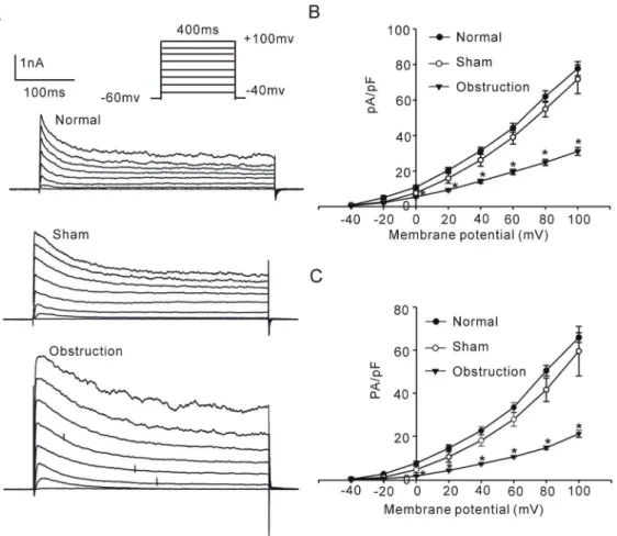
2. IKVdensities and properties in hypertrophic intestinal smooth muscle cells
IKVcan be separated into two components by its sensitivities to
TEA and 4-AP, two traditional potassium channel blockers. We studied changes of the two IKVcomponents in the hypertrophic
intestinal smooth muscle cells. Fig. 3A showed that incremental 20 mV depolarizing steps with 400 ms duration from a constant holding potential of 260 mV to test voltages as positive as +100 mV elicited families of outward currents in intestinal smooth muscle cells of normal, sham and obstruction groups. The average densities of peak potassium currents from beginning to100 ms (IKVpeak) and the mean currents from 300 ms to the end
(IKVsustained) elicited at each test potential were calculated to
compare current-voltage (I–V) relationships in normal, sham and obstruction groups. The densities of IKVpeak and IKVsustainedin
hypertrophied smooth muscle cells was higher than that in normal and sham groups at every commend membrane potential from 0 mV to+100 mV (Fig. 3B). For example, the current densities of IKVpeak elicited by step depolarizing pulses to +40 mV and
+60 mV were 31.2560.978 pA/pF, 44.1762.57 pA/pF in nor-mal group, 26.0663.51 pA/pF, 38.8963.84 pA/pF in sham group and 14.1861.07 pA/pF, 19.4161.45 pA/pF in obstruction group, respectively (Fig. 3B, nnormal= 13, nsham= 11 and n obstruc-tion= 12, respectively, P,0.05 vs normal or sham group). The
current densities of IKVsustainedelicited by step depolarizing pulses
to +40 mV and +60 mV were 22.5761.83 pA/pF, 33.3662.26 pA/pF in normal group, 17.9762.65 pA/pF, 27.7963.17 pA/pF in sham group and 7.1060.43 pA/pF, 10.3860.58 pA/pF in obstruction group, respectively (Fig. 3C, n = 13, 11 and 12, respectively,P,0.05vsnormal or sham group). The results suggest that the IKVfunction is down-regulated in the
hypertrophic intestinal smooth muscle cells induced by partial obstruction.
In succession the steady-state activation and steady-state inactivation curves were obtained in the normal, sham and obstructed intestinal smooth muscle cells to determine whether the down regulation of current densities is associated with alteration of channel voltage sensitivity (Fig. 4). The shifts of activation curves of IKVpeak and IKVsustainedshowed different tendency in
hyper-trophic smooth muscle cells. In comparison to sham and normal groups, the half-activation voltage (V0.5 act) of IKVpeak in
hypertrophic smooth muscle cells shifted to more negative potential (V0.5 act, normal =22.7862.64 mV,
sham =24.363.63 mV and obstruction =215.7463.8 mV, n = 12, 11, 13, respectively, P,0.05, Fig. 4A, a, b). On the contrary, the half-activation voltage of IKVsustainedshifted to more
positive potential (V0.5 act, normal =28.6562.78 mV,
sham =28.9963.8 mV and obstruction = 5.1462.61 mV, n = 12, 11, 13, respectively, P,0.05, Fig. 4A, c). However, the half-inavtivation voltage (V0.5inact) of IKVpeak slightly shifted to
Figure 1. The hypertrophy of smooth muscle cells induced by partial intestinal obstruction.A shows normal (a) and hypertrophic smooth muscle cells (b), the cell size was significantly enlarged in obstruction group. B shows the mean values of cell capacitances among normal, sham and obstruction groups. Data showed the means6SE *P,0.01 versus sham and normal groups, *P,0.05 versus normal and sham group, Bar = 100mm.
positive in hypertrophic smooth muscle cells but there were no significant difference among normal, sham and obstruction groups (Fig. 4B, a, b, V0.5 inact, normal =261.262.27 mV,
sham =260.162.88 mV and obstruction =253.5563.79 mV, n = 12, 11, 12, respectively). Besides of this, no significant alteration was observed in the slop factors and IKVsustainedinactive
curves among obstruction, normal and sham groups (Fig. 4 A, a, b, c, B, a, b, c, d, V0.5inact (normal) =246.3863.94 mV, V0.5inact
(sham) =247.5263.58 mV and V0.5inact
(obstruc-tion) =247.2464.08 mV, n = 10, 9, 11, respectively).
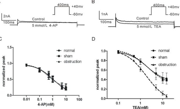
3. Effect of TEA and 4-AP on IKV
4-AP and TEA are two kinds of voltage-dependent potassium channel blockers that have been widely used in studying KV
channel function. In order to examine the sensitivities of IKVpeak
and IKVsustainedto 4-AP and TEA, we compared the dose-response
curves of 4-AP and TEA in intestinal smooth muscle cells of normal, sham and obstruction groups, respectively. The results demonstrated that IKVpeakwas more sensitive to 4-AP (5 mM) and
IKVsustained was more sensitive to TEA (5 mM) when the
membrane potential was depolarized from 260 mV of holding potential to +40 mV (Fig. 5A and B). There was no significant difference in IKV sensitivity to 4-AP among obstruction, normal
and sham groups (Fig. 5C;P.0.05). However, the IC
50of TEA on
IKV in hypertrophic smooth muscle cells was slightly decreased than that in normal and sham groups (Fig. 5D, P.0.05). Interestingly, when the membrane potential was depolarized from 260 mV of holding potential to+40 mV, TEA (15 mM) almost completely blocked the IKVsustainedin hypertrophic smooth muscle
cells (95.360.04%), but TEA only blocked 55.460.05% and 52.760.05% of the IKVsustained in normal and sham groups
(Fig. 5D). These results suggest that the sensitivity of IKVsustainedto
TEA is significantly increased in hypertrophic smooth muscle cells.
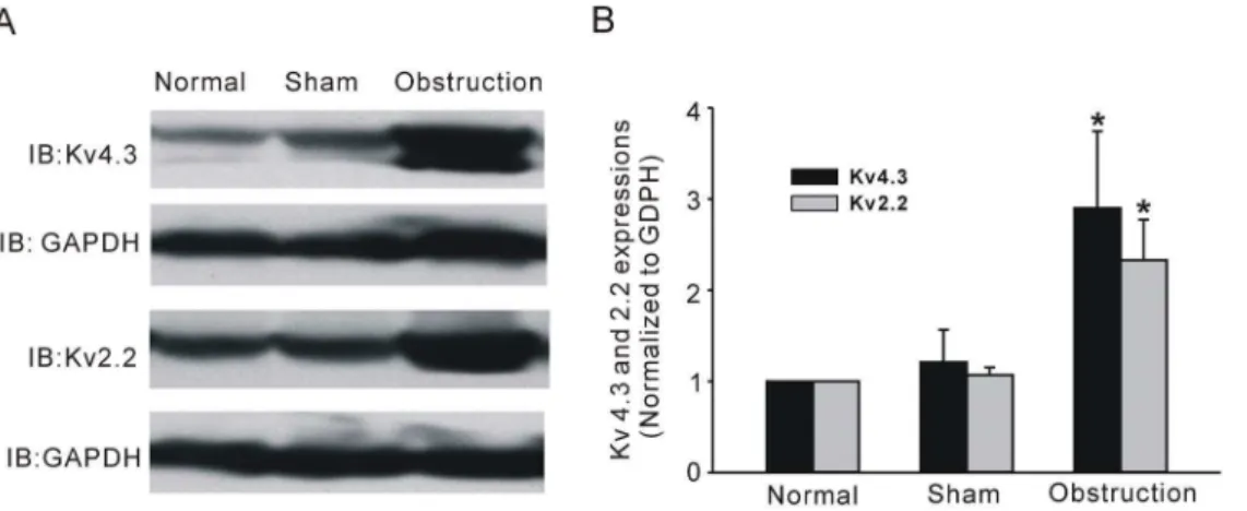
4. Alteration of KVchannel subunits
In further experiments, we examined whether KVchannel pore
forming a subunit alteration could be correlated with partial obstruction-induced electric remodeling. Western blot analysis was performed to observe alteration of channel subunit protein expression. Two immunoreactive bands were detected at
<71 kD and at<102 kD respectively, the former is corresponding
to the long isoform of Kv4.3 channel protein and the latter is the Kv2.2 band (Fig. 6A). The expression levels of both Kv4.3 and Kv2.2 were up-regulated in hypertrophic intestine smooth muscle tissues compared with sham and normal tissues (Fig. 6B, n = 6, P,0.05). The result was quite opposite to the change in IK
V
densities. To further confirm the Western blot result, we next compared the protein expression in situ with the immunofluores-cence method. Similar results of increased Kv4.3 and Kv2.2 immunoreactivities were observed in frozen sections (Fig. 7 and Fig. 8). In the normal and sham groups, weak Kv4.3 and Kv2.2 immunoreactivities were detected in the smooth muscle regions of small intestine, but the change in mucosa layer was not found. Two weeks after ileum obstruction, Kv4.3 and Kv2.2 fluorescence intensity was significantly increased compared with the normal and sham groups (Fig. 7 and Fig. 8). In addition, with the cell membrane staining, we found that the most channel immuno-staining was merged with plasmalemma of longitudinal muscle Figure 2. Changes of slow wave and resting membrane potential (RMP) in hypertrophic smooth muscles.Electrical slow waves were recorded from small intestinal muscle stripes of normal group (Aa), sham group (Ab), and obstruction group (Ac). The RMP (Ba), amplitudes (Bb) and frequencies (Bc) of slow wave were significantly changed in normal, sham and obstruction groups. Data showed the means6SE *P,0.01 versus sham and normal groups.
cells (Fig. 7 and Fig. 8). This told us that channel proteins were still in the plasmalemma other than blocked in the ion channel trafficking pathway.
Protein phosphorylation in particular plays a significant role in regulating protein function. Thus, we examined changes in channel protein phosphorylation level for Kv4.3 and Kv2.2. Because no commercial antibodies against Kv4.3 specific phos-phorylation sites are available and there are no reports of phosphorylation about the Kv2.2, we pulled down the channel proteins from the intestinal smooth muscles tissue with immuno-precipitation method and used the threonine or anti-p-serine antibodies to detect the whole phosphorylation levels of immunoprecipitated proteins. As shown in Fig. 9, in the protein pulled down by anti-Kv2.2 antibody, the ratios of phosphorylated threonine protein and phosphorylated serine protein to total protein were up-regulated significantly in obstruction group compared with other two groups (Fig. 9A and B, n = 5,P,0.05). The ratios of phosphorylated protein to total protein of Kv4.3 were also increased in hypertrophic smooth muscle tissue (Fig. 9A and B). These results suggested that there were great alterations of KV channel phosphorylation in hypertrophic segment of small
intestine.
Discussion
In the present study, we investigated the voltage dependent potassium channel remodeling in hypertrophic smooth muscle cells induced by partial intestinal obstruction in mouse. Our experiments demonstrated that partial obstruction-induced smooth muscle hypertrophy in the proximally located intestine after 14 days of obstruction was accompanied by electric remodeling. The amplitude and frequency of slow waves of intestinal smooth muscles were significantly reduced, whilst the resting membrane potentials were depolarized compared with normal and sham animals. The current density of IKV was
profoundly reduced. The steady-state activation curve of IKVpeak
was shifted to the negative potential and the IKVsustained was
shifted to the positive potential in hypertrophic smooth muscle. However, no significant difference was observed in the inactive curves of IKV. The sensitivity of IKVsustained to TEA was also
increased in hypertrophic smooth muscle cells. Interestingly, the expressions of the main isoforms, pore-forming Kv4.3 and Kv4.2 subunits were up-regulated in the hypertrophic smooth muscle cells membrane. In addition, both threonine and serine phos-phorylation levels in Kv4.3 and Kv4.2 channel proteins were obviously up-regulated. The data implies that phosphorylation of the channel proteins may be involved in electric remodeling of hypertrophic smooth muscle cells induced by partial intestinal obstruction.
Figure 3. Comparison of IKVin normal sham and obstruction groups.A shows representative current traces elicited from a holding potential of260 mV using voltage steps of 400 ms from240 mV to+100 mV in 20 mV increments. Interval between each pulse is 10 s. Averaged current density-voltage relation of IKVpeak(B) and IKVsustained(C) plotted for the smooth muscle cells from normal, sham, and obstruction groups. Data showed the means6SE *P,0.01 versus sham and normal groups.
Figure 4. Comparisons of voltage-dependent activation and inactivation of IKVin normal, sham and obstruction groups.Aa shows representative raw traces were elicited by a series of voltage champs from a holding potential of260 mv to selected test potentials ranging from
250 to+60 mV for 400 ms. Interval between each pulse is 10 s. Voltage-dependent activation of IKVpeak(Ab) and IKVsustained(Ac) were respectively converted into conductivity using the Goldman-Hodgkin-Katz current equation. The conductivies were the normalized and plotted as a function of test potential and fitted with a Boltzmann function. Ba shows representative current traces of IKVwere elicited by a series of the conditioning potential ranging from2100 to 0 mV for 500 ms following a 0 mV test potential for 400 ms. Bc shows current traces of IKVwere elicited by a series of the conditioning potential ranging from2100 to 0 mV for 20 s following 40 mV test potential to adequately activate the IKVsustained. Interval between each pulse is 10 s (Ba c). Voltage-dependent inactivation curves of IKVpeak(Bb) and IKVsustained(Bd) were plotted as a function of the conditioning potential and fitted with a Boltzmann function. Data showed the means6SE(Ab c, Bb c).
Chang et al [6] reported that the RMP of smooth muscles is decreased after obstruction in mouse small intestine. In the present study, we obtained similar results. This phenomenon suggests that RMP reduction may be a common and important pathophysio-logical alteration in the hypertrophic smooth muscle cells induced by obstruction. It is generally accepted that voltage-dependent potassium channels are ubiquitously expressed in the whole GI tract and generate outward currents in the smooth muscle cells. This current regulates gastroineestinal motility by setting the RMP, influencing slow waves and action potential configuration [24–27]. As described above, the RMP became more positive in hypertrophic smooth muscle cells, so we proposed a hypothesis that KV channels may be involved in the RMP alteration of
hypertrophic smooth muscle cells in the obstruction model. In the present study, we found that IKV density was significantly
decreased in the hypertrophic smooth muscle cells. Many GI motility disorders were reported in the hypertrophic GI tract of mouse, rat and guinea pig [6–9]. However, the molecular mechanism of the RMP alteration is still unclear. In this regard, this finding apparently provided the first evidence to link the alteration of IKV with the motility disorder in hypertrophic GI
tract. In generally, IKVpeak has a more negative activation
threshold (,250 mv) than IKVsustained; that is to say IKVpeak
can participate in setting RMP but not IKVsustainedunder resting
condition [11], despite their tendency to undergo rapid inactiva-tion at depolarized potentials. Because of this, IKVpeakmay be the
key player in the decrease of the RMP other than IKVsustained,
although the density of IKVsustained diminished. The decrease of
IKV can enhance the excitability of smooth muscle cell, and
enhance the tension of muscle stripe and facilitate the muscle contraction. Then in the partial obstruction model, reduced IKV
endows hypertrophic intestinal smooth muscles with higher excitability to fit for the high tension environment.
In present study, we also observed that the value for half-activation voltage (V0.5, act) of IKVsustainedbecame more positive
and the value of IKVpeak showed opposite tendency in
hypertro-phic smooth muscle cells. These findings suggest that voltage-sensitivity of IKVpeakis increased while IKVsustainedis reduced in
hypertrophic smooth muscle cells. Increase of voltage sensitivity of IKVpeak can not contribute to the decrease of RMP in
hypertrophic smooth muscle cells but not the activation kinetics alteration of IKVsustained. To some extent. The alteration of KV
channel pharmacological properties also provided additional supportive evidence to comprehend the complicated IKV
remod-eling. Under normal conditions, the direct binding of TEA or 4-AP to potassium channels blocks the ion passing the channels [28– 30]. In our present study we found that the sensitivity of IKVpeakto
4-AP in hypertrophic cells did not change in comparison with normal and sham group but the sensitivity of IKVpeak to TEA
slightly decreased (Fig. 5). Interestingly, high concentration of TEA (15 mM) blocked much more component of the IKVsustained
in hypertrophic smooth muscle cells than normal and sham cells. The results suggest that the sensitivity of IKVsustainedto TEA but
not 4-AP significantly was up-regulated in hypertrophic smooth muscle cells induced by partial intestinal obstruction.
Base on their kinetic and pharmacological characters, it was reported that the Kv4 subfamily makes a great contribution to IKVpeakin murine myocytes of small intestine and colon [14]. And
the Kv4.3 subunit is thought to be the main isoform of the Kv4 subfamily in mouse ileum [14]. Although we did not find any reports about Kv subfamily corresponding to the IKVsustained in
mouse, in canine GI smooth muscle the KV2.2 may contribute to
IKVsustained and Kv2.2 has similar kinetic and pharmacological
properties with IKVsustained[15]. We examined the expressions of
KV4.3 and KV2.2 to explain the molecular mechanisms of IKV
remodeling. Surprisingly, both KV4.3 and KV2.2 expressions were
Figure 5. Comparison of the sensitivities of IKVto 4-AP and TEA in normal, sham and obstruction groups.Membrane currents elicited by 400 ms steps pulse from260 mV to+40 mV in control or in 4-AP 5 mM (A) or TEA 5 mM (B). Interval between each pulse is 10 s. Average dose-response curves of 4-AP (C) and TEA (D) where each point was the averaged Itest/Icontroland error bars were means6SE. IC50values were obtained through the software GraphPad Prism 5.
up-regulated in the hypertrophic smooth muscle tissues. It was an opposite result to decrease of IKVdensity. In succession we further
confirmed the Western blot results by using immunofluorescence experiments. Meanwhile, we stained the cell membrane to detect whether the two channel proteins were still in the plasma membrane. As we know, channel protein stability in the cell membrane is regulated by many events such as channel trafficking, protein degradation, endocytosis, and post-translational modifica-tion. Many reports indicated that over expression of K channel interacting protein (KChIP) facilitated the surface-expression Kv4 family in vitro and the KChIPs were also detected in the GI smooth muscles [14,31,32]. Our finding showed that the Kv4.3
and Kv2.2 proteins merged with plasma membrane and the protein quantity in the hypertrophic longitudinal muscle cell surface was much more than that in normal and sham groups. Thus, we excluded the possibility that the channel proteins stably existing in the cell surface were decreased in the hypertrophic smooth muscle cells.
Ion channels are the target of many signal pathways, including protein phosphorylation and dephosphorylation. Generally, pro-tein kinases especially the serine/threonine kinases regulate most type of ion channels in normal or pathological conditions. Dixion et al. and Serodio et al. speculated that there were 8 possible phosphorylation sites for PKA and 14 sites for PKC in KV4.3
Figure 6. Kv4.3 and Kv2.2 expressions of intestinal smooth muscle tissues in normal, sham, and obstruction groups.A shows the westernblot bands performed with anti-Kv4.3 or anti- Kv2.2 antibodies to detect the Kv4.3 and Kv2.2 expression levels. GAPDH was used as internal control to normalize for difference in loading. Corresponding bands were scanned and the Kv4.3 or Kv2.2 band optical density was normalized by the GAPDH protein density. Data showed the means6SD, n = 6, *P,0.05 versus sham groups (B).
doi:10.1371/journal.pone.0086109.g006
Figure 7. Immunofluorescence staining of KV4.3 in the mice small intestine frozen section.The cell membrane was stained by Fm 4-64 membrane stain (red fluorescence) and cell nuclear was stained by DAPI (blue fluorescence). The green fluorescence was KV4.3 immunofluorescence. Scale bar = 20mm, the experiments repeated six times.
channel protein with bioinformatics method in which Thr503 was identified as an exact site for PKC in human KV4.3 and the
phosphorylation of this site resulted in the decreasing of PKC activity in human ventricular myocytes [18,33,34]. Little is known about the information of Kv2.2 phosphorylation, but there are 83 serine and 52 threonine residues in Kv2.2 amino acid sequence that provide possible phosphorylation sites for protein kinases. A number of reports have suggested that Kv2.1, another Kv2 family
subunit, is highly phosphorylated on numerous serine and threonine residues in mammalian neurons [35–37]. In the present study, we also found Kv4.3 and Kv2.2 were phosphorylated in mouse GI smooth muscles. The total phosphorylation levels of Kv4.3 and Kv2.2 were both increased in the hypertrophic smooth muscle cells compared with normal and sham groups, which implied that phosphorylation/dephosphorylation may affect the channels function in hypertrophic process. Although we can not Figure 8. Immunofluorescence staining of KV2.2 in the mice small intestine frozen section.The cell membrane was stained by Fm 4-64 membrane stain (red fluorescence) and cell nuclear was stained by DAPI (blue fluorescence). The green fluorescence was KV2.2 immunofluorescence. Scale bar = 20mm, the experiments repeated six times.
doi:10.1371/journal.pone.0086109.g008
Figure 9. Phosphorylation levels of KV4.3 and KV2.2 in normal, sham, and obstruction groups.Phosphorylation of Kv4.3 and Kv2.2 were examined by immunoprecipitation (IP) with ant-Kv4.3 antibody followed by IB with antibody against p-threonine, p-serine and Kv4.3 or IP with anti-Kv2.2 antibody followed by IB with antibody against p-threonine, p-serine and anti-Kv2.2 (A). Corresponding bands were scanned and the phosphorylation band optical density was normalized by the total protein density. Data were the means6SD and were expressed as folds versus normal. *P,0.05 versus normal and sham, n = 5 (B).
provide direct evidence to link changes of phosphorylation level with Kv4.3 and Kv2.2 activities, in fact there is a high possibility that many protein kinases are activated during the hypertrophic process. The similar hypothesis has been proved to be true in hypertrophic cardiomyocytes of heart failing [38,39]. Up to now, there is not enough information about regulation of Kv4.3 or Kv2.2 phosphorylation, more basic study about their upstream signaling molecules and phosphorylation sites are needed to be done especially in the GI tract. In the future work, we will try to explore the possible mechanism of Kv4.3 and Kv2.2 phosphor-ylation in the hypertrophic smooth muscle cells induced by partial intestinal obstruction.
In the present study, we found the current density of IKV
significantly decreased in the hypertrophic smooth muscle cells. Many reasons may contribute to the electric remodeling process. On one hand, IKV density may be diluted by enlarging cell
membrane area in hypertrophic smooth muscle cells compared with normal or sham cells. On the other hand, the electric remodeling also due to changes in KVchannel properties. As we
know, the amplitude of a macroscopic current is governed by the product of number of channels (N), its open probability (Po) and the single channel current (I = NPoi). In this study, Western blot and immunohistochemistry results revealed that channel protein expressions were increased in hypertrophic smooth muscle tissues, so the reduction of IKVdensity is not due to decrease of channel
number. It should be noted, however, that we did not test other possible channels expression levels in the cell surface, and, hence, can not exclude the possibility that other channels protein decreased. Previous studies also reported similar phenomenon that the changes in protein expression of calcium channel did not correlate with alteration of current density in colonic inflammation model [40,41]. As a kind of important post-translational modifi-cation, phosphorylation influences the inherent potassium channel properties including channel open probability and single channel currents. In our present study, the phosphorylation levels of serine
and threonine, the KV4.3 and KV2.2 residues, were obviously
up-regulated in hypertrophic smooth muscle cells which implied that the reduction of IKV density may due to enhanced
phosphory-lation of channel proteins in hypertrophic smooth muscles. We should acknowledge that we did not do the single channel analysis to test alteration of the channel open probability and unitary amplitude of KV channels in our animal model. The unitary
amplitude of IKV is too small to detect, only 1,2 pA. Previous
studies only detected a fast active current corresponding to IKVpeak
in guinea pig colonic myocytes but not IKVsustained[11,42].
In summary, the present study demonstrates that the two components of IKV densities are significantly decreased in the
intestinal obstruction-induced hypertrophic smooth muscle cells. This electric remodeling leads to more positive RMP in hypertrophic smooth muscle cells which is beneficial to maintain high excitability of hypertrophic smooth muscle to overcome the high resistance of intestinal luminal pressure induced by obstruc-tion. Interestingly, expressions of the channel proteins correspond-ing to IKVin the cell surface are increased in hypertrophic smooth
muscle cells, which is contradictory to the decrease of IKVdensity.
Furthermore, we found that the phosphorylation levels of Kv4.3 and Kv2.2 are enhanced in the hypertrophic smooth muscles tissues, which suggest that the impaired channel function may be due to the phosphorylation of channel proteins. Future studies should explore what factors or signal pathway involved in electric remodeling, whether there is a ‘‘channelopathies’’ in hypertrophic gastrointestinal smooth muscle, and what is the up stream mechanism of electric remodeling mechanism of ‘‘channelopa-thies’’ in partial intestinal obstruction model.
Author Contributions
Conceived and designed the experiments: WX DL. Performed the experiments: DL XH XG XM. Analyzed the data: DL CZ. Contributed reagents/materials/analysis tools: YW HL YK. Wrote the paper: DL.
References
1. Oue T, Puri P (1999) Smooth muscle cell hypertrophy versus hyperplasia in infantile hypertrophic pyloric stenosis. Pediatric Research 45: 853–857. 2. Lane PW (1966) Association of megacolon with two recessive spotting genes in
the mouse. Journal of Heredity 57: 29–31.
3. Webster W (1973) Embryogenesis of the enteric ganglia in normal mice and in mice that develop congenital aganglionic megacolon. Journal of Embryology and Experimental Morphology 30: 573–585.
4. Friesed DL, Henderson RD, Hanna W (1983) Ultrastructure of the esophageal muscle in achalasia and diffuse esophageal spasm. American Journal of Clinical Pathology 79: 319–325.
5. Smith B (1982) The neuropathology of pseudo-obstruction of the intestine. Scandinavian Journal of Gastroenterology Supplement 71: 103–109. 6. Chang IY, Glasgow NJ, Takayama I, Horiguchi K, Sanders KM, et al. (2001)
Loss of interstitial cells of Cajal and development of electrical dysfunction in murine small bowel obstruction. J physiol 536.2: 555–568.
7. Guo X, Huang X, Wu YS, Liu DH, Lu HL, et al. (2012) Down-Regulation of Hydrogen Sulfide Biosynthesis Accompanies Murine Interstitial Cells of Cajal Dysfunction in Partial Ileal Obstruction. Plos One 7: e48249.
8. Wade GR, Laurier LG, Preiksaitis HG, Sims SM (1999) Delayed rectifier and Ca2+
dependent K+
currents in human esophagus: roles in regulating muscle contraction. Am J Physiol 277: G885–G895.
9. Zhao J, Liao D, Yang J, Gregersen H (2010) Biomechanical remodelling of obstructed guinea pig jejunum. Journal of Biomechanics 43: 1322–1329. 10. Bertoni S, Gabella G, Ghizzardi P, Ballabeni V, Impicciatore M, et al. (2004)
Motor responses of rat hypertrophic intestine following chronic obstructiom. Neurogastroenterol Motil 16: 365–374.
11. Vogalis F (2000) Potassium channels in gastrointestinal smooth muscle. J Auton Pharmacol 20(4):207–19.
12. Mollen A, Thuneberg L, Huizinga JD (1993) Characterization of the outward rectifying potassium channels in a novel mouse intestinal smooth muscle cell preparation. J physiol 470: 211–229.
13. Koh SD, Ward SM, Dick GM, Epperson A, Bonner HP, et al. (1999) Contribution of delayed rectifier potassium currents to the electrical activity of murine colonic smooth muscle. J physiol 515.2: 475–487.
14. Amberg GC, Koh SD, Hatton WJ, Murray KJ, Monaghan K, et al. (2002) Contribution of Kv4 channels toward the A-type potassium current in murine colonic myocytes. J physiol 544.2: 403–41531.
15. Schmalz F, Kinsella J, Koh SD, Vogalis F, Schneider A, et al. (1998) Molecular identification of a component of delayed rectifier current in gastrointestinal smooth muscles. Am J Physiol Gastrointest Liver Physiol 274: G901–G911. 16. Schrader LA, Anderson AE, Mayne A, Pfaffinger PJ, Sweatt JD (2002) PKA
modulation of Kv4.2-encoded A-type potassium channels requires formation of a supramolecular complex. J Neurosci 22: 10123–33.
17. Anderson AE, Adams JP, Qian Y, Cook RG, Pfaffinger PJ, et al. (2000) Kv4.2 phosphorylation by cyclic AMP-dependent protein kinase. J Biol Chem 275: 5337–46.
18. Po SS, Wu RC, Juang GJ, Kong W, Tomaselli GF (2001) Mechanism of a-adrenergic regulation of expressed hKv4.3 currents. Am J Physiol Heart Circ Physiol 281: H2518–27.
19. Apkon M, Nerbonne JM (1988) a1-Adrenergic agonists selectively suppress
voltage-dependent K+
currents in rat ventricular myocytes. Proc Natl Acad Sci U S A 85: 8756–60.
20. Adams JP, Anderson AE, Varga AW, Dinelay KT, Cook RG, et al. (2000) The A-type potassium channel Kv4.2 is a substrate for the mitogen-activated protein kinase ERK. J Neurochem 75: 2277–87.
21. Akbarali HI, G Hawkins E, Ross GR, Kang M (2010) Ion channel remodeling in gastrointestinal inflammation. Neurogastroenterol Motil 22: 10451055. 22. Liu XR, Rush NJ, Striessnig J, Sarna SK (2001) Down-regulation of L-type
calcium channels in inflamed circular smooth muscle cells of the canine colon. Gastroenterology 120: 480–489.
23. Ward SM, Burns AJ, Torihashi S, Sanders KM (1994) Mutation of the proto-oncognen c-kit blocks development of interstitial cells and electrical rhythmicity in murine intestine. Journal of Physiology 480, 91–97.
24. Sanders KM (1992) Ionic mechanisms of electrical rhythmicity in gastrointes-tinal smooth muscles. Annu Rev Physiol 54: 439–453.
26. Kito Y, Suzuki H (2007) Role of potassium channels in the regulation of electrical spontaneous activity of the mouse small intestine. Pfugers Arch 455: 505–514.
27. Wade GR, Laurier LG, Preiksaitis HG, Sims SM (1999) Delayed rectifier and Ca2+
dependent K+
currents in human esophagus: roles in regulating muscle contraction. Am J Physiol 277: G885–G895.
28. Tseng GN (1999) Different State Dependencies of 4-Aminopyridine Binding to rKv1.4 and rKv4.2: Role of the Cytoplasmic Halves of the Fifth and Sixth Transmembrane Segments. J Pharmacol Exp Ther 290:569–577.
29. Judge SIV, Bever CT (2006) Potassium channel blockers in multiple sclerosis: Neuronal Kv channels and effects of symptomatic treatment. Pharmacol Ther 111: 224–59.
30. Leung YM (2012) Involvement of C-type inactivation gating in the actions of voltage-gated K+channel inhibitors. Pharmacol Ther 133 (2):151–158. 31. An WF, Bowlby MR, Betty M, Cao J, Ling HP, et al. (2000) Modulation of
A-type potassium channels by a family of calcium sensors. NATURE 403: 554– 556.
32. Birnbaum SG, Varga AW, Yuan LL, Anderson AE, Sweatt JD, et al. (2004) Structure and function of Kv4 family transient potassium channels. Physiol Rev 84: 803–833.
33. Dixon JE, Shi W, Wang HS, McDonald C, Yu H, et al. (1996) Role of the Kv4.3 K+
channel in ventricular muscle: a molecular correlate for the transient outward current. Circ Res 79: 659–68.
34. Sero’dio P, Vega-Saenz de Miera E, Rudy B (1996) Cloning of a novel component of A-type K+
channels operating at subtreshold potentials with unique expression in heart and brain. J Neurophysiol 75: 2174–9.
35. Mohapatra DP, Park KS, Trimmer JS (2007) Dynamic regulation of the voltage-gated Kv2.1 potassium channel by multisite phosphorylation. Biochem Soc Trans 35(Pt 5): 1064–8.
36. Ikematsu N, Dallas ML, Ross FA, Lewis RW, Rafferty JN, et al. (2011) Phosphorylation of the voltage-gated potassium channel Kv2.1 by AMP-activated protein kinase regulates membrane excitability. Proc Natl Acad Sci U S A 108(44): 18132–7.
37. Cerda O, Trimmer JS (2011) Activity-dependent phosphorylation of neuronal Kv2.1 potassium channels by CDK5. J Biol Chem 286(33): 28738–48. 38. Bers DM, Grandi E (2009) CaMKII Regulation of Cardiac Ion Channels.
J Cardiovasc Pharmacol 54(3): 180–187.
39. Ashpole NM, Herren AW, Ginsburg KS, Brogan JD, Johnson DE, et al. (2012) Ca2+/calmodulin-dependent protein kinase II (CaMKII) regulates cardiac sodium channel NaV1.5 gating by multiple phosphorylation sites. J Biol Chem 287(24): 19856–69.
40. Akbarali HI, Pothoulakis C, Castagliuolo I (2000) Altered ion channel activity in murine colonic smooth muscle myocytes in an experimental colitis model. Biochem Biophys Res Commun 275: 637–42.
41. Kang M, Morsy N, Jin X, Lupu F, Akbarali HI (2004) Protein and gene expression of Ca2+
channel isoforms in murine colon: effect of inflammation. Pfugers Arch 449: 288–97.