Determining Antioxidant Activities of
Lactobacilli Cell-Free Supernatants by Cellular
Antioxidant Assay: A Comparison with
Traditional Methods
Jiali Xing1, Gang Wang1*, Qiuxiang Zhang1, Xiaoming Liu1, Zhennan Gu1, Hao Zhang1, Yong Q. Chen1, Wei Chen1,2*
1State Key Laboratory of Food Science and Technology, School of Food Science and Technology, Jiangnan University, Wuxi, China,2Synergistic Innovation Center for Food Safety and Nutrition, Wuxi, China
*[email protected](GW); [email protected](WC)
Abstract
Antioxidant activity of lactic acid bacteria is associated with multiple health-protective fects. Traditional indexes of chemical antioxidant activities poorly reflect the antioxidant ef-fects of these bacteria in vivo. Cellular antioxidant activity (CAA) assay was used in this study to determine the antioxidant activity of cell-free supernatants (CFSs) of 10 Lactobacil-lusstrains. The performance of the CAA assay was compared with that of four chemical an-tioxidant activity assays, namely, DPPH radical scavenging, hydroxyl radical scavenging (HRS), reducing power (RP), and inhibition of linoleic acid peroxidation (ILAP). Results of the CAA assay were associated with those of DPPH and ILAP assays, but not with those of RP and HRS assays. The inter- and intra-specific antioxidant activities of CFS were charac-terized by chemical and CAA assays.L.rhamnosusCCFM 1107 displayed a high antioxida-tive effect similar to posiantioxida-tive controlL.rhamnosus GGATCC 53103 in all of the assays. The CAA assay is a potential method for the detection of antioxidant activities of lactobacilli CFSs.
Introduction
Increasing scientific evidence suggests that oxidative stress is involved in the pathogenesis of various disorders and diseases, such as alcohol-induced liver injury, non-alcoholic fatty liver disease, ageing, and cancer [1–4]. Oxidative stress is a result of an imbalance between produc-tion and eliminaproduc-tion of reactive oxygen species (ROS) and free radicals, which are primarily re-moved by the endogenous antioxidant defense system [5]. Consumption of antioxidants, which can quench free radicals and ROS, may be beneficial to human health. Synthetic antioxi-dants are effective in slowing oxidation, but pose concerns in regard to the safety and toxicity of the antioxidants [6,7].
OPEN ACCESS
Citation:Xing J, Wang G, Zhang Q, Liu X, Gu Z, Zhang H, et al. (2015) Determining Antioxidant Activities of Lactobacilli Cell-Free Supernatants by Cellular Antioxidant Assay: A Comparison with Traditional Methods. PLoS ONE 10(3): e0119058. doi:10.1371/journal.pone.0119058
Academic Editor:Karl X Chai, University of Central Florida, UNITED STATES
Received:August 6, 2014
Accepted:January 9, 2015
Published:March 19, 2015
Copyright:© 2015 Xing et al. This is an open access
article distributed under the terms of theCreative
Commons Attribution License, which permits unrestricted use, distribution, and reproduction in any medium, provided the original author and source are credited.
Data Availability Statement:All relevant data are within the paper and its Supporting Information files.
Funding:This work was supported by the National High Technology Research and Development Program of China (863 Program Nos.
Lactic acid bacteria (LAB), which are widely used in the food industry, are generally recog-nized as safe [8]. LAB, particularly lactobacilli, have recently received increasing attention be-cause of their specific role in maintaining human health and in decreasing the risk of ROS accumulation [9,10]. The benefits of lactobacilli have been consistently supported by epidemio-logical reports which indicate that lactobacilli are associated with a reduced risk of developing chronic diseases [11,12]. These abilities are mainly focused on viable LAB and cell-free extracts [13,14]. Furthermore, cell-free supernatant (CFS) is a well-known good source of antioxidants [15,16]. Bing et al. suggested that culture supernatant ofLactobacillus acidophiluscontains anti-ulcer and anti-oxidative metabolites [17]. Wang et al. reported thatLactobacillus rhamno-susGG culture supernatant ameliorated acute alcohol-induced intestinal permeability and liver injury [18]. Shen et al. found that CFS ofBifidobacterium animalis01 exhibited strong antioxi-dant activities (AAs) [19]. Wang et al. discovered that CFSs from 35 LAB exhibit stronger AAs than bacterial suspensions and cell-free extracts [20].
Chemical AA assays are currently available to study AAs of CFS. Commonly used chemical assays include DPPH radical scavenging, hydroxyl radical scavenging (HRS) method, reducing power (RP) and inhibition of linoleic acid peroxidation (ILAP) [19,21,22]. These assays depend on the capacity of antioxidants to quench and/or reduce free radicals. However, these chemical assays present several limitations. For instance, the DPPH assay is a fast and simple way to de-termine AA, however, this method does not consider certain parameters in complex cell envi-ronments, such as bioavailability and membrane permeability. Moreover, the DPPH assay is affected by light intensity, oxygen concentration, and solvent type [23]. In addition, the mecha-nisms of antioxidants are not only by scavenging free radicals, but also by inhibiting produc-tion of free radicals and improving levels of endogenous antioxidants [24]. Therefore, the traditional indexes of chemical antioxidant capacity poorly reflect the antioxidant effects asso-ciated with a particular sample in vivo [25].
Although studies involving animal models and humans are more appropriate than in vitro studies, they are expensive and time consuming [25]. Meanwhile, cellular AA (CAA) assay is an extremely attractive intermediate testing method to support antioxidant research prior to animal studies and human clinical trials [26]. This method shows high physiological quality in AA measurement, thus, it has been applied to study several natural product extracts, foods, and dietary supplements [25]. Data obtained from a cell-based assay may be comprehensively interpreted given the multiple health-protective effects of lactobacilli CFS.
The present study aims (i) to evaluate the toxicity of CFS using HepG2 cell line, (ii) to apply CAA assay to detect AAs of CFS, (iii) to analyze the difference of inter and intra-specific of CFS by comparing the antioxidant activities of 10Lactobacillusstrains from seven species which are used as probiotic bacteria, and (iv) to study the correlations between chemical and CAA assays to provide theoretical guidance in rationally screening LAB.
Materials and Methods
Bacterial Strains and Culture Conditions
AllLactobacillusstrains used in this study are listed inTable 1, includingL.rhamnosus GG
ATCC 53103 (LGG), which was used in all of experiments as a reference strain [12,27].L. aci-dophilus,L.fermentum,L.rhamnosus,L.casei,L.plantarumandL.reuteriare known species that display AAs [12,28–32].L.farciminiswhich is rarely reported to exhibit AA was also adopted as a reference strain. These strains were maintained as frozen stocks (−80°C) in deMan, Rogosa, and Sharpe (MRS) broth (Hopebio Company, Qingdao, China) supplemented with 30% (v/v) glycerol. All strains were consecutively transferred at least three times using 1% (v/v) inoculum in MRS broth at 37°C for 20 h prior to use.
2012BAD12B08, 2012BAD28B07), the 111 project B07029, and a project funded by the Priority Academic Program Development of Jiangsu Higher Education Institutions. The funders had no role in study design, data collection and analysis, decision to publish, or preparation of the manuscript.
Solution Preparation
A: Linoleic acid emulsion (20 mL) was prepared by mixing 0.1 mL of linoleic acid (Sigma— Aldrich, St. Louis, MO), 0.2 mL of Tween 20, and 19.7 mL of deionized water, and the emulsion was stored in the dark.
B: A 200 mM stock solution of 2’, 7’-dichlorofluorescin diacetate (DCFH-DA) (Sigma— Aldrich, St. Louis, MO) in dimethyl sulfoxide (Sigma—Aldrich, St. Louis, MO) was prepared, aliquoted, and stored at -20°C.
C: A 200 mM stock solution of 2, 2-azobis (2-amidinopropane) dihydrochloride (ABAP) (Sigma—Aldrich, St. Louis, MO) in Hanks’Balanced Salt Solution (HBSS) (Sigma—Aldrich, St. Louis, MO) was prepared and stored at -40°C.
Preparation of lactobacilli CFS
Lactobacilli CFSs were prepared as described by Chen and Wang [12,18] with slight modifica-tion. Cultures of the 10Lactobacillusstrains were adjusted to approximately 109CFU/mL. Sub-sequently, the aliquots of the culture were transferred to 5 mL polypropylene tubes and centrifuged (10,000g, 10 min, 4°C). The pH value of the supernatant was continuously neutral-ized with 1 M NaOH [33]. The resulting supernatant was filtered (0.22μm pore size). LGG
CFS was used as a positive control. MRS broth medium (pH 7.0, filtered with a 0.22μm pore
size filter) served as a negative control.
Chemical Assays to Determine AAs of Lactobacilli CFSs
DPPH Radical Scavenging Activity Assay. The scavenging effect of the CFSs of 10 Lacto-bacillusstrains on the free radical DPPH was measured in accordance with the slightly modi-fied method of Lin and Chang [28]. A Sample (CFS, MRS broth, 1 mL) and a freshly prepared DPPH solution (0.2 mM, 1 mL, Sigma—Aldrich, St. Louis, MO) were mixed. The mixture was vigorously shaken and left to react for 30 min in the dark at room temperature. The control sample contained deionized water instead of the sample solution. The scavenged DPPH was then monitored by determining the absorbance at 517 nm using SpectraMax M5 microplate reader (Molecular Devices, Sunnyvale CA). The radical scavenging activity was quantified as Table 1. Lactic acid bacteria used in this study.
LactobacillusStrain Characteristics Source or Reference
L.rhamnosus GGATCC 53103 Healthy human intestinalflora Valio Ltd., Helsinki, Finland
L.rhamnosusCCFM-JU 1107 Pickles CCFM-JU*
L.rhamnosusCCFM-JU 7469 Pickles CCFM-JU
L.casei2W Commercial yoghurt TCBD*
L.plantarumCCFM-JU 8661 Pickles CCFM-JU
L.reuteriCCFM-JU 14 Pickles CCFM-JU
L.acidophilusCCFM-JU 137 Human feces CCFM-JU
L.farciminisCCFM—JU 419 Fish tea CCFM-JU
L.fermentiCCFM-JU 381 Old leavened dough CCFM-JU
L.fermentiCCFM-JU 424 Acid kidney bean CCFM-JU
CCFM-JU*, Culture Collection of Food Microorganisms of Jiangnan University (Wuxi, China); TCBD*, Technology Center of Bright Dairy and Food Co., Ltd. (Shanghai, China).
units/mL (U/mL) by using following formula [34]:
DPPH activityðU=mLÞ ¼ ðABSC ABSSÞ=S100
whereABSCandABSSare the absorbance of the control and test samples at 517 nm, respective-ly, andSis the volume (mL) of the sample.
HRS Activity of Lactobacilli CFSs. The HRS activity of the CFSs was analyzed as previ-ously described modified method [13,19]. A sample (CFS, MRS broth, 1 mL), 1, 10-phenan-throline (2.5 mM, 1 mL; Sigma—Aldrich, St. Louis, MO), PBS (pH 7.4, 1 mL), and FeSO4(2.5
mM, 1 mL) were mixed. The reaction was initiated by adding H2O2(20 mM, 1 mL) and
incu-bating at 37°C for 90 min. HRS activity was monitored by identifying the increase in absor-bance at 536 nm by using an SpectraMax M5 microplate reader (Molecular Devices, Sunnyvale CA). HRS activity was calculated using the following equation:
HRS activityð%Þ ¼ ðAS ACÞ=ðAb ACÞ 100%
whereASis the absorbance of the sample,ACis the absorbance of the control solution (deion-ized water was used instead of the sample at the same amount), andAbis the absorbance of the solution without samples and H2O2.
RP of Lactobacilli CFSs. The CFS reducing activity of the CFSs was determined as de-scribed by Lin and Yen [35] with slight modification. A sample (CFS, MRS broth, 0.5 mL) was briefly mixed with potassium ferricyanide (1%, 0.5 mL) and PBS (pH 6.6, 0.5 mL). Subsequent-ly, the mixture was heated at 50°C for 20 min and allowed to cool. Upon cooling, 0.5 mL of 10% trichloroacetic acid (TCA) was added to the mixture and then centrifuged at 3000g for 5 min. The upper layer (1 mL) was mixed with ferric chloride (0.1%, 1 mL) and allowed to react for 10 min. The absorbance of the mixture was obtained at 700 nm by using an Spectra-Max M5 microplate reader (Molecular Devices, Sunnyvale CA). Higher absorbance of the mix-ture indicated higher reducing activity. The reducing activity of cysteine served as the standard.
ILAP of Lactobacilli CFSs. The anti-lipid peroxidation activity of the CFSs was assessed by using the thiobarbituric acid (TBA) method [22] with slight modifications. PBS (pH 7.4, 0.5 mL), linoleic acid emulsion (1 mL), FeSO4(0.01% w/v, 0.2 mL), ascorbic acid (0.02%, w/v,
0.2 mL), and sample (CFS, MRS broth, 0.5 mL) were mixed and then incubated at 37°C for 12 h. Subsequently, 2.0 mL of the reaction mixture was mixed with butylated hydroxytoluene (0.4% w/v, 0.2 mL), TCA (4% w/v, 0.2 mL) and TBA (0.8% w/v, 2 mL). The mixture was incu-bated at 100°C for 30 min and then allowed to cool, 2 mL of chloroform was then added for ex-traction. The upper extract was obtained and absorbance was determined at 532 nm by using an SpectraMax M5 microplate reader (Molecular Devices, Sunnyvale CA). The samples were substituted with deionized water in the control group. The inhibition rate was calculated by using the following equation:
Inhibition effectð%Þ ¼ ð1 AS=ACÞ 100%
whereASis the absorbance of the sample, andACis the absorbance of control solution that
sample solution, in which the sample solution was replaced with same amount of deionized water.
Assessment of Antioxidant Effects of Lactobacilli CFSs using CAA
Assay
supplemented with 10% fetal bovine serum (Gibco, Grand Island, NY, USA), penicillin (100 U/mL), streptomycin (100μg/mL) (Sigma—Aldrich, St. Louis, MO), and 10 mM
4(2-hydroxyethyl)-1-piperazineethanesulfonic acid (HEPES) (Sigma—Aldrich, St. Louis, MO). The cells were maintained at 37°C in an incubator with 5% CO2. The cells used in this study
were at passage 10 to 20.
Cell Cytotoxicity Assay. Cytotoxicity of the lactobacilli CFSs was measured according to the modified methylene blue assay [36]. Briefly, HepG2 cells were seeded at a density of 4×10⁴ cells/well on a 96-well microplate in 100μL of DMEM for 24 h at 37°C. After washing with
PBS, HepG2 cells were treated with 100μL of the sample (CFS, MRS) or deionized water
(con-trol) for up to 24 h at 37°C. To assess cell viability, the cells were washed with PBS and then in-cubated with 50μL/well methylene blue (98% HBSS, 0.67% glutaraldehyde, and 0.6%
methylene blue) for 1 h at 37°C. After the incubation, the cells were washed with PBS until the PBS was clear, and 100μL/well elution (49% PBS, 50% ethanol, and 1% acetic acid) was then
added, the microplate was then placed on a table oscillator (Thermomixer Comfort, Eppendorf AG, Hamburg, Germany) for 20 min. The absorbance was obtained at 570 nm by using an SpectraMax M5 microplate reader (Molecular Devices, Sunnyvale CA). Different samples were compared with the control. A CFS with more than 10% less absorbance than the control, was considered cytotoxic [37].
CAA Assay. CAA assay was used to evaluate the lactobacilli CFSs as previously described [26,38,39]. Briefly, HepG2 cells, the most commonly used in CAA assay [26], were seeded at a density of 6×10⁴cells/well on a black 96-well microplate (with transparent bottoms) in 100μL
of DMEM for 24 h at 37°C. After rinsing with PBS, HepG2 cells were treated with 100μL of the
sample (CFS, MRS broth), which includes 25μM of DCFH-DA for up to 1 h at 37°C. The cells
were washed with PBS and then treated with 100μL of 600 mM ABAP solution. Fluorescence
was obtained using an SpectraMax M5 microplate reader (Molecular Devices, Sunnyvale CA) for 13 cycles at 5 min intervals (λex = 485 andλem = 538). After blank subtraction from the
fluorescent readings, the area under the curve of fluorescence versus time was integrated to cal-culate the CAA value of each sample as follows:
CAAðunitÞ ¼ f1 ðRSA=RCAÞg 100
Where SA is the area of the sample, and CA is the integrated area in the control curve.
Statistical Analysis
All of the tests were performed in triplicate. Data were presented as mean ± standard deviation (SD). One-way ANOVA was performed with SPSS (Version 13.0, SPSS Inc., Chicago, IL), fol-lowed by Fisher’s least significant difference, to verify significant differences between samples. The results were considered significant whenp<0.05. The Pearson correlation test was con-ducted to determine correlation between variables.
Results and Discussion
Chemical AA Assay of Lactobacilli CFSs
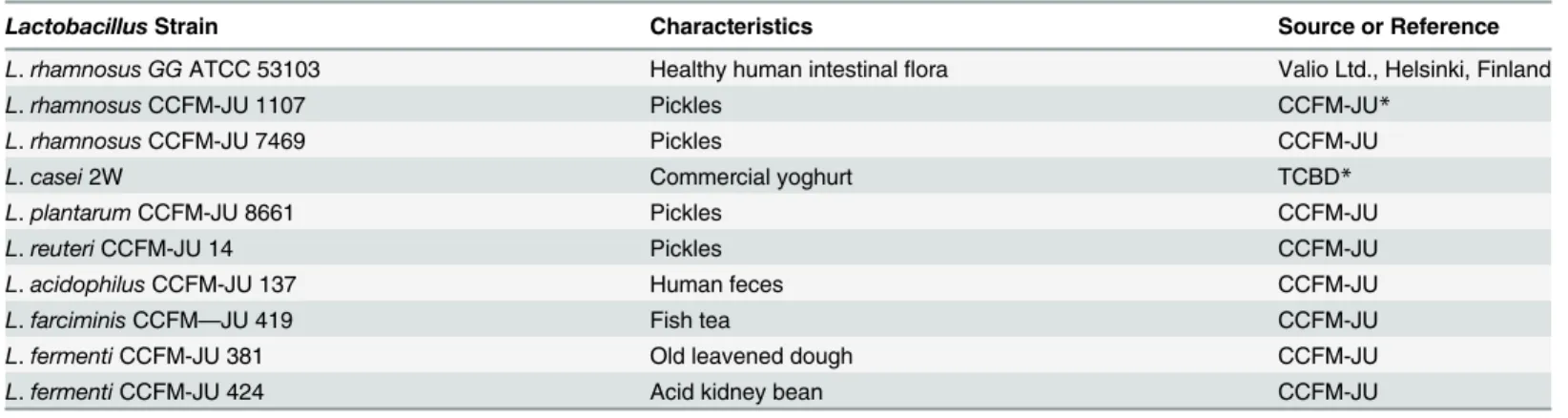
CFSs (Fig. 1). Significant differences in AA values were observed between MRS broth and CFSs, except for CCFM 8661, CCFM381, and CCFM 424 in scavenging hydroxyl radicals (Fig. 1,p<0.05). Both the HRS and DPPH methods indicated that the CFSs of CCFM 1107 and CCFM 7469 exhibited strong AAs.
The CFSs of CCFM8661 exhibited strong DPPH radical scavenging activities (75.94 ± 1.05 U/mL) that were not significantly different from that of the positive control LGG (77.29 ± 1.51 U/mL) (p>0.05). However, CCFM8661 demonstrated significantly weaker HRS activity than LGG in scavenging hydroxyl radical (p<0.05). Furthermore, the effect ofL.casei2W on DPPH and hydroxyl radical contradicted that of CCFM8661. Intact cells and intracellular cell-free extracts fromL.caseisubsp.caseiSY13 andL.delbrueckiisubsp.bulgaricusLJJ also differ in DPPH and HRS activities [13]. The different mechanisms involved in the
radical-antioxidant reactions may explain the different in scavenging potentials of the compounds [40]. Moreover, the DPPH and hydroxyl radical activities of bothL.rhamnosusCCFM 1107 and CCFM 7469 were significantly stronger than those ofL.fermentiCCFM381 and CCFM 424 (p<0.05). Liu et al. [8] reported that 12Lactobacillusstrains exhibit varying capabilities in DPPH radical scavenging. Therefore, a certain degree of inter-specific difference in radical scavenging activities could exist among the 10 testedLactobacillusstrains.
RP of Lactobacilli CFSs. An earlier report showed that AAs and RP are directly related [41]. The RP of lactobacilli CFS is based on kinetics of the reduction of Fe3+to Fe2+to prevent the oxidation reaction and control transition metal ions [41]. The RP activities of lactobacilli Fig 1. DPPH and HRS activities of the CFSs (mean±SD, n = 3).MRS broth control sample withoutLactobacillus. The letters a, b, c, d, and e indicate statistically significant difference atp<0.05 within each row comparison between the DPPH radical scavenging groups. The letters a’, b’, c’, d’, and e’
indicate statistically significant difference atp<0.05 within each row comparison between the HRS groups. Bars with no common letters are significantly different (p<0.05).
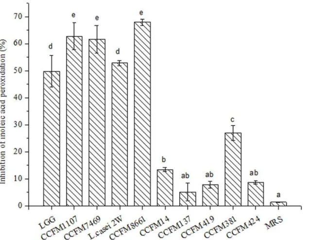
CFS are shown inFig. 2. The CFSs of LGG, CCFM1107, CCFM7469, CCFM14, CCFM137, CCFM419, CCFM381, and CCFM424 showed significantly higher RP values than that of MRS broth (p<0.05). Meanwhile, the CFSs ofL.casei 2Wand CCFM 8661 increased, but not sig-nificantly, compared with that of MRS broth (p>0.05). In all of the tests, CCFM1107 showed a high reducing activity that was close to that of the positive control LGG and equivalent to that of 62.68μML-cysteine. The results are consistent with the findings of a previous report
[8], which revealed that 12 strains show varied abilities in reducing RP activity and thatL. aci-dophilusBCRC 14079 displays six-fold stronger activity thanB.infantisBCRC 14602. There-fore, an inter-specific difference exists in the RP of the test CFSs.
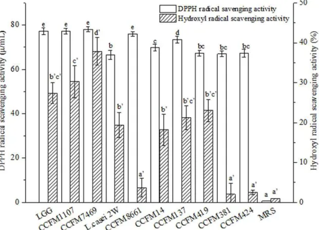
ILAP of Lactobacilli CFSs. ILAP is commonly used to analyze AAs. Linoleic acid has been used as a source of unsaturated fatty acids [42]. In the present study, the AAs of lactoba-cilli CFSs were measured by ILAP assay. As shown inFig. 3, the inhibitory rates of test lactoba-cilli CFSs on linoleic acid peroxidation ranged from 5.1 ± 0.22% to 67.93 ± 1.22% (Fig. 3). The inhibitory rates of the lactobacilli CFSs were significantly higher than those of MRS broth (p<0.05), except for CCFM137, CCFM419, and CCFM424 (p>0.05). The CFSs of
CCFM1107, CCFM7469, and CCFM 8661 showed high inhibitory effects on ILAP compared with the positive control LGG (p>0.05). Meanwhile,L.rhamnosusCCFM1107 and
CCFM7469 demonstrated significantly stronger activities on ILAP than didL.fermenti
CCFM381 and CCFM424. These observations indicated an inter-specific difference in ILAP among the lactobacilli CFSs. Moreover, the higher inhibitory effect of CCFM381 CFS than of CCFM424 CFS indicated an intra-specific difference in ILAP between these two CFSs. These results agree with the findings of an earlier report [42], which revealed that the inhibitory rates of six strains ofL.acidophiluson ILAP range from 34.9% to 46.3%.
Correlations among the Four Chemical AA Assays. Results indicated that the lactobacilli CFSs, which were measured by using chemical methods, exhibited certain antioxidant proper-ties. Among the tested CFSs, LGG and CCFM1107 had the highest AA in all of the assays. In addition, lactobacilli CFSs exerted different AAs in various chemical antioxidant models. For example, the CFS of CCFM8661, CCFM7469, andL.casei2W displayed different AA values in radical scavenging, RP, and ILAP. In a previous study, Zhu et al. [43] found that the water ex-tracts of okara koji and the water extract of soybean koji show different AAs according to dif-ferent in vitro antioxidant models. In this case, four chemical assays were used and compared to determine the antioxidant capacities of the CFSs, and their correlations are shown in
Table 2. A significant association was found between scavenging of DPPH and hydroxyl radi-cals (r= 0.511,p<0.01, n = 11). Meanwhile, the DPPH radical scavenging of the CFSs corre-lated significantly with ILAP (r= 0.604,p<0.01, n = 11). HRS also had a significant
association with RP (r= 0.494,p<0.01, n = 11). However, ILAP showed no significant correla-tion with HRS and RP because of the diverse chemical aspects of potential antioxidant com-pound(s) explored [25]. The lack of correlation between results obtained through these different assays can be attributed to reasons related to instrument limitations, mechanisms, endpoint, quantification method, and biological relevance [44]. Therefore, the use of a single chemical method to screen strains with high AAs is difficult due to the different mechanisms of CFSs in vivo.
CAA Assessment of the AAs of Lactobacilli CFSs
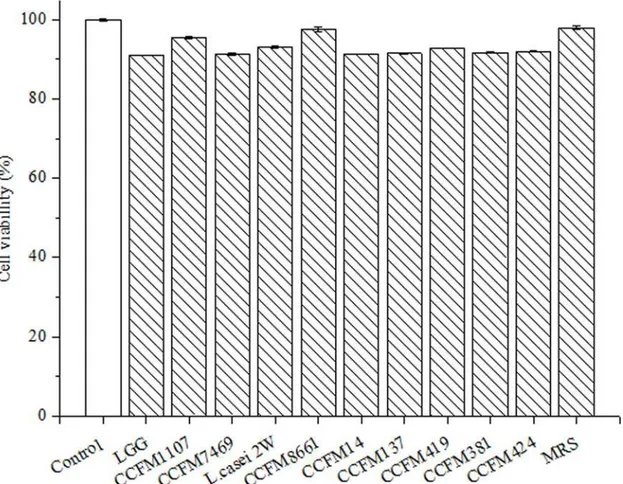
to stain the HepG2 cells. Results showed that incubation with CFS and MRS broth inhibited less than 10% of HepG2 cells (Fig. 4). This result indicates that both CFS and MRS broth do not show significant cytotoxicity to HepG2 cells after 24 h incubation [37]. Previous research showed that feijoada whole meal, apple extracts, and vegetables inhibit less than 10% of HepG2 cells [39,46,47]. Therefore, we deduced that the CFSs are not cytotoxic, which is a typical obser-vation for such functional products. Moreover, Maudsdotter et al. [48] found that the CFS of LGG can reduce cell cytotoxicity caused byStreptococcus pyogenesby producing lactic acid. Li et al. [49] showed that the CFSs ofL.acidophilusnot only display non- cytotoxicity, but also stimulate proliferation of embryonic, endothelial, and inflammatory cells in vivo. These obser-vations suggested that the CFSs of theLactobacillusstrains are non-cytotoxic.
CAA Assay. In the CAA assay developed by Wolfe and Liu [26], the ABAP-induced oxi-dation of dye DCFH-DA to fluorescent DCF and the fluorescence intensity of DCF were mea-sured to represent the oxidation rate, thereby enabling the CAA assay to measure the
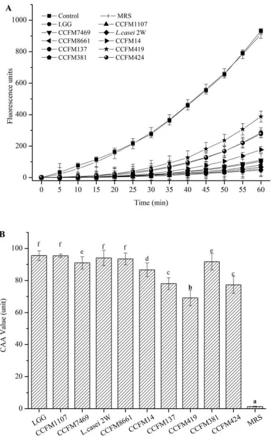
antioxidation capacity of the antioxidants [26,50]. In the present study, we applied the CAA assay to determine the AAs of lactobacilli CFSs. The positive control showed fluorescence in-tensity of ABAP oxidation over time. The negative control group contained the cells without the addition of ABAP. These cells were used to illustrate the conditions devoid of any oxidation inducer. The CFSs of all 10Lactobacillusstrains inhibited oxidation, with higher fluorescence intensities than the negative control (p<0.05) (Fig. 5A). However, these intensities were lower Fig 2. RP values of the CFSs (mean±SD, n = 3).MRS control sample without lactobacilli. RP values are expressed asμM of cysteine. Different letters
indicate statistically significant differences atp<0.05. Bars with no common letters are significantly different (p<0.05).
than that of the positive control over time, except for the MRS broth, which showed similar in-tensities to the positive control. The trends of inhibiting DCFH-DA to DCFH of CFS are simi-lar to those of pure compounds, such as theacrine, sugarcane molasses, andCrataegus azarolus
[51–53]. Puertollano et al. previously reported that concentrated supernatants fromL. plan-tarumreduce ROS accumulation in HL-60 cells [54]. In addition, the present results showed that the ROS in HepG2 cells could be reduced by lactobacilli CFSs. Similar to other methods used for AA assessment, the CAA method reveals the total antioxidative capacity of the ana-lyzed sample rather than the capacity of the individual components of the system. However, Fig 3. ILAP of the CFSs (mean±SD, n = 3).MRS control sample without lactobacilli. Different letters mean statistically significant differences atp<0.05. Bars with no common letters are significantly different (p<0.05).
doi:10.1371/journal.pone.0119058.g003
Table 2. Correlations among the four chemical antioxidant activity assays.
Correlation coefficient DPPH radical scavenging HRS RP ILAP
DPPH radical scavenging 1
HRS 0.511** 1
RP 0.377 0.494** 1
ILAP 0.604** 0.273 −0.012 1
*p<0.05 **p<0.01
the CAA also differs in other aspects. It is the only method capable of predicting antioxidant re-sponse at the cellular level. It also allows the analysis of samples activity to change the redox cellular state. Moreover, the participation of different component cells is critical to develop an antioxidant response [25].
CAA units were calculated on the basis of the area under the curve of the fluorescence inten-sities of the CFS and ABAP-treated cells over time. A smaller area denotes higher CAA units and higher AAs of the sample [51]. All of the lactobacilli CFSs possessed significantly higher CAA values than the MRS broth (p<0.05) (Fig. 5B). The cellular AA of quercetin was also de-termined by CAA assay in HepG2 cells (S1 Fig.) as a standard. The calculation results of CAA values (units) of the CFSs ofLactobacillusstrains to the equivalent amount of quercetin with the same AA (μM) present more intuitive view of the AA of the CFSs of theLactobacillus
strains compared with this established antioxidant with known clinical efficacy (S1 Table). The CAA values of the CFSs ranged from 69.14 ± 4.87 to 95.55 ± 2.99 CAA units. The positive con-trol LGG also exhibited a strong antioxidant property (95.55 ± 2.99 CAA units) in the CAA assay, which was stronger than that ofCrataegus azarolusaqueous extract (79.62 CAA units in 800μg/mL) [53]. The difference essentially depends on the bioavailability of the specific
mix-ture of the available compounds and their synergistic interactions to yield final antioxidant re-sponses at the cellular level [25]. CCFM1107,L.casei2W, and CCFM8661 showed the highest AAs (95.40, 94.06, and 93.48 CAA units, respectively). No significant difference was found Fig 4. Cytotoxicity of the CFSs on human hepatocellular carcinoma HepG2 cells.Negative control was untreated cells (mean±SD, n = 3).
Fig 5. AAs of the CFSs evaluated by the CAA method.(A) Peroxyl radical-induced oxidation of DCFH to DCF in HepG2 cells, and inhibition of oxidation by CFS and MRS (mean±SD, n = 3). (B) CAA units of CFS and MRS broth. CAA unit was calculated as the difference in the area under the curve between the tested samples and control wells. Data represent the mean±SD values that were obtained from six wells in each group. Different letters mean statistically significant differences atp<0.05.
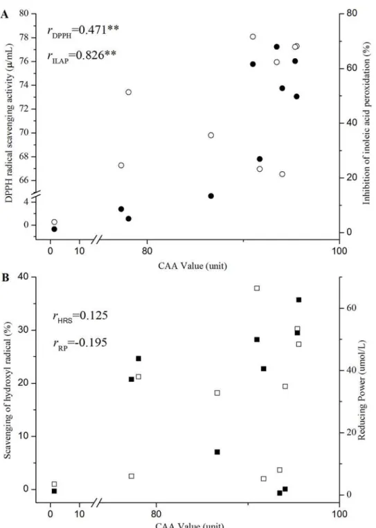
Fig 6. DPPH radical scavenging, HRS, RP, and ILAP values vs. CAA indexes for the different CFSs and MRS broth.(A) CAA values vs DPPH radical scavenging and ILAP indexes for CFSs and MRS broth. (B) CAA values vs HRS and RP indexes for CFSs and MRS broth. () represents DPPH radical scavenging values; (●) represents HRS values; (☐) represents RP values; and (■) represents ILAP values.*and**
mean statistically significant difference atp<0.05 andp<0.01, respectively. Abbreviations: HRS, hydroxyl radical scavenging; RP, reducing power; ILAP, inhibition of linoleic acid peroxidation; CFSs, cell-free supernatants; CAA, cellular antioxidant activity; rDPPH, correlation between the CAA and DPPH radical
scavenging activity assay; rILAP, correlation between the CAA and ILAP assay; rHRS, correlation between the
CAA and HRS assay; and rRP, correlation between the CAA and RP assay.
with LGG (p>0.05). However, CCFM419 provided the lowest CAA value, which was signifi-cantly different from that of the other strains (p<0.05). Consequently, the 10Lactobacillus
strains showed significant interspecific differences in CAA.L.rhamnosusshowed a higher CAA value than that ofL.fermenti. CCFM1107 and CCFM7469 originating from the same
L.rhamnosus, but the two strains had significantly different CAA values (p<0.05). Similar re-sults were observed betweenL.fermentiCCFM381 andL.fermentiCCFM424. These observa-tions indicate intra-specific differences of test CFSs in the CAA assays. Various researchers have reported similar findings which were obtained using different methods [19,55] that is, intra- and inter-specific differences exist for both intact bacteria and CFS.
Correlations between CAA and the Four Chemical AA Assays. The AA values of lacto-bacilli CFSs assessed with the four chemical assays did not correlate well with their abilities to inhibit the radical-mediated damage on HepG2 cells (CAA index). The CFSs of several Lacto-bacillusstrains that exhibited low chemical AAs showed high responses in the CAA assay (Fig. 6). For instance, the CFSs ofL.casei2W and CCFM8661 showed weak AAs in HRS and RP, but were proven efficacious in the CAA assay. CCFM419 had low antioxidant efficacy in the CAA assay, but exhibited high HRS activity. No significant association was found among the results of the HRS, RP, and CAA assays (rHRS= 0.125, rRP=−0.195,p>0.05,Fig. 6A). This observation agrees with the report of Huang et al. [56], in which the antioxidant values of Chi-nese bayberry obtained from ABTS, FRAP, DPPH, ORAC, and CAA assays do not correlate significantly with one another. In this case, the results are not surprising because the CAA assay monitors oxidative stress in cells that are related to cellular uptake, distribution, and me-tabolism of antioxidants. In chemical assays, antioxidants directly react with radicals [44]. Con-versely, CCFM8661 has a high value in CAA, DPPH, and ILAP. The CAA assay revealed a significant association with DPPH and ILAP (rDPPH= 0.471,rILAP= 0.826,p<0.01,Fig. 6B). In addition, LGG and CCFM1107 exhibited high AAs in the four chemical assays and CAA assay, whereas MRS showed a low AA. Considering the inter- and intra-species difference among numerous bacteria strains, the AAs evaluated by CAA assay show accordance with that got by the four traditional chemical assays to some extent. For the mammalian cells engaged in CAA assay, the AAs evaluated by this assay may be more correlated with the actual situation in organisms than that got with chemical assays. Thus, the CAA assay is a potential method for the detection of AAs of lactobacilli CFSs.
Conclusion
This study shows that lactobacilli CFSs exhibit AAs that can be assessed quantitatively in HepG2 cells. The CAA method is a relatively reliable and sensitive method compared with the four chemical assays in screening the AAs of CFS. Oxidative stress is involved in numerous chronic degenerative diseases. CFSs that show AAs can be evaluated by the CAA method as promising candidates in the prevention and control of several free radical-related disorders. Future studies will be conducted to elucidate the possible mechanisms of CFS and to verify the specific functions of CFSs screened by CAA assay in animal models or human studies.
Supporting Information
S1 Fig. Calibration curves with nonlinear fitting of AAs of quercetin evaluated by the CAA method.The CAA value was calculated based on the difference in the area under the curve be-tween the tested samples and control wells.
S1 Table. CAA values of the CFSs of the 10 Lactobacillus strains and MRS broth.The CAA value is expressed as an equivalent amount of quercetin (μM). Different letters indicate
statisti-cally significant differences at p<0.05. (DOCX)
Author Contributions
Conceived and designed the experiments: JX GW QZ. Performed the experiments: JX ZG. An-alyzed the data: JX ZG QZ. Contributed reagents/materials/analysis tools: HZ GW YQC WC. Wrote the paper: JX GW XL QZ.
References
1. Cederbaum AI, Lu Y, Wu D. Role of oxidative stress in alcohol-induced liver injury. Arch Toxicol. 2009; 83: 519–548. doi:10.1007/s00204-009-0432-0PMID:19448996
2. Nobili V, Parola M, Alisi A, Marra F, Piemonte F, Mombello C, et al. Oxidative stress parameters in pae-diatric non-alcoholic fatty liver disease. Int J Mol Med. 2010; 26: 471–476. PMID:20818484
3. Beckman KB, Ames BN. The free radical theory of aging matures. Physiol Rev. 1998; 78: 547–581. PMID:9562038
4. Yu JH, Kim H. Oxidative stress and cytokines in the pathogenesis of pancreatic cancer. J Cancer Pre. 2014; 19: 97–102. doi:10.15430/JCP.2014.19.2.97PMID:25337577
5. Halliwell B, Gutteridge J. Free radicals in biology and medicine. Oxford Clarendon Press; 1985. pp. 23–30.
6. Sies H. Strategies of Antioxidant Defense. Eur J Biochem. 1993; 215: 213–219. PMID:7688300
7. Velioglu YS, Mazza G, Gao L, Oomah BD. Antioxidant activity and total phenolics in selected fruits, vegetables, and grain products. J Agric Food Chem. 1998; 46: 4113–4117.
8. Liu C, Pan T. In Vitro Effects of Lactic Acid Bacteria on Cancer Cell Viability and Antioxidant Activity. J Food Drug Anal. 2010; 18: 77–86.
9. Choi SS, Kim Y, Han KS, You S, Oh S, Kim SH. Effects of Lactobacillus strains on cancer cell prolifera-tion and oxidative stress in vitro. Lett Appl Microbiol. 2006; 42: 452–458. PMID:16620202
10. Lee J, Hwang KT, Heo MS, Lee JH, Park KY. Resistance of Lactobacillus plantarum KCTC 3099 from Kimchi to oxidative stress. J Med Food. 2005; 8: 299–304. PMID:16176138
11. Forsyth CB, Farhadia A, Jakate SM, Tang YM, Shaikh M, Keshavarzian A. Lactobacillus GG treatment ameliorates alcohol-induced intestinal oxidative stress, gut leakiness, and liver injury in a rat model of alcoholic steatohepatitis. Alcohol. 2009; 43: 163–172. doi:10.1016/j.alcohol.2008.12.009PMID: 19251117
12. Chen P, Zhang Q, Dang H, Liu X, Tian F, Zhao J, et al. Screening for potential new probiotic based on probiotic properties andα-glucosidase inhibitory activity. Food Control. 2014; 35: 65–72.
13. Zhang S, Liu L, Su Y, Li H, Sun Q, Liang X, et al. Antioxidative activity of lactic acid bacteria in yogurt. Afr J Microbiol Res. 2011; 5: 5194–5201.
14. Lin M-Y, Yen C-L. Antioxidative Ability of Lactic Acid Bacteria. J Agric Food Chem. 1999; 47: 1460–
1466. PMID:10563999
15. Zhang L, Liu C, Li D, Zhao Y, Zhang X, Zeng X, et al. Antioxidant activity of an exopolysaccharide isolat-ed from Lactobacillus plantarum C88. Int J Biol Macromol. 2013; 54: 270–275. doi:10.1016/j.ijbiomac. 2012.12.037PMID:23274679
16. Zhang J, Wang D, Zhang J, Wang Y-H. Extraction and antioxidant activity of the soluble protein from Lactobacillus rhamnosus broth. Food Sci Technol. 2012; 37: 235–239.
17. Bing SR, Kinouchi T, Kataoka K, Kuwahara T, Ohnishi Y. Protective effects of a culture supernatant of Lactobacillus acidophilus and antioxidants on ileal ulcer formation in rats treated with a nonsteroidal antiinflammatory drug. Microbiol Immunol. 1998; 42: 745–753. PMID:9886147
18. Wang YH, Liu YL, Sidhu A, Ma ZH, McClain C, Feng WK. Lactobacillus rhamnosus GG culture super-natant ameliorates acute alcohol-induced intestinal permeability and liver injury. Am J Physiol-Gastroint Liver Physiol. 2012; 303: G32–G41. doi:10.1152/ajpgi.00024.2012PMID:22538402
20. Wang X, Luo X, Xu X, Yu M, Yu C, Jiang N, et al. Comparative Studies on Antioxidant Activities of Dif-ferent Lactic Acid Bacterial Strains. Food science. 2010; 31: 197–201.
21. Kanno T, Kuda T, An C, Takahashi H, Kimura B. Radical scavenging capacities of saba-narezushi, Japanese fermented chub mackerel, and its lactic acid bacteria. LWT-Food Sci Technol. 2012; 47: 25–30.
22. Lin MY, Yen CL. Reactive Oxygen Species and Lipid Peroxidation Product-Scavenging Ability of Yo-gurt Organisms. J Dairy Sci. 1999; 82: 1629–1634. PMID:10480088
23. Apak R, Gueclue K, Demirata B, Oezyuerek M, Celik SE, Bektasoglu B, et al. Comparative evaluation of various total antioxidant capacity assays applied to phenolic compounds with the CUPRAC assay. Molecules. 2007; 12: 1496–1547. PMID:17909504
24. Niki E. Assessment of antioxidant capacity in vitro and in vivo. Free Radic Biol Med. 2010; 49: 503–
515. doi:10.1016/j.freeradbiomed.2010.04.016PMID:20416370
25. López-Alarcón C, Denicola A. Evaluating the antioxidant capacity of natural products: A review on chemical and cellular-based assays. Anal Chim Acta. 2013; 763: 1–10. doi:10.1016/j.aca.2012.11.051 PMID:23340280
26. Wolfe KL, Liu RH. Cellular antioxidant activity (CAA) assay for assessing antioxidants, foods, and die-tary supplements. J Agric Food Chem. 2007; 55: 8896–8907. PMID:17902627
27. Sun J, Hu XL, Le GW, Shi YH. Lactobacilli prevent hydroxy radical production and inhibit Escherichia coli and Enterococcus growth in system mimicking colon fermentation. 2010; 50: 264–269. doi:10. 1111/j.1472-765X.2009.02786.xPMID:20059670
28. Lin M, Chang F. Antioxidative effect of intestinal bacteria Bifidobacterium longum ATCC 15708 and Lactobacillus acidophilus ATCC 4356. Dig Dis Sci. 2000; 45: 1617–1622. PMID:11007114
29. Persichetti E, De Michele A, Codini M, Traina G. Antioxidative capacity of Lactobacillus fermentum LF31 evaluated in vitro by oxygen radical absorbance capacity assay. Nutrition. 2014; 30: 936–938. doi:10.1016/j.nut.2013.12.009PMID:24985014
30. Lee J, Hwang KT, Chung MY, Cho DH, Park CS. Resistance of Lactobacillus casei KCTC 3260 to reac-tive oxygen species (ROS): Role for a metal ion chelating effect. J Food Sci. 2005; 70: M388–M391.
31. Wang J, Ji HF, Wang SX, Zhang DY, Liu H, Shan DC, et al. Lactobacillus plantarum ZLP001: In vitro Assessment of Antioxidant Capacity and Effect on Growth Performance and Antioxidant Status in Weaning Piglets. Asian Australas J Anim Sci. 2012; 25: 1153–1158. doi:10.5713/ajas.2012.12079 PMID:25049675
32. Hathout AS, Mohamed SR, El-Nekeety AA, Hassan NS, Aly SE, Abdel-Wahhab MA. Ability of Lactoba-cillus casei and LactobaLactoba-cillus reuteri to protect against oxidative stress in rats fed
aflatoxins-contaminated diet. Toxicon. 2011; 58: 179–186. doi:10.1016/j.toxicon.2011.05.015PMID:21658402
33. Bermudez-Brito M, Munoz-Quezada S, Gomez-Llorente C, Matencio E, Bernal MJ, Romero F, et al. Cell-Free Culture Supernatant of Bifidobacterium breve CNCM I-4035 Decreases Pro-Inflammatory Cytokines in Human Dendritic Cells Challenged with Salmonella typhi through TLR Activation. PLoS One. 2013; 8:1–8.
34. Gazi MR, Yokota M, Tanaka Y, Kanda S, Itabashi H. Effects of protozoa on the antioxidant activity in the ruminal fluid and blood plasma of cattle. Anal Chim Acta. 2007; 78: 34–40.
35. Lin MY, Yen CL. Antioxidative ability of lactic acid bacteria. 1999; 47: 1460–1466. PMID:10563999
36. Wang L, Chen J, Xie H, Ju X, Liu RH. Phytochemical profiles and antioxidant activity of adlay varieties. J Agric Food Chem. 2013; 61: 5103–5113. doi:10.1021/jf400556sPMID:23647066
37. Felice DL, Sun J, Liu RH. A modified methylene blue assay for accurate cell counting. J Funct Food. 2009; 1: 109–118.
38. Malta LG, Tessaro EP, Eberlin M, Pastore GM, Liu RH. Assessment of antioxidant and antiproliferative activities and the identification of phenolic compounds of exotic Brazilian fruits. Food Res Int. 2013; 53: 417–425.
39. Faller ALK, Fialho E, Liu RH. Cellular Antioxidant Activity of Feijoada Whole Meal Coupled with an in Vitro Digestion. J Agric Food Chem. 2012; 60: 4826–4832. doi:10.1021/jf300602wPMID:22509740
40. Huang DJ, Ou BX, Prior RL. The chemistry behind antioxidant capacity assays. J Agric Food Chem. 2005; 53: 1841–1856. PMID:15769103
41. Meir S, Kanner J, Akiri B, Philosoph-Hadas S. Determination and Involvement of Aqueous Reducing Compounds in Oxidative Defense Systems of Various Senescing Leaves. J Agric Food Chem. 1995; 43: 1813–1819.
43. Zhu YP, Fan JF, Cheng YQ, Li LT. Improvement of the antioxidant activity of Chinese traditional fer-mented okara (Meitauza) using Bacillus subtilis B2. Food Control. 2008; 19: 654–661.
44. Tabart J, Franck T, Kevers C, Pincemail J, Serteyn D, Defraigne JO, et al. Antioxidant and anti-inflammatory activities of Ribes nigrum extracts. Food Chemistry. 2012; 131: 1116–1122.
45. Yi Y, Huang F, Zhang MW, Zhang RF, Deng YY, Wei ZC, et al. Solution Properties and in Vitro Anti-Tumor Activities of Polysaccharides from Longan Pulp. Molecules. 2013; 18: 11601–11613. doi:10. 3390/molecules180911601PMID:24051475
46. Yang J, Liu RH. Synergistic Effect of Apple Extracts and Quercetin 3-beta-D-Glucoside Combination on Antiproliferative Activity in MCF-7 Human Breast Cancer Cells in Vitro. J Agric Food Chem. 2009; 57: 8581–8586. doi:10.1021/jf8039796PMID:19694432
47. Song W, Derito CM, Liu MK, He XJ, Dong M, Liu RH. Cellular Antioxidant Activity of Common Vegeta-bles. J Agric Food Chem. 2010; 58: 6621–6629. doi:10.1021/jf9035832PMID:20462192
48. Maudsdotter L, Jonsson H, Roos S, Jonsson AB. Lactobacilli reduce cell cytotoxicity caused by Strep-tococcus pyogenes by producing lactic acid that degrades the toxic component lipoteichoic acid. Anti-microb Agents Chemother. 2011; 55: 1622–1628. doi:10.1128/AAC.00770-10PMID:21245448
49. Li WI, Brackett BG, Halper J. Culture supernatant of Lactobacillus acidophilus stimulates proliferation of embryonic cells. Exp Biol Med. 2005; 230: 494–500. PMID:15985625
50. Hirawan R, Diehl-Jones W, Beta T. Comparative Evaluation of the Antioxidant Potential of Infant Cere-als Produced from Purple Wheat and Red Rice Grains and LC-MS Analysis of Their Anthocyanins. J Agric Food Chem. 2011; 59: 12330–12341. doi:10.1021/jf202662aPMID:22035073
51. Li WX, Li YF, Zhai YJ, Chen WM, Kurihara H, He RR. Theacrine, a Purine Alkaloid Obtained from Ca-mellia assamica var. kucha, Attenuates Restraint Stress-Provoked Liver Damage in Mice. J Agric Food Chem. 2013; 61: 6328–6335. doi:10.1021/jf400982cPMID:23678853
52. Asikin Y, Takahashi M, Mishima T, Mizu M, Takara K, Wada K. Antioxidant activity of sugarcane molas-ses against 2,20-azobis(2-amidinopropane) dihydrochloride-induced peroxyl radicals. Food Chemistry.
2013; 141: 466–472. doi:10.1016/j.foodchem.2013.03.045PMID:23768381
53. Mustapha N, Bouhlel I, Chaabane F, Bzeouich IM, Ghedira K, Hennebelle T, et al. Aqueous extract of Crataegus azarolus protects against DNA damage in human lymphoblast Cell K562 and enhances anti-oxidant activity. Appl Biochem Biotechnol. 2014; 172: 2266–2275. doi:10.1007/s12010-013-0667-3 PMID:24347159
54. Puertollano MA, Puertollano E, Alvarez de Cienfuegos G, de Pablo MA. Culture Supernatants from Lactobacillus plantarum Induce Necrosis on a Human Promyelocytic Leukemia Cell Line. P Nutr Soc. 2013; 13: 195–203(9).
55. Ramesh V, Kumar R, Singh RRB, Kaushik JK, Mann B. Comparative evaluation of selected strains of lactobacilli for the development of antioxidant activity in milk. Dairy Sci Technol. 2012; 92: 179–188.