Estrogen Receptor
β
Agonists Differentially
Affect the Growth of Human Melanoma Cell
Lines
Monica Marzagalli1, Lavinia Casati2, Roberta M. Moretti1, Marina Montagnani Marelli1, Patrizia Limonta1*
1Department of Pharmacological and Biomolecular Sciences, Universitàdegli Studi di Milano, Milano, Italy, 2Department of Medical Biotechnologies and Translational Medicine, Universitàdegli Studi di Milano, Milano, Italy
Abstract
Background
Cutaneous melanoma is an aggressive malignancy; its incidence is increasing worldwide and its prognosis remains poor. Clinical observations indicate that estrogen receptorβ
(ERβ) is expressed in melanoma tissues and its expression decreases with tumor progres-sion, suggesting its tumor suppressive function. These experiments were performed to investigate the effects of ERβactivation on melanoma cell growth.
Methods and Results
Protein expression was analyzed by Western blot and immunofluorescence assays. Cell proliferation was assessed by counting the cells by hemocytometer. ERβtranscriptional activity was evaluated by gene reporter assay. Global DNA methylation was analyzed by restriction enzyme assay and ERβisoforms were identified by qRT-PCR. We demonstrated that ERβis expressed in a panel of human melanoma cell lines (BLM, WM115, A375, WM1552). In BLM (NRAS-mutant) cells, ERβagonists significantly and specifically inhibited cell proliferation. ERβactivation triggered its cytoplasmic-to-nuclear translocation and tran-scriptional activity. Moreover, the antiproliferative activity of ERβagonists was associated with an altered expression of G1-S transition-related proteins. In these cells, global DNA was found to be hypomethylated when compared to normal melanocytes; this DNA hypo-methylation status was reverted by ERβactivation. ERβagonists also decreased the prolif-eration of WM115 (BRAF V600D-mutant) cells, while they failed to reduce the growth of A375 and WM1552 (BRAF V600E-mutant) cells. Finally, we could observe that ERβ iso-forms are expressed at different levels in the various cell lines. Specific oncogenic muta-tions or differential expression of receptor isoforms might be responsible for the different responses of cell lines to ERβagonists.
a11111
OPEN ACCESS
Citation:Marzagalli M, Casati L, Moretti RM, Montagnani Marelli M, Limonta P (2015) Estrogen ReceptorβAgonists Differentially Affect the Growth of Human Melanoma Cell Lines. PLoS ONE 10(7): e0134396. doi:10.1371/journal.pone.0134396
Editor:Alessandro Weisz, University of Salerno, Faculty of Medicine and Surgery, ITALY
Received:April 2, 2015
Accepted:July 8, 2015
Published:July 30, 2015
Copyright:© 2015 Marzagalli et al. This is an open access article distributed under the terms of the Creative Commons Attribution License, which permits unrestricted use, distribution, and reproduction in any medium, provided the original author and source are credited.
Data Availability Statement:All relevant data are within the paper.
Funding:Funding was provided by Fondazione Banca del Monte di Lombardia 2012,http://www.fbml. it/(P.L., L.C., R.M.M., M.M.M.); PRIN-MIUR 2010-20112010, NFEB9L_004,http://prin.miur.it/(P.L., R.M. M., M.M.M.). The funders had no role in study design, data collection and analysis, decision to publish, or preparation of the manuscript.
Conclusions
Our results demonstrate that ERβis expressed in melanoma cell lines and that ERβ ago-nists differentially regulate the proliferation of these cells. These data confirm the notion that melanoma is a heterogeneous tumor and that genetic profiling is mandatory for the develop-ment of effective personalized therapeutic approaches for melanoma patients.
Introduction
The incidence of cutaneous melanoma is increasing worldwide [1] and its prognosis is still poor [2]. Cytotoxic drugs, dacarbazine or temozolomide, were reported to be associated with serious side effects and with development of resistance. Interleukin-2 or interferon-αyielded limited response rates with no benefit on overall survival or progression-free survival [3]. Patients treated with either mutated BRAF or MEK inhibitors, despite initial excellent response rates, showed a rapid relapse [4]. The anti-CTLA-4 (cytotoxic T-lymphocyte antigen 4) mono-clonal antibody ipilimumab, despite its effectiveness, has side effects that can be non-reversible (autoimmune responses, bowel perforation) [5]. Thus, the elucidation of the molecular mecha-nisms of melanoma growth and progression is urgently needed for the identification of novel targets of intervention for the prevention and therapy of this disease [6].
The association of estrogens with tumor development has been investigated for many years. Estrogens exert their effects through the binding to two estrogen receptor (ER) subtypes, ERα
and ERβ. These receptors are structurally similar, however they differ in the ligand binding domain and this confers them selectivity for different ligands [7]. After being activated by the binding of 17β-estradiol (E2) or of synthetic compounds these receptors exert their effects at
the nuclear level through the binding to estrogen response elements on DNA to regulate the expression of specific target genes [7,8].
Both ER subtypes are expressed in different cells/tissues where they are involved in the con-trol of specific physiological functions [9]. In addition, the activation of the two receptor sub-types elicits opposite effects on cancer growth and progression. ERαis associated with a proliferative activity while ERβexerts a significant antitumor effect, being considered a protein with tumor suppressive functions [7,10,11]. These observations indicate that the actions of estrogens on cancer growth might depend on the relative ERα/ERβratio in a given tumor cell/ tissue [12]. The expression of ERβwas found to be reduced in several cancer cells [13,14]. Moreover, overexpression of ERβor its activation by means of agonistic ligands were reported to inhibit cell proliferation in different tumor cells, both classically related (breast, ovarian, and prostate cancer) [15–17] and unrelated (colon cancer, mesothelioma, cholangiocarcinoma, lymphoma) [18–21] to the reproductive system. Research is now focusing on the development and evaluation of selective ERβligands that might increase the activity of this receptor in tumors [8]. The expression of the different variants of this receptor (ERβ1, corresponding to ERβ, ERβ2 and ERβ5) and their specific role in tumor growth are also under investigation [22].
Alterations of DNA methylation, histone modifications, and modified expression of micro-RNAs are well-established epigenetic mechanisms of cell neoplastic transformation. In particu-lar, melanoma cells present aberrant DNA methylation patterns with DNA hypermethylation at the level of CpG islands in the promoter of tumor suppressor genes (leading to their inactiva-tion) and global DNA hypomethylation (contributing to genomic instability). Hypomethyla-tion of specific genes was also shown, leading to the overexpression of normally silenced oncogenes [28,29]. Global DNA hypomethylation was reported to correlate with melanoma progression to the most aggressive phase and with less favourable clinical outcomes [28,30]. Epigenetic modifications and their reversibility by means of pharmacologic interventions might offer promising novel therapies for melanoma patients.
In this paper, we demonstrate that ERβ(but not ERα) is expressed in a panel of human mel-anoma cell lines (BLM, WM115, A375, WM1552). In BLM (NRAS-mutant) cells, activation of this receptor, by means of agonistic ligands, induces its translocation into the nucleus and initi-ation of transcriptional activity, thereby significantly and specifically decreasing melanoma cell proliferation. This antitumor activity is accompanied by an altered expression of the proteins involved in the G1-S progression of the cell cycle, but not by triggering of the apoptosis path-way. Moreover, in these cells, ERβagonists increase global DNA methylation, reverting the observed DNA hypomethylation status of melanoma cells compared to normal melanocytes.
ERβagonists also exert a tumor suppressor activity in WM115 (BRAF V600D-mutant) cells, while they fail to reduce cell proliferation in A375 and WM1552 (BRAF V600E-mutant) cells. Moreover, ERβisoforms show different levels of expression in the various cell lines. The possible reasons for the differential effects of ERβagonists on the growth of the melanoma cells (oncogenic mutations, receptor isoform expression) are discussed.
Materials and Methods
Cell culture and reagents
The human BLM (NRAS-mutant, BRAF-wild type) melanoma cell line was provided by Dr. G. N. van Muijen (Department of Pathology, Radbound University Nijmegen Medical Center, Nijmegen, The Netherlands). This cell line is a subline of BRO melanoma cells isolated from lung metastases after subcutaneous inoculation of nude mice with BRO cells [31]. This cell line was previously utilized in the authors' laboratory to study the antitumor activity of gonadotro-pin-releasing hormone receptors in melanoma cells [32,33]. The human WM115 (BRAF V600D-mutant) and WM1552 (BRAF V600E-mutant) melanoma cell lines, kindly provided by Dr. R. Giavazzi (Department of Oncology, Mario Negri Institute for Pharmacological Research, Milano, Italy) were originally from Dr. M. Herlyn (Wistar Institute, Philadelphia, PA) [34,35]. The human IGR-39 (BRAF V600E-mutant) melanoma cell line, kindly provided by Dr. C.A. La Porta, was originally from Leibniz-Institut DSMZ-Deutsche Sammlung von Mikroorganismen and Zellkulturen GmbH (38124 Braunschweig, Germany). The human MCF-7 breast cancer, A375 (BRAF V600E-mutant) and embryonic kidney (HEK) 293 cells were from American Type Culture Collection (ATCC, Manassas, VA, USA). Primary human melanocytes were provided by Dr. F. Crovato (Regional Reference Centre for Human Epider-misin vitroCulture and Bank for Tissue Crypreservation, Niguarda Hospital, Milano, Italy). Stocks of cells were stored frozen in liquid nitrogen and kept in culture for no more than 10–12 weeks.
MCF-7 and WM115 cells were routinely grown in RPMI-1640 medium supplemented with 10% FBS, glutamine (1 mmol/l) and antibiotics (100 IU/ml, penicillin G sodium and 100μg/ml
cultured in DMEM medium supplemented with 10% FBS, glutamine and antibiotics, as described above. Cells were cultured in humidified atmosphere of 5% CO2/95% air at 37°C.
17β-Estradiol was purchased from Sigma-Aldrich (Milano, Italy); the ER antagonist ICI 182,780 and the ERβagonist DPN (diarylpropionitrile) were from Tocris Biosciences (Bristol, UK). The selective ERβagonists KB1, KB2, and KB4 were kindly provided by Dr. S. Nilsson (Karo Bio AB, Novum, SE-141 57 Huddinge, Sweden). KB1 (also known as KB9520) was previ-ously utilized in different studies as the specific ligand of ERβto investigate the antitumor activity of the receptor in cancer cells [14,20,21,36]. The selective activity of this class of com-pounds as ERβligands was previously demonstrated [37]. The compounds can be obtained fol-lowing contact with Karo Bio AB and after signing of a Material Transfer Agreement together with a detailed protocol of planned study. A fee covering the cost of compound synthesis will be charged.
ER
β
overexpression
The plasmid pCMV5-hERbeta, expressing human wild type ERβ, was kindly provided by Dr. A. Maggi (Department of Pharmacological and Biomolecular Sciences, University of Milano, Milano, Italy). BLM cells were plated (8 x10⁴cells/dish) in 6-well plates in DMEM complete medium. After 48 h, the medium was replaced with DMEM medium and the cells were tran-siently transfected using Lipofectamine 2000 (Invitrogen, Monza, Italy), according to the man-ufacturer’s protocol. After 24–72 h of transfection the cells were lysed in RIPA buffer for protein extraction.
Western blot analysis
Cells were lysed in RIPA buffer (0.05 M Tris-HCl pH 7.7, 0.15 M NaCl, 0.8% SDS, 10 mM EDTA, 100μM NaVO4, 50 mmol/L NaF, 0.3 mM PMSF, 5 mM iodoacetic acid) containing
leupeptin (50μg/ml), aprotinin (5μl/ml) and pepstatin (50μg/ml). Protein concentration in
lysates was determined using the BCA method. Protein extracts (20–30μg) were resuspended
in Sample buffer (0.5 M Tris.HCl pH 6.8, 20% glycerol, 10% SDS, 0.2% 2β-mercaptoethanol, 0.05% blue bromophenol) and heated at 95°C for 5 min. Following electrophoretic separation by 10% SDS-PAGE, proteins were transferred onto nitrocellulose membranes. Membranes were blocked in nonfat dry milk (7.5% for ERβand 5% for ERα) prior to incubation at 4°C overnight with the primary antibodies (1:1000): rabbit polyclonal antibody SC 8974 (clone H-150, Santa Cruz Biotechnology, Santa Cruz, CA) and mouse monoclonal antibody ab288 (clone 14C8, Abcam, Cambridge, MA) for ERβ; rat monoclonal antibody Ab-21 (clone H222, Thermo Scientific, Waltham, MA) for ERα. Detection was done using a horseradish-peroxi-dase-conjugated secondary antibody and enhanced chemiluminescence reagents (GE Health-care, Life Sciences, Milano, Italy).
(Asp175; 1:500, clone 5A1E). All these primary antibodies were from Cell Signaling Technol-ogy (Danvers, MA). Detection was done using a horseradish peroxidase-conjugated anti-mouse or a horseradish peroxidase-conjugated anti-rabbit secondary antibody (Santa Cruz Biotechnology), according to the primary antibody used, and enhanced chemiluminescence ECL-Prime reagents (GE Healthcare, Life Sciences). In each experiment, actin expression was evaluated as a loading control, using the goat polyclonal anti-human antibody (1:1000; I-19, sc-1616, Santz Cruz Biotechnology) as the primary antibody. Detection was done using a horseradish peroxidase-conjugated anti-goat secondary antibody (Santa Cruz Biotechnology) and enhanced chemiluminescence reagents, as described above. The experiments were repeated three times.
Cell proliferation assays
BLM melanoma cells were plated (15x103cells/dish) in 6-cm dishes in DMEM complete medium. After 48 h, the medium was replaced with phenol red free medium supplemented with 10% charcoal stripped FBS. Cells were then treated as follows: i) DPN (10−9, 5x10⁻⁹, 10−8, 5x10⁻⁸, 10−7M); E2(10−9,10−8, 10−7M); KB1, KB2, or KB4 (10−9, 10−8, 10−7M); KB1 (10−9,
5x10⁻⁹, 10−8, 5x10⁻⁸, 10−7M) every 48 h, for three times; ii) ICI 182,780 (10−6M) for 1 h, fol-lowed by DPN, E2, or K1 (10−8M) every 48 h, for three times. Cells were then harvested and
counted by hemocytometer.
Experiments were also performed on IGR-39 (expressing almost undetectable levels of ERβ) and on A375, WM1552 and WM115 melanoma cells. IGR-39, A375 and WM1552 cells were treated with DPN (10−9, 10−8, 10−7M); WM115 cells were treated with DPN, E2or KB1 (10−10,
10−9, 10−8, 10−7M) as described above. Each proliferation assay was repeated three-five times.
Immunofluorescence assay
BLM cells (2x10⁴cells) were seeded on 13-mm diameter coverslips in DMEM complete medium. After 48 h, the medium was replaced with phenol red free medium supplemented with 10% charcoal stripped FBS. Cells were then treated with either DPN or E2(10−8M) for 24
h, fixed with 4% paraformaldehyde in 2% sucrose-PBS for 15 min and permeabilized with 0.2% PBS/Triton buffer (1 mM PBS, 300 mM sucrose, 50 mM NaCl, 3 mM MgCl2, 0.5% Triton
X-100) for 20 min at room temperature and stained with the primary rabbit anti-human ERβ
polyclonal antibody SC 8974 (1:50; clone H-150, Santa Cruz Biotechnology), followed by FITC-conjugated goat anti-rabbit secondary antibody Alexa Fluor 488 (1:2000; Molecular Probes Inc., Eugene, OR). Labeled cells were examined under a Zeiss Axiovert 200 microscope with a 63x/1.4 objective lens linked to a Coolsnap Es CCD camera (Roper Scientific-Crisel Instruments, Roma, Italy). The experiment was repeated three times.
ER
β
transcriptional activity assay
BLM cells, seeded in 24-well plates (5x10⁴cells/well) in phenol red free DMEM medium sup-plemented with 10% charcoal stripped FBS, were transfected using Lipofectamine 2000 (Life Technologies, Monza, Italy), according to the manufacturer’s protocol [38]. The following con-structs were cotransfected: pVERE-tk-Luc (1μg), the reporter plasmid encoding the firefly
luciferase reporter gene under the control of the estrogen response element (ERE; kindly pro-vided by Dr. A. Maggi, Department of Pharmacological and Biomolecular Sciences, University of Milano, Milano, Italy), to evaluate the transcriptional activity of ERβ, and pCMVβ(0.4μg),
ERβtranscriptional activity was measured using the neolite system (PerkinElmer, Boston, MA), according to the manufacturer’s protocols. Briefly, 6 h after transfection, BLM cells were treated with DPN (10−8M), E2(10−8M), or ethanol (controls) for 24 h. Cell medium was
removed and 175μl of fresh medium were added to each well. Neolite luciferase reagent
(175μl) was added to each sample and firefly luminescence was read after 10 min. At the end
of this step, 100μl of the lysate were added in a microcentrifuge tube containing the assay
buffer with ortho-Nitrophenyl-β-galactoside (ONPG). Ifβ-galactosidase is present, it hydro-lyzes the ONPG molecule into galactose and ortho-nitrophenol (yellow color). The samples were transferred into a 96-well plate and analyzed in an EnSpire Multimode Plate reader (Per-kinElmer) at 420 nm. Data are expressed as the mean of the ratio (± SE) between luminescence of the experimental reporter (firefly luciferase activity) and that of the control reporter (β -galactosidase activity) and are the mean values from three replicates of three distinct experiments.
Methylation analysis of GC-rich regions
DNA from human melanocytes and BLM cells was extracted using Qiagen column methods (Qiagen, Milano, Italy) according to the manufacturer's protocol. DNA quality and concentra-tion was evaluated by measuring the 260/280 nm optical ratio using Nanodrop 2000 (OD260/ 280).
Digestion of genomic DNA with restriction enzymes RsaI, MspI and HpaII (Euroclone, Pero, Milano, Italy) was performed, as previously described [39]. For each DNA sample, 2 restriction digests were performed: one with RsaI and MspI, and one with RsaI and HpaII. RsaI is methylation insensitive, while MspI and HpaII are sensitive to DNA methylation and are able to cut only unmethylated restriction sites. In general, MspI will not cut if the external cyto-sine is methylated, and HpaII will not cut if any of the two cytocyto-sines is methylated [39,40]. Global DNA digestion was performed o/n at 37°C. Restriction digests were performed with 1μg of DNA and 5 units of RsaI in Roche buffer L. After 1 h incubation at 37°C, 2.5 unit
ali-quots of MspI or HpaII were added, 2 h apart. Total incubation time was 18 h. The enzymes were inactivated by 10-min incubation at 65°C, and the digests were used for PCR using a sin-gle primer (50-AACCCTCACCCTAACCCCGG-30) that arbitrarily binds within GC-rich regions of DNA. Samples were resolved on 1% agarose gel. The intensity of the band was deter-mined using ImageLab (Chemidoc Imager, Bio-Rad, Segrate, Milano, Italy). Data are expressed as the MspI/RsaI or HpaII/RsaI ratios relative to the intensity of the bands.
To investigate the effects of ERβactivation on the global DNA methylation profile of mela-noma cells, BLM cells, seeded in 6-well plates (3x10⁴cell/well), in phenol red free DMEM medium supplemented with 10% charcoal stripped FBS, were treated with DPN (10−8M), E2
(10−8M), or ethanol (controls) for 24 or 48 h. The analysis of DNA methylation profile was per-formed as described above. Each experiment was repeated three times.
ER
β
isoform expression by quantitative PCR
Total RNA from BLM, A375 and WM115 cells was isolated by RNeasy MINI kit (Qiagen) for the evaluation of the expression of ERβisoforms; RNA pellets were dissolved in sterile distilled water and their concentrations were assessed using Nanodrop 2000 (OD260/280). Specific set of primers for each ERβisoform were synthesized (Sigma-Aldrich) and utilized, as previously described by Collins and coworkers [41]. Real time-DNA amplification for ERβwas performed in CFX96 Bio-Rad using 20μl of total volume. The efficiency of each set of primer was
RNA were retrotranscribed using the IScript Supermix kit (Bio-Rad), according to the manu-facturer’s protocol. The reaction was carried out on 40 ng of total cDNA using SYBR chemistry (iTAQ Universal SYBR green supermix, Bio-Rad) according to the manufacturer’s protocol. Real-time PCR was run according to the following protocol: an initial step of 30 sec at 95°C fol-lowed by 40 cycles of 5 sec at 95°C and 30 sec at 60°C. A dissociation stage with a melt curve analysis was also performed. Three replicates were performed for each experimental point and experiments were repeated three times. Gene expression was quantified using the comparative threshold-cycle (Ct) method considering that the targets and the reference genes have the same amplification efficiency (near to 100%) [42]. In each cell line,ΔCt values (difference between target and reference gene Ct) for ERβ2 or ERβ5 were compared with ERβ1ΔCt value in the same cell line.
Statistical analysis
When appropriate, data were analyzed by Dunnet's or Bonferroni's test, after one-way analysis of variance. APvalue<0.05 was considered statistically significant.
Results
Expression of ER
β
in human melanoma cell lines
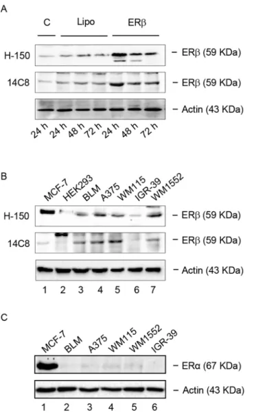
The expression of ERβin human melanoma cell lines was analyzed by Western blot assay uti-lizing two primary antibodies: H-150 (Santa Cruz Biotechnology) and 14C8 (Abcam). First, in order to obtain an appropriate positive control, ERβwas evaluated in BLM melanoma cells engineered to overexpress the receptor protein.Fig 1Ashows that a protein band, correspond-ing to the molecular weight of 59 kDa, is expressed in BLM cells either in normal culture condi-tions (C) or in the presence of Lipofectamine (Lipo), at 24–72 h. More importantly,Fig 1Aalso shows that the expression levels of this protein band are sharply increased in BLM cells overex-pressing the receptor (ERβ) at 24 after transfection, and slightly decrease at 48 and 72 h. These data, obtained with the two different antibodies, confirm that the molecular weight of this receptor subtype corresponds to 59 kDa, as previously reported [43,44].
ERβexpression was then analyzed, utilizing the two primary ERβantibodies (H-150 and 14C8) in a panel of human melanoma cell lines.Fig 1Bshows that a specific band correspond-ing to the molecular weight of 59 kDa, is expressed in BLM (lane 3; confirmcorrespond-ing the data reported inFig 1A), A375 (lane 4), WM115 (lane 5) and WM1552 (lane 7) human melanoma cells.Fig 1Balso shows that in the human IGR-39 melanoma cell line (lane 6) the receptor is expressed at almost undetectable levels. The molecular weight of the protein band detected in the melanoma cell lines corresponds to that found in human MCF-7 breast cancer cells (posi-tive control; lane 1). As expected, no band of this size could be detected in the HEK293 cells (negative control; lane 2), confirming previous observations [45]. It should be underlined that, when evaluated with the 14C8 primary antibody, the level of expression of the receptor in MCF-7 cells was found to be low, and this agrees with previous data in the literature [46,47]. On the other hand, in these cells the receptor seems to be expressed at higher levels when evalu-ated with the H-150 antibody. At present, the reason for this discrepancy is unclear; however, it might be due to a different degree of specificity of the two antibodies.
ER
β
agonists inhibit the proliferation of BLM melanoma cells
Experiments were first performed to investigate the effects of ERβactivation on the growth of BLM melanoma cells, expressing ERβ. The selective ERβagonist DPN decreased BLM cell pro-liferation at the concentrations of 10−9and 5x10⁻⁹M, being significantly effective at the dose of
Fig 1. ERβ, but not ERα, is expressed in human melanoma cells. (A)As a positive control, the expression of ERβwas evaluated by Western blot analysis in human BLM melanoma cells engineered to overexpress the receptor subtype protein, utilizing two primary antibodies: H-150 (Santa Cruz) and 14C8 (Abcam). A band corresponding to the receptor protein (59 kDa) was detected in basal conditions, both in control (C) and in Lipofectamine (Lipo) treated BLM cells. As expected, the intensity of this band was found to be significantly increased after ERβoverexpression (24–72 h), with the highest level of expression at 24 h.(B)By Western
blot analysis, utilizing the two primary antibodies H-150 and 14C8, ERβwas found to be expressed at high levels in human BLM, A375, WM115, WM1552 melanoma cell lines (lanes 3, 4, 5, 7), while the human IGR-39 melanoma cell line expressed the receptor at almost undetectable levels (lane 6). ERβwas also expressed in human MCF-7 breast cancer cells, utilized as a positive control (lane 1), but it was not expressed in the human HEK293, utilized as a negative control.(C)On the other hand, all the human melanoma cells lines tested (lanes 2–6) did not express the ERαreceptor isoform, which was expressed only
in the control cell line (MCF-7, lane 1).β-actin was utilized as a loading control. For each analysis, one representative of three different experiments, which gave similar results, is shown.
10−8M (Fig 2A). This significant effect was followed by a decline at concentrations of 5x10⁻⁸ and 10−7M. Accordingly, the natural estrogenic ligand E2exerted a significant antiproliferative
effect on BLM cell proliferation at the concentration of 10−8M (Fig 2B). The antiproliferative activity of both DPN and E2(10−8M) was completely counteracted by cotreatment of the cells
with the ER antagonist ICI 182,780 (10−6M) (Fig 2C).
Experiments were also performed with different ERβagonists (KB1, KB2, KB4). We found that all these compounds significantly inhibit BLM cell proliferation at the concentration of 10−8M (Fig 2D); the dose-response curve obtained after treating the cells with different con-centrations of KB1 was similar to that obtained with DPN (Fig 2Evs.Fig 2A). This effect was completely counteracted by the ER antagonist ICI 182,780 (10−6M) (Fig 2F).
These data demonstrate that ERβactivation is associated with antiproliferative activity in BLM melanoma cells, with 10−8M being the most effective dose, as previously reported for dif-ferent tumor cells [14,18,20,21,49–51]. A curve of the dose-response effect of ERβagonists on cancer cell proliferation, similar to that here shown, has been previously reported for
Fig 2. ERβagonists significantly and specifically inhibit the proliferation of BLM melanoma cells. (A)BLM cells were treated with different doses of the classical ERβagonist DPN every 48 h for three times. DPN significantly decreased cell proliferation at the dose of 10−8M.(B)Similar results were
obtained when the cells were treated with E2.(C)The antiproliferative effect of both ERβligands (10−8M) was found to be specific since it was completely abrogated by cotreatment of the cells with the ER antagonist ICI 182,780 (10−6M).(D)BLM cells were treated with KB1, KB2, or KB4 (10−9, 10−8, 10−7M)
every 48 h for three times. All three ERβligands significantly reduced cell proliferation at the dose of 10−8M.(E)BLM cells were treated with KB1, at different
doses (109
−10−7M). The ERβagonists decreased cell growth, being significantly effective at the dose of 10−8M.(F)The antiproliferative activity of KB1 (10−8
M) was found to be specific since it was completely abrogated by cotreatment of the cells with the ER antagonist ICI 182,780. Each experimental group consisted of six replicates and each experiment was repeated three-five times. Results are given as cell number/plate. Data represent mean values±SEM. *P<0.05. C, controls.
cholangiocarcinoma and mesothelioma cells [14,20]. On the basis of these results, the concen-tration of 10−8M was selected for the subsequent studies.
Activation of ER
β
induces its cytoplasmic-to-nuclear translocation and
transcriptional activity in BLM cells
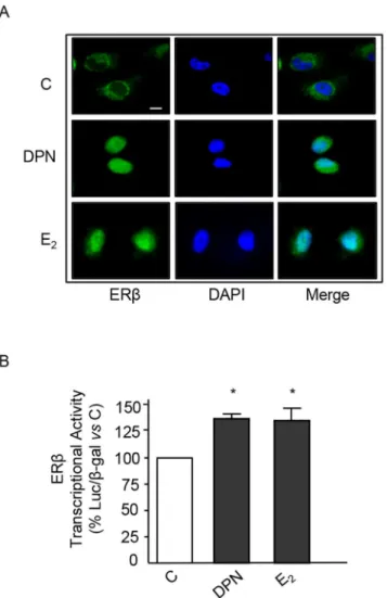
Experiments were performed to verify whether, in BLM melanoma cells, ERβmight function according to the classical model of estrogen action [11]. By immunofluorescence analysis, we could show that, in BLM cells, most of the ERβstaining was confined in the cytoplasm (Fig 3A); treatment of melanoma cells (24 h) with both DPN and E2(10−8M) induced its nuclear
translocation (Fig 3A). Then, we analyzed the effects of ERβligands on the transcriptional activity of the receptor in melanoma cells.Fig 3Bshows that treatment of BLM cells with either
Fig 3. ERβligands trigger cytoplasmic-to-nuclear translocation of ERβand induce its transcriptional acitivity in BLM melanoma cells. (A)Immunofluorescence assay of ERβintracellular localization. In control BLM melanoma cells, ERβis mainly localized at the cytoplasmic level. Treatment of the cells with either DPN or E2(10−8M, for 24 h) induces ERβtranslocation into the nucleus. A representative picture from three
experiments done independently, which gave the same results, is reported.(B)The transcriptional activity of the ERβprotein in BLM cells was analyzed using the pVERE-tk-LUC plasmid (cotransfected with pCMVβ). The results were normalized forβ-galactosidase activity. Treatment of the cells with either DPN or E2(10−8M, for 24 h) significantly increased ERβtranscriptional activity. Each experimental group consisted of three replicates and each experiment was repeated three times. Data represent mean values±SEM.*P<0.05. C, controls.
DPN or E2for 24 h significantly increased the transcriptional activation of the ERE-Luc
reporter plasmid (normalized forβ-galactosidase) indicating that, in BLM melanoma cells, ERβis associated with the classical transcriptional activity at the nuclear level.
ER
β
agonists affect the expression of cell cycle-related proteins in BLM
cells
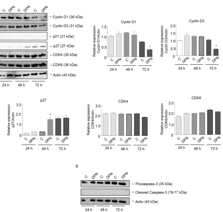
Estrogens have been shown to affect cancer cell growth through the regulation of proteins involved in cell cycle progression [52]. Experiments were performed to investigate whether ERβagonists might affect melanoma cell proliferation through alteration of the expression of cell cycle-related proteins. BLM cells were treated with the ERβagonist DPN (10−8M) for dif-ferent time intervals (24–72 h). By Western blot assay, we could demonstrate that treatment with DPN induced a significant reduction in the expression of G1 cyclins, such as cyclin D1 and D3 (at 72 h of treatment), and a significant increase in the expression of the CDK inhibitor p27 (at 48 h of treatment) (Fig 4A). On the other hand, the expression of the cyclin D partners CDK4 and CDK6 was not modified by the treatment; the CDK inhibitor p21 was found to be expressed at almost undetectable levels in BLM cells and its expression was not affected by DPN treatment (Fig 4A). Interestingly, DPN did not modify the expression of procaspase-3 as well as that of the cleaved (active) form of caspase-3 (Fig 4B). Taken together, these results indicate that ERβactivation in melanoma cells decreases cell proliferation, through the modu-lation of the expression of proteins involved in the G1-S progression of the cell cycle, and that the apoptosis pathway is not involved in this activity.
ER
β
activation induces global DNA methylation reprogramming in BLM
cells
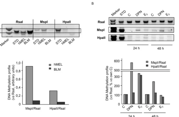
Experiments were first carried out to analyze the global DNA methylation status of human BLM melanoma cells when compared to that of human melanocytes. To this purpose, a restric-tion enzymatic assay, utilizing the two methylarestric-tion sensitive restricrestric-tion enzymes MspI and HpaII, was performed. These enzymes recognize the same tetranucleotide sequence (5'-CCGG-3') but display different sensitivity to DNA methylation. In particular, MspI does not cut when the external cytosine is methylated while HpaII does not cut when any of the two cytosines is methylated [39,40].Fig 5Ashows that BLM cells are globally hypomethylated when compared to human melanocytes (hMEL), when both MspI and HpaII restriction enzymes are utilized. These data confirm that melanoma cells are characterized by an aberrant global DNA hypo-methylation, which is known to be associated with genome instability.
We then evaluated whether activation of ERβmight affect the DNA methylation status of melanoma cells. BLM cells were treated with either DPN or E2(10−8M) for 24 or 48 h; the
DNA methylation status was analyzed as described above.Fig 5Bshows that DPN significantly increased DNA methylation at 24 and 48 h of treatment, when the MspI restriction enzyme was utilized. On the other hand, E2significantly increased the methylation degree of CG-rich
regions at 24 h of treatment, when both restriction enzymes were utilized. These data indicate that ERβactivation reverts the DNA hypomethylation status in melanoma cells and suggest that different ERβligands might increase the methylation of the different cytosines of the CG-rich regions (internalvs. external) in a specific way.
ER
β
agonists differentially affect the proliferation of melanoma cell lines
of ERβactivation on the proliferation of different melanoma cell lines, either lacking the expression of ERβor expressing ERβwhile harboring different oncogenic mutations (e.g., BRAF). Specifically, the effects of ERβagonists were assessed on the proliferation of the follow-ing human melanoma cell lines: IGR-39 cells (expressfollow-ing almost undetectable levels of ERβ),
Fig 4. The specific ERβligand DPN affects the expression of cell cycle-related proteins in BLM melanoma cells.BLM cells were treated with DPN (10−8M) for 24, 48, or 72 h. Western blot analysis was performed on whole cell extracts by using specific antibodies against cell cycle-related proteins, such
as cyclin D1, cyclin D3, p21, p27, CDK4, CDK6(A), procaspase-3 and cleaved caspase-3(B). Actin expression was evaluated as a loading control. The treatment with DPN reduced the expression of cyclin D1 and cyclin D3 and increased that of p27, while the levels of cleaved (active) caspase-3 were not affected by the treatment. One representative of three different experiments, which gave similar results, is shown. A statistical evaluation has been performed on the densitometric analysis of the results obtained from the three Western blot experiments performed on cell cycle-related proteins(A).
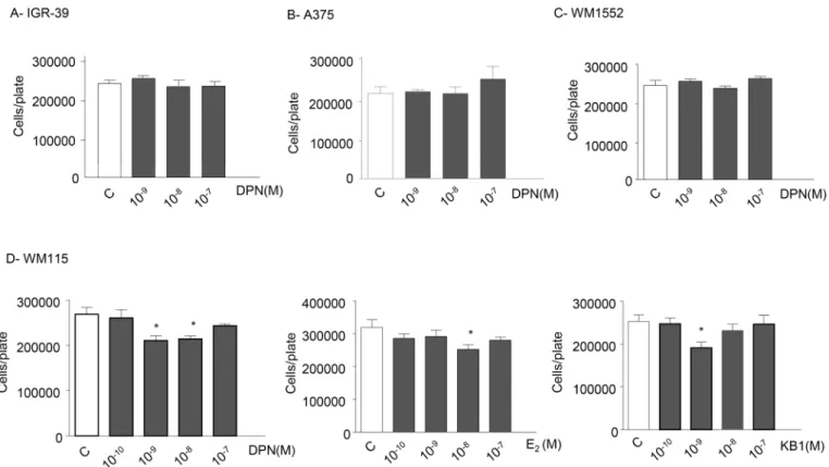
A375 and WM1552 cells (expressing ERβand harboring the BRAF V600E mutation), and WM115 cells (expressing ERβand harboring the rare BRAF V600D mutation). IGR-39, A375 and WM1552 cells were treated with DPN (10−9–10−7M) while WM115 cells were treated with DPN, E2and KB1 (10−10–10−7M), as described for BLM cells. As expected, we found that
DPN does not affect the proliferation of IGR-39 cells, lacking ERβexpression (Fig 6A). Unex-pectedly, and interestingly, the ERβagonist also failed to affect the growth of A375 and WM1552 melanoma cells, expressing the receptor subtype (Fig 6B and 6C). On the other hand, the proliferation of WM115 cells was reduced by the treatment with DPN, E2and KB1, with a
dose-response curve similar to that observed in BLM cells (Fig 6D). Taken together, these results indicate that ERβactivation differentially affects the proliferation of melanoma cell lines. The reasons for these observations are still unclear; however, we might speculate that the efficacy of ERβagonists in reducing melanoma growth might depend not only on the presence of the receptor but also on other particular features of each melanoma, such as the oncogenic mutation status (NRAS, BRAF) of the tumor.
Expression of ER
β
isoforms in melanoma cell lines
So far, five alternatively spliced transcript variants of the ERβgene have been described (ERβ1–
5) in humans [53]. ERβwild type, also referred to as ERβ1, is the main variant and ERβ2 and
Fig 5. ERβactivation induces global DNA methylation reprogramming in BLM melanoma cells. (A)Preliminary experiments were carried out to analyze the global DNA methylation status of BLM cells when compared to that of human normal melanocytes (hMel). To this purpose, a restriction enzymatic assay was employed. For each DNA sample, two restriction digests were performed: one with RsaI and MspI, and one with RsaI and HpaII. RsaI is methylation insensitive, while MspI and HpaII are sensitive to DNA methylation and are able to cut only unmethylated restriction sites. The digests were then amplified by PCR. Data are expressed as the MspI/RsaI or HpaII/RsaI ratios relative to the intensity of the bands. BLM melanoma cells were found to be globally hypomethylated when compared to normal melanocytes, when both MspI and HpaII restriction enzymes were utilized. One representative of three different experiments, which gave similar results, is reported.(B)Experiments were performed to evaluate whether activation of ERβmight affect the global DNA hypomethylation status observed in melanoma cells. BLM cells were treated with either DPN or E2(10−8M) for 24 or 48 h; the DNA methylation status was then evaluated as described above. Both DPN (at 24 and 48 h) and E2(at 24 h) increased the DNA methylation profile of BLM cells, indicating that ERβ activation reverts the DNA hypomethylation status in melanoma cells. One representative of three different experiments, which gave similar results, is reported.
ERβ5 are the most studied splice variants [22]. The expression of these variants has been shown to be tissue-specific and to differentially modulate E2signaling [54].
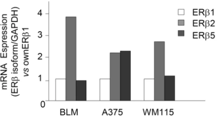
As discussed above, the reasons for the differential effects of ERβagonists on the prolifera-tion of melanoma cell lines are still unclear. In addiprolifera-tion to the proposed correlaprolifera-tion with spe-cific oncogenic mutations, these effects might also be related to the differential expression of ERβisoforms in the various melanoma cell lines. Based on these observations, by quantitative RT-PCR we analyzed the expression of ERβ1, ERβ2, and ERβ5 in BLM, A375 and WM115 mel-anoma cell lines.
Fig 7shows that the pattern of expression of the ERβisoforms is similar in BLM and in WM115 cells, with ERβ1and ERβ5 being expressed at similar levels and ERβ2 showing a higher level of expression. On the other hand, in A375 cells, both ERβ2 and 5 are expressed at higher levels than the ERβ1 isoform.
Discussion
Increasing evidence strongly suggests that ERβplays a fundamental role in the development and progression of melanoma. In particular, the expression of ERβwas shown to inversely cor-relate with melanoma progression, being significantly lower in thick melanoma compared with thin melanoma tissues [23,26,27,55]. These observations point toward a potential role of ERβ
as a protein associated with suppressive function in this tumor.
Fig 6. ERβagonists differentially affect the proliferation of melanoma cell lines. (A)IGR-39, A375 and WM1552 melanoma cells were treated with DPN (10−9
–10−7M) every 48 h for three times. No effect on cell proliferation could be observed in any cell line tested.(B)WM115 cells were treated with DPN, E2, or KB1 (10−10
–10−7M) every 48 h for three times. DPN was significantly effective in decreasing cell proliferation at the doses of 10−9and 10−8M. On the other
hand, both E2and KB1 significantly reduced cell proliferation at the dose of 10−8M. Each experimental group consisted of six replicates and each experiment was repeated three times. Results are given as cell number/plate. Data represent mean values±SEM.*P<0.05. C, controls.
In this study, we first investigated the expression of ERβin a panel of melanoma cell lines; we demonstrated that this estrogen receptor subtype (but not the ERαsubtype) is expressed in most of these cell lines.
Then, we analyzed the effects and the possible mechanisms of action of ERβactivation in BLM cells. We could demonstrate that activation of ERβ, achieved by treating the cells with E2
or ERβsubtype-selective agonists (the classical ERβagonist, DPN, or more recently synthesized agonists, KB1, KB2, and KB4) significantly decreased BLM melanoma cell proliferation. This tumor cell inhibitory activity was found to be target protein specific since it was completely abrogated by cotreatment of the cells with the ER antagonist ICI 182,780.
In these experiments, the ERβagonists displayed bell-shaped responses with growth inhibi-tion at low doses and opposite effect at high doses, as previously reported for cholangiocarci-noma and mesothelioma cells [14,20]. As underlined by Pinton and coworkers [14], this kind of response in not unusual for hormones. The term 'hormesis' has been widely used to describe a biphasic dose response phenomenon characterized by a low dose stressful stimulation and a high dose adaptive response that increases the resistance of the cell to evoked stress [56,57]. A possible explanation could be that cells may increase the production of cytoprotective and restorative proteins which can mediate their adaptive response to the stress induced by ERβ
agonists.
In BLM cells, activation of ERβinduced its translocation from the cytoplasm into the nucleus and triggered its transcriptional activity. These data demonstrate that, in these mela-noma cells expressing ERβ, this receptor subtype exerts its repressive activity through the clas-sical genomic action of steroid receptors at the nuclear level.
In this paper, we could also show that, in BLM melanoma cells, ERβagonists exert their antiproliferative activity through the modulation of cell cycle progressing factors (cyclin D1, cyclin D3, p27), without triggering the apoptosis pathway. These data suggest that ERβ activa-tion may inhibit melanoma growth by blocking the G1-S transiactiva-tion phase. Specifically, we could observe that the ERβagonist DPN significantly reduces cyclin D1 and cyclin D3 protein expression at 72 h of treatment, while increasing the expression of p27 at 48 h of treatment. Since ERβnuclear translocation and transcriptional activity occur 24 h after treatment of the cells with DPN, we hypothesize that these cell cycle-related proteins might not be directly regu-lated by ERβbut, more likely, they might be the target of the activity of other direct ERβ Fig 7. ERβisoforms (1, 2, and 5) are differentially expressed in melanoma cell lines.The relative expression of ERβ1, 2, and 5 isoforms was evaluated by quantitative RT-PCR, utilizing specific sets of primers. BLM and WM115 cells showed a similar expression of ERβ1 and 5, while expressing higher levels of ERβ2. On the other hand, a high expression of both ERβ2 and 5 isoforms (when compared to ERβ1) was observed in A375 cells. One representative of three different experiments, which gave similar results, is shown.
downstream proteins. For instance, Wu and coworkers [58] have recently reported that, in bladder cancer cells, the ERβligand resveratrol inhibits cell growth through decreased phos-phorylation, nuclear translocation and transcription of STAT3, resulting in the downregulation of the expression of STAT3 downstream genes (cyclin D1, survivin, c-Myc and VEGF). More-over, Nakamura and coworkers demonstrated that, in prostate cancer cells, activation of ERβ
induces cyclin D1 expression through increased expression of FOS and JUN; however, accord-ing to the data reported, the authors conclude that the interaction of ERβwith the two tran-scription factors is not direct and likely involves early responsive genes which still need to be identified [59].
Taken together, our results obtained in BLM cells agree with the concept that the antitumor effect of ERβis associated with altered expression of proteins involved in the cell cycle progres-sion [10,11]. In agreement with the data here reported, ERβagonists, as well as phytoestrogens (such as apigenin, resveratrol) have been shown to arrest breast cancer cell growth by causing a cell cycle arrest, through the regulation of cell cycle-related proteins, such as cyclin D1 and the CDK inhibitors p21 and p27 [60–62]; more recently, it has been reported that preferential ERβ
ligands reduce the expression of the antiapoptotic protein Bcl-2 to increase autophagy in hor-mone-resistant breast cancer cells [63]. In prostate cancer cells, ERβagonists inhibit the prolif-eration rate and the invasive behavior [64,65]. Moreover, ERβagonists impede prostate cancer epithelial-to-mesenchymal transition, by repressing VEGF-A expression [66]. ERβligands were also shown to exert suppressive effects, through modulation of the expression of cell cycle progression proteins, on the growth of tumor cells classically unrelated to the reproductive sys-tem, such as colon [18], malignant pleural mesothelioma [14,19,36], lymphoma [21], glioma [67], and cholangiocarcinoma [20] cells. More recently, ERβagonists have been reported to prevent the development of UVB-induced nonmelanoma skin cancer in mice [68].
It is now well accepted that epigenetic mechanisms play a central role in tumor develop-ment. In particular, melanoma cells have been reported to present global DNA hypomethyla-tion, contributing to the genomic instability of tumor cells, when compared to normal cells [28,30]. Thus, reversibility of these epigenetic modifications might represent an effective strat-egy of treatment for this aggressive form of cancer. In this study, we first confirmed that DNA is globally hypomethylated in human BLM melanoma cells when compared to normal human melanocytes. Then, we could show that treatment of BLM cells with both DPN and E2
signifi-cantly increased global DNA methylation.
Taken together, our data demonstrate that, in BLM melanoma cells, ERβactivation reduces cell growth, through the modulation of cell cycle related proteins, and that this antitumor activ-ity is associated with the reversal of the global DNA hypomethylation status of these cancer cells.
In this paper, as expected, we could show that ERβagonists did not affect the proliferation of melanoma cells expressing almost undetectable levels of ERβ(IGR-39).
On the other hand, suprisingly, we found that ERβagonists were also ineffective in reducing the proliferation of A375 and WM1552 melanoma cells, shown to express the estrogen receptor isoform. At present, the reason for these unexpected results is unclear. However, a possible explanation is that ERβagonists differentially affect the proliferation of various cell lines, expressing ERβ, according to the cell line-specific oncogenic mutation status. Actually, NRAS and BRAF mutations are very frequently found in melanoma tumors; in particular, BLM cells are NRAS-mutant (a mutation present in about 30% of patients), while both A375 and
BRAF mutations, only the MEK/ERK pathway results to be overactivated. Interestingly, ERβ
agonists have been shown to exert their significant antitumor/proapoptotic effect through RAS inactivation and specific inhibition of its downstream PI3K/Akt pathway in different cancer cells [11,72,73]. Wang and coworkers [74] have recently reported that ERβexpression inversely correlate with PTEN/PI3K/Akt pathway in triple-negative breast cancer. Moreover, in breast cancer cells, calycosin-induced ERβactivation was associated with a decreased activity of the PI3K/Akt pathway, while the ERK1/2 cascade was not affected by the natural compound [72]. Based on our results as well as on these recently reported observations, we hypothesize that ERβagonists might effectively reduce the proliferation of melanoma cells harboring the NRAS mutation, through the specific inhibition of the activity of one of the two downstream signaling pathways: the PI3K/Akt cascade. On the other hand, ERβagonists will not reduce the growth of melanoma cells harboring the BRAF (V600E) mutation, which is associated with the overac-tivation of the MEK/ERK signaling pathway. Studies are ongoing in our laboratory to confirm this hypothesis.
Taken together, these data would suggest that, in melanoma patients harboring the NRAS mutation, ERβmight represent a novel molecular target for personalized therapeutic strategies, based on ERβagonists, either alone or in combination with a specific inhibitor of the MEK pathway (i.e., trametinib). Moreover, these results support the notion that not only the expres-sion of ERβ, but also the genetic analysis of the concurrent oncogenic mutations should be con-sidered to predict the possible response of melanomas to ERβtargeted therapeutic approaches.
In this paper we could also show that ERβagonists are able to decrease the proliferation of WM115 melanoma cells harboring the BRAF V600D mutation. However, no hypothesis can be suggested in this case, since this is considered a very rare BRAF mutation and very little is known about its associated intracellular signaling alterations; it has actually been reported that, in melanoma cells, BRAF mutations can be associated to different intracellular pathways, in addition to the MEK/ERK cascade [75]. Moreover, whether BRAF inhibitors might have the same effectiveness in patients with this rare BRAF mutation still has to be evaluated [76].
The differential effect of ERβagonists on the proliferation of the various melanoma cell lines here reported might also be associated with the relative expression of the ERβisoforms in each cell line. We found that BLM and WM115 cells show a similar pattern of expression of the isoforms with similar levels of ERβ1 and ERβ5, but higher expression of ERβ2. On the other hand, in A375 cells both ERβ2 and ERβ5 are expressed at higher levels than ERβ1. The possible correlation between the expression of the ERβisoforms and the differential effects of ERβagonists on melanoma cells is at present unclear. ERβisoforms have been shown to be co-expressed in various types of tumors, (including breast, ovarian, endometrial, prostate, colon and lung cancers); however, conflicting results have been so far reported on the potential col-lective effect of their co-existence [41,43,77–81]. In agreement with Hapangama and coworkers [82], we believe that the lack of commercially available specific antibodies for the different receptor isoforms represents a major obstacle in the investigation and clarification of their functions. Based on this observation, most of the functional data so far reported in the litera-ture are only related to ERβ(ERβ1), with little reference to the other alternatively spliced variants.
melanoma cell lines might be related either to the specific oncogenic mutational status (NRAS, BRAF) or to the relative expression of receptor isoforms in each cell line.
These data confirm that melanoma is a very heterogeneous tumor and support the concept that genetic profiling is mandatory for the development of novel and effective personalized therapeutic strategies for melanoma patients.
Acknowledgments
The authors thank Dr. S. Nilsson (Karo Bio AB, Novum, SE-141 57 Huddinge, Sweden) for kindly providing KB1, KB2, and KB4 compounds.
Author Contributions
Conceived and designed the experiments: MM LC RMM MMM PL. Performed the experi-ments: MM LC RMM MMM. Analyzed the data: MM LC RMM MMM. Wrote the paper: PL.
References
1. Siegel R, Ma J, Zou Z, Jemal A. Cancer statistics, 2014. CA Cancer J Clin. 2014; 64: 9–29. doi:10.
3322/caac.21208PMID:24399786
2. DeSantis CE, Lin CC, Mariotto AB, Siegel RL, Stein KD, Kramer JL, et al. Cancer treatment and survi-vorship statistics, 2014. CA Cancer J Clin. 2014; 64: 252–271. doi:10.3322/caac.21235PMID:
24890451
3. Jilaveanu LB, Aziz SA, Kluger HM. Chemotherapy and biologic therapies for melanoma: do they work? Clin Dermatol. 2009; 27: 614–625. doi:10.1016/j.clindermatol.2008.09.020PMID:19880049
4. Pasquali S, Spillane A. Contemporary controversies and perspectives in the staging and treatment of patients with lymph node metastasis from melanoma, especially with regards positive sentinel lymph node biopsy. Cancer Treat Rev. 2014; 40: 893–899. doi:10.1016/j.ctrv.2014.06.008PMID:25023758
5. Leung AM, Lee AF, Ozao-Choy J, Ramos RI, Hamid O, O'Day SJ, et al. Clinical benefit from ipilimumab therapy in melanoma patients may be associated with serum CTLA4 levels. Front Oncol. 2014; 4: 110. doi:10.3389/fonc.2014.00110PMID:24904825
6. Voskoboynik M, Arkenau HT. Combination therapies for the treatment of advanced melanoma: a review of current evidence. Biochem Res Int. 2014; 2014: 307059. doi:10.1155/2014/307059PMID: 24693430
7. Nilsson S, Gustafsson J-Å. Estrogen receptors: therapies targeted to receptor subtypes. Clin Pharma-col Ther. 2011; 89: 44–55. doi:10.1038/clpt.2010.226PMID:21124311
8. Nilsson S, Koehler KF, Gustafsson J-Å. Development of subtype-selective oestrogen receptor-based therapeutics. Nat Rev Drug Discov. 2011; 10: 778–792. doi:10.1038/nrd3551PMID:21921919
9. Dahlman-Wright K, Cavailles V, Fuqua SA, Jordan VC, Katzenellenbogen JA, Korach KS, et al. Interna-tional Union of Pharmacology. LXIV. Estrogen receptors. Pharmacol Rev. 2006; 58: 773–781. PMID:
17132854
10. Thomas C, Gustafsson J-Å. The different roles of ER subtypes in cancer biology and therapy. Nat Rev Cancer. 2011; 11: 597–608. doi:10.1038/nrc3093PMID:21779010
11. Dey P, Barros RP, Warner M, Ström A, Gustafsson J-Å. Insight into the mechanisms of action of estro-gen receptor beta in the breast, prostate, colon, and CNS. J Mol Endocrinol. 2013; 51: T61–T74. doi:
10.1530/JME-13-0150PMID:24031087
12. Warner M, Gustafsson J-Å. The role of estrogen receptor beta (ERbeta) in malignant diseases-a new potential target for antiproliferative drugs in prevention and treatment of cancer. Biochem Biophys Res Commun. 2010; 396: 63–66. doi:10.1016/j.bbrc.2010.02.144PMID:20494112
13. Yang GS, Wang Y, Wang P, Chen ZD. Expression of oestrogen receptor-alpha and oestrogen recep-tor-beta in prostate cancer. Chin Med J (Engl). 2007; 120: 1611–1615.
14. Pinton G, Manente AG, Daga A, Cilli M, Rinaldi M, Nilsson S, et al. Agonist activation of estrogen recep-tor beta (ERbeta) sensitizes malignant pleural mesothelioma cells to cisplatin cytotoxicity. Mol Cancer. 2014; 13: 227. doi:10.1186/1476-4598-13-227PMID:25277603
15. Leung YK, Gao Y, Lau KM, Zhang X, Ho SM. ICI 182,780-regulated gene expression in DU145 prostate cancer cells is mediated by estrogen-receptorβ/NFκB crosstalk. Neoplasia. 2006; 8: 242–249. PMID:
16. McPherson SJ, Hussain S, Balanathan P, Hedwards SL, Niranjan B, Grant M, et al. Estrogen receptor-beta activated apoptosis in benign hyperplasia and cancer of the prostate is androgen independent and TNFalpha mediated. Proc Natl Acad Sci U S A. 2010; 107: 3123–3128. doi:10.1073/pnas.0905524107
PMID:20133657
17. Leung YK, Lee MT, Lam HM, Tarapore P, Ho SM. Estrogen receptor-beta and breast cancer: translat-ing biology into clinical practice. Steroids. 2012; 77: 727–737. doi:10.1016/j.steroids.2012.03.008
PMID:22465878
18. Hartman J, Edvardsson K, Lindberg K, Zhao C, Williams C, Ström A, et al. Tumor repressive functions of estrogen receptor beta in SW480 colon cancer cells. Cancer Res. 2009; 69: 6100–6106. doi:10.
1158/0008-5472.CAN-09-0506PMID:19602591
19. Pinton G, Thomas W, Bellini P, Manente AG, Favoni RE, Harvey BJ, et al. Estrogen receptor beta exerts tumor repressive functions in human malignant pleural mesothelioma via EGFR inactivation and affects response to gefitinib. PLoS One. 2010; 5: e14110. doi:10.1371/journal.pone.0014110PMID: 21124760
20. Marzioni M, Torrice A, Saccomanno S, Rychlicki C, Agostinelli L, Pierantonelli I, et al. An oestrogen receptor beta-selective agonist exerts anti-neoplastic effects in experimental intrahepatic cholangiocar-cinoma. Dig Liver Dis. 2012; 44: 134–142. doi:10.1016/j.dld.2011.06.014PMID:21782536
21. Yakimchuk K, Hasni MS, Guan J, Chao MP, Sander B, Okret S. Inhibition of lymphoma vascularization and dissemination by estrogen receptor beta agonists. Blood. 2014; 123: 2054–2061. doi:10.1182/
blood-2013-07-517292PMID:24470591
22. Leung YK, Mak P, Hassan S, Ho SM. Estrogen receptor (ER)-βisoforms: a key to understanding ER-β signaling. Proc Natl Acad Sci USA. 2006; 103: 13162–13167. PMID:16938840
23. Schmidt AN, Nanney LB, Boyd AS, King LE Jr., Ellis DL. Oestrogen receptor-beta expression in mela-nocytic lesions. Exp Dermatol. 2006; 15: 971–980. PMID:17083364
24. Scoggins CR, Ross MI, Reintgen DS, Noyes RD, Goydos JS, Beitsch PD, et al. Gender-related differ-ences in outcome for melanoma patients. Ann Surg. 2006; 243: 693–698; discussion 698–700. PMID:
16633005
25. Joosse A, de Vries E, Eckel R, Nijsten T, Eggermont AM, Holzel D, et al. Gender differences in mela-noma survival: female patients have a decreased risk of metastasis. J Invest Dermatol. 2011; 131: 719–726. doi:10.1038/jid.2010.354PMID:21150923
26. de Giorgi V, Gori A, Gandini S, Papi F, Grazzini M, Rossari S, et al. Oestrogen receptor beta and mela-noma: a comparative study. Br J Dermatol. 2013; 168: 513–519. doi:10.1111/bjd.12056PMID:
23013061
27. de Giorgi V, Gori A, Grazzini M, Rossari S, Scarfi F, Corciova S, et al. Estrogens, estrogen receptors and melanoma. Expert Rev Anticancer Ther. 2011; 11: 739–747. doi:10.1586/era.11.42PMID:
21554049
28. Ecsedi SI, Hernandez-Vargas H, Lima SC, Herceg Z, Adany R, Balazs M. Transposable hypomethyla-tion is associated with metastatic capacity of primary melanomas. Int J Clin Exp Pathol. 2013; 6: 2943–2948. PMID:24294382
29. Besaratinia A, Tommasi S. Epigenetics of human melanoma: promises and challenges. J Mol Cell Biol. 2014; 6: 356–367. doi:10.1093/jmcb/mju027PMID:24895357
30. Ecsedi S, Hernandez-Vargas H, Lima SC, Vizkeleti L, Toth R, Lazar V, et al. DNA methylation charac-teristics of primary melanomas with distinct biological behaviour. PLoS One. 2014; 9: e96612. doi:10. 1371/journal.pone.0096612PMID:24832207
31. Van Muijen GN, Cornelissen LM, Jansen CF, Figdor CG, Johnson JP, Brocker EB, et al. Antigen expression of metastasizing and non-metastasizing human melanoma cells xenografted into nude mice. Clin Exp Metastasis. 1991; 9: 259–272. PMID:2060184
32. Moretti RM, Montagnani Marelli M, Van Groeninghen JC, Limonta P. Locally expressed LHRH recep-tors mediate the oncostatic and antimetastatic activity of LHRH agonists on melanoma cells. J Clin Endocrinol Metab. 2002; 87: 3791–3797. PMID:12161512
33. Moretti RM, Mai S, Montagnani Marelli M, Bani MR, Ghilardi C, Giavazzi R, et al. Dual targeting of tumor and endothelial cells by gonadotropin-releasing hormone agonists to reduce melanoma angio-genesis. Endocrinology. 2010; 151: 4643–4653. doi:10.1210/en.2010-0163PMID:20685877
34. Satyamoorthy K, DeJesus E, Linnenbach AJ, Kraj B, Kornreich DL, Rendle S, et al. Melanoma cell lines from different stages of progression and their biological and molecular analyses. Melanoma Res. 1997; 7 Suppl 2: S35–42. PMID:9578415
36. Manente AG, Valenti D, Pinton G, Jithesh PV, Daga A, Rossi L, et al. Estrogen receptor beta activation impairs mitochondrial oxidative metabolism and affects malignant mesothelioma cell growth in vitro and in vivo. Oncogenesis. 2013; 2: e72. doi:10.1038/oncsis.2013.32PMID:24061575
37. Barkhem T, Carlsson B, Nilsson Y, Enmark E, Gustafsson J-Å, Nilsson S. Differential response of estrogen receptor alpha and estrogen receptor beta to partial estrogen agonists/antagonists. Mol Phar-macol. 1998; 54: 105–112. PMID:9658195
38. Martini PG, Delage-Mourroux R, Kraichely DM, Katzenellenbogen BS. Prothymosin alpha selectively enhances estrogen receptor transcriptional activity by interacting with a repressor of estrogen receptor activity. Mol Cell Biol. 2000; 20: 6224–6232. PMID:10938099
39. Xie Y, Liu J, Benbrahim-Tallaa L, Ward JM, Logsdon D, Diwan BA, et al. Aberrant DNA methylation and gene expression in livers of newborn mice transplacentally exposed to a hepatocarcinogenic dose of inorganic arsenic. Toxicology. 2007; 236: 7–15. PMID:17451858
40. Lewies A, Van Dyk E, Wentzel JF, Pretorius PJ. Using a medium-throughput comet assay to evaluate the global DNA methylation status of single cells. Front Genet. 2014; 5: 215. doi:10.3389/fgene.2014. 00215PMID:25071840
41. Collins F, MacPherson S, Brown P, Bombail V, Williams ARW, Anderson RA, et al. Expression of oes-trogen receptors, ERα, ERβ, and ERβvariants, in endometrial cancers and evidence that prostaglandin F may play a role in regulating expression of ERα. BMC Cancer. 2009; 9: 330. doi: 10.1186/1471-2407-9-330PMID:19758455
42. Casati L, Celotti F, Negri-Cesi P, Sacchi MC, Castano P, Colciago A. Platelet derived growth factor (PDGF) contained in Platelet Rich Plasma (PRP) stimulates migration of osteoblasts by reorganizing actin cytoskeleton. Cell Adh Migr. 2014; 8: 595–602. doi:10.4161/19336918.2014.972785PMID:
25482626
43. Dey P, Jonsson P, Hartman J, Williams C, Ström A, Gustafsson J-Å. Estrogen receptorsβ1 andβ2 have opposing roles in regulating proliferation and bone metastasis genes in the prostate cancer cell line PC3. Mol Endocrinol. 2012; 26:1991–2003. doi:10.1210/me.2012.1227PMID:23028063
44. Dey P, Ström A, Gustafsson J-Å. Estrogen receptorβupregulates FOXO3a and causes induction of apoptosis through PUMA in prostate cancer. Oncogene. 2014; 33: 4213–4225. doi:10.1038/onc.2013.
384PMID:24077289
45. Zhao C, Putnik M, Gustafsson J-Å, Dahlman-Wright K. Microarray analysis of altered gene expression in ERβoverexpressing HEK293 cells. Endocrine. 2009; 36: 224–232. doi:10.1007/s12020-009-9233-8
PMID:19680825
46. Charn TH, Liu ET-B, Chang EC, Lee YK, Katzenellenbogen JA, Katzenellenbogen BS. Genome-wide dynamics of chromatin binding of estrogen receptrorsαandβ: mutual restriction and competitive site selection. Mol Endocrinol. 2010; 24: 47–59. doi:10.1210/me.2009-0252PMID:19897598
47. Rizza P, Barone I, Zito D, Giordano F, Lanzino M, De Amicis F, et al. Estrogen receptor beta as a novel target of androgen receptor action in breast cancer cell lines. Breast Cancer Res. 2014; 16: 21. 48. Zhou JH, Kim KB, Myers JN, Fox PS, Ning J, Bassett RL, et al. Immunohistochemical expression of
hormone receptors in melanoma of pregnant women, non-pregnant women and men. Am J Dermato-pathol. 2014; 36: 74–79. doi:10.1097/DAD.0b013e3182914c64PMID:23812018
49. Paruthiyil S, Parmar H, Kerekatte V, Cunha GR, Firestone GL, Leitman DC. Estrogen receptor beta inhibits human breast cancer cell proliferation and tumor formation by causing a G2 cell cycle arrest. Cancer Res. 2004; 64: 423–428. PMID:14729654
50. Ström A, Hartman J, Foster JS, Kietz S, Wimalasena J, Gustafsson J-Å. Estrogen receptor beta inhibits 17beta-estradiol-stimulated proliferation of the breast cancer cell line T47D. Proc Natl Acad Sci U S A. 2004; 101: 1566–1571. PMID:14745018
51. Docquier A, Garcia A, Savatier J, Boulahtouf A, Bonnet S, Bellet V, et al. Negative regulation of estro-gen signaling by ERbeta and RIP140 in ovarian cancer cells. Mol Endocrinol. 2013; 27: 1429–1441.
doi:10.1210/me.2012-1351PMID:23885094
52. Foster JS, Henley DC, Ahamed S, Wimalasena J. Estrogens and cell-cycle regulation in breast cancer. Trends Endocrinol Metab. 2001; 12: 320–327. PMID:11504672
53. Moore JT, Mckee DD, Slentz-Kesler K, Moore LB, Jones SA, Horne EL, et al. Cloning and characteriza-tion of human estrogen receptor beta isoforms. Biochem Biophys Res Commun. 1998; 247: 75–78.
PMID:9636657
54. Ramsey TL, Risinger KE, Jernigan SC, Mattingly KA, Klinge CM. Estrogen receptor beta isoforms exhibit differences in ligand-activated transcriptional activity in an estrogen response element sequence-dependent manner. Endocrinology. 2004; 145: 149–160. PMID:14500565
immunohistochemical study. Arch Dermatol. 2009; 145: 30–36. doi:10.1001/archdermatol.2008.537
PMID:19153340
56. Calabrese EJ. Cancer biology and hormesis: human tumor cell lines commonly display hormetic (biphasic) dose response. Crit Rev Toxicol 2005; 35: 463–582. PMID:16422392
57. Calabrese EJ. Biphasic dose responses in biology, toxicology and medicine accounting for their gener-alizability and quantitative features. Environ Pollut. 2013; 182: 452–460. doi:10.1016/j.envpol.2013.07.
046PMID:23992683
58. Wu ML, Li H, Yu LJ, Chen XY, Kong QY, Song X, et al. Short-term resveratrol exposure causes in vitro and in vivo growth inhibition and apoptosis of bladder cancer cells. PLoS One. 2014; 9: e89806. doi: 10.1371/journal.pone.0089806PMID:24587049
59. Nakamura Y, Felizola SJ, Kurotaki Y, Fujishima F, McNamara KM, Suzuki T, et al. Cyclin D1 (CCND1) expression is involved in estrogen receptor beta (ERbeta) in human prostate cancer. Prostate. 2013; 73: 590–595. doi:10.1002/pros.22599PMID:23060014
60. Nair HB, Kirma NB, Ganapathy M, Vadlamudi RK, Tekmal RR. Estrogen receptor-beta activation in combination with letrozole blocks the growth of breast cancer tumors resistant to letrozole therapy. Ste-roids. 2011; 76: 792–796. doi:10.1016/j.steroids.2011.02.038PMID:21477609
61. Bilal I, Chowdhury A, Davidson J, Whitehead S. Phytoestrogens and prevention of breast cancer: The contentious debate. World J Clin Oncol. 2014; 5: 705–712. doi:10.5306/wjco.v5.i4.705PMID:
25302172
62. Harrison ME, Power Coombs MR, Delaney LM, Hoskin DW. Exposure of breast cancer cells to a subcy-totoxic dose of apigenin causes growth inhibition, oxidative stress, and hypophosphorylation of Akt. Exp Mol Pathol. 2014; 97: 211–217. doi:10.1016/j.yexmp.2014.07.006PMID:25019465
63. Ruddy SC, Lau R, Cabrita MA, McGregor C, McKay BC, Murphy LC, et al. Preferential estrogen recep-tor beta ligands reduce Bcl-2 expression in hormone-resistant breast cancer cells to increase autop-hagy. Mol Cancer Ther. 2014; 13: 1882–1893. doi:10.1158/1535-7163.MCT-13-1066PMID:
24785256
64. Dondi D, Piccolella M, Biserni A, Della Torre S, Ramachandran B, Locatelli A, et al. Estrogen receptor beta and the progression of prostate cancer: role of 5alpha-androstane-3beta,17beta-diol. Endocr-Relat Cancer. 2010; 17: 731–742. doi:10.1677/ERC-10-0032PMID:20562232
65. Piccolella M, Crippa V, Messi E, Tetel MJ, Poletti A. Modulators of estrogen receptor inhibit proliferation and migration of prostate cancer cells. Pharmacol Res. 2014; 79: 13–20. doi:10.1016/j.phrs.2013.10.
002PMID:24184124
66. Mak P, Leav I, Pursell B, Bae D, Yang X, Taglienti CA, et al. ERbeta impedes prostate cancer EMT by destabilizing HIF-1alpha and inhibiting VEGF-mediated snail nuclear localization: implications for Glea-son grading. Cancer Cell. 2010; 17: 319–332. doi:10.1016/j.ccr.2010.02.030PMID:20385358
67. Sareddy GR, Nair BC, Gonugunta VK, Zhang QG, Brenner A, Brann DW, et al. Therapeutic signifi-cance of estrogen receptor beta agonists in gliomas. Mol Cancer Ther. 2012; 11: 1174–1182. doi:10.
1158/1535-7163.MCT-11-0960PMID:22442308
68. Chaudhary SC, Singh T, Talwelkar SS, Srivastava RK, Arumugam A, Weng Z, et al. Erb-041, an estro-gen receptor-beta agonist, inhibits skin photocarcinoestro-genesis in SKH-1 hairless mice by downregulating the WNT signaling pathway. Cancer Prev Res (Phila). 2014; 7: 186–198.
69. Ekedahl H, Cirenajwis H, Harbst K, Carneiro A, Nielsen K, Olsson H, et al. The clinical significance of BRAF and NRAS mutations in a clinical-based metastatic melanoma cohort. Br J Dermatol. 2013; L169: 1049–1055.
70. Lo JA, Fisher DE. The melanoma revolution: from UV carcinogenesis to a new era in therapeutics. Sci-ence. 2014; 346: 945–949. doi:10.1126/science.1253735PMID:25414302
71. Vujic I, Sanlorenzo M, Posch C, Esteve-Puig R, Yen AJ, Kwong A, et al. Metformin and trametinib have synergistic effects on cell viability and tumor growth inNRASmutant cancer. Oncotarget. 2014; 6: 969–978.
72. Chen J, Hou R, Zhang X, Ye Y, Wang Y, Tian J. Calycosin suppresses breast cancer cell growth via ERβ-dependent regulation of IGF-1R, p38 MAPK and PI3K/Akt pathways. PLoS One. 2014; 9: e01245. 73. Liu X, Wang L, Chen J, Ling Q, Wang H, Li S, et al. Estrogen receptorβagonist enhances
temozolo-mide sensitivity of glioma cells by inhibiting PI3K/AKT/mTOR pathway. Mol Med Rep. 2015; 11: 1516–1522. doi:10.3892/mmr.2014.2811PMID:25351348
75. Liu D, Liu X, Xing M. Activities of multiple cancer-related pathways are associated withBRAFmutation
and predict the resistance to BRAF/MEK inhibitors in melanoma cells. Cell Cycle. 2014; 13: 208–219.
doi:10.4161/cc.26971PMID:24200969
76. Heinzerling L, Kühnapfel S, Meckbach D, Baiter M, Kaempgen E, Keikavoussi P, et al. Rare BRAF mutations in melanoma patients: implications for molecular testing in clinical practice. Br J Cancer. 2013; 108: 2164–2171. doi:10.1038/bjc.2013.143PMID:23579220
77. Campbell-Thompson M, Lynch IJ, Bhardwaj B. Expression of estrogen receptor (ER) subtypes and ERβisoforms in colon cancer. Cancer Res. 2001; 61: 632–640. PMID:11212261
78. Leung YK, Lam HM, Wu S, Song D, Levin L, Cheng L, et al. Estrogen receptorβ2 andβ5 are associ-ated with poor prognosis in prostate cancer, and promote cancer cell migration and invasion. Endocr-Relat Cancer. 2010; 17: 675–689. doi:10.1677/ERC-09-0294PMID:20501637
79. Leung YK, Lee MT, Lam HM, Tarapore P, Ho SM. Estrogen-receptor-beta and breast cancer: translat-ing biology into clinical pratice. Steroids. 2012; 77: 727–737. doi:10.1016/j.steroids.2012.03.008
PMID:22465878
80. Liu Z, Liao Y, Tang H, Chen G. The expression of estrogen receptorsβ2, 5 identifies and is associated with prognosis in non-small cell lung cancer. Endocrine. 2013; 44:517–524. doi:
10.1007/s12020-013-9916-zPMID:23475473
81. Ciucci A, Zannoni GF, Travaglia D, Petrillo M, Scambia G, Gallo D. Prognostic significance of the estro-gen receptor beta (ERβ) isoforms ERβ1, ERβ2, and ERβ5 in advanced serous ovarian cancer. Gynecol Oncol. 2014; 132: 351–359. doi:10.1016/j.ygyno.2013.12.027PMID:24378878