Cop
yright
© ABE&M t
odos os dir
eit
os r
eser
vados
.
New mutation in the
PTEN
gene in a Brazilian patient
with Cowden’s syndrome
Nova mutação no gene PTEN em um paciente
brasileiro com síndrome de Cowden
Erika U. de Lima1, Iberê C. Soares2, Debora L. S. Danilovic1, Suemi Marui1
SUMMARY
Cowden syndrome is characterized by hamartomatous polyps, trichilemmomas, increased risk of developing neoplasms, and is associated with germline mutations in the PTEN gene. We searched for germline mutations in PTEN in a 49-year-old female patient who presented trichilemmoma with previous history of breast carcinoma, and thyroidectomy for a thyroid nodule. We also searched for somatic mutations in breast and thyroid tumoral tissues. DNA was extrac ted from peripheral leukocytes, parafin samples of breast carcinoma, and cytological smears of thyroid nodule ine-needle aspiration biopsy, whose inal histopathological diagnosis was adenomatous goiter. PTEN was ampliied and sequenced. We identiied a novel mutation, due to a T>A inversion at position 159 and A>T inversion at position 160, leading to valine-to-aspartic acid substitution at position 53. The p.Val53Asp was also found in homozygous state in samples obtained from ad-enocarcinoma breast and thyroid biopsy, denoting loss of heterozygosity. Here, we demonstrated a novel germline mutation in PTEN, as well as somatic loss of the wild-type PTEN allele in breast and thyroid tumors in a patient with Cowden syndrome. Arq Bras Endocrinol Metab. 2012;56(8):592-6
SUMÁRIO
A síndrome de Cowden é caracterizada por pólipos de hamartoma, triquelomomas, risco au-mentado em desenvolver neoplasias e está associada a mutações germinativas no gene PTEN. Procuramos por mutação germinativa no PTEN de uma paciente de 49 anos que apresentou triquilemomas com história pregressa de carcinoma de mama e realizou tireoidectomia devido a nódulo de tireoide. Investigamos também uma mutação somática em tecidos tumorais de mama e tireoide. O DNA foi extraído de leucócitos periféricos, de amostras de paraina de carcinoma de mama e exame citológico de nódulo de tireoide obtido de biópsia por agulha ina, cujo diagnóstico histopatológico foi de bócio adenomatoso. O PTEN foi ampliicado e sequenciado. Identiicou-se uma nova mutação em decorrência de uma inversão de T>A na posição 159 e A>T na posição 160, levando à substituição de valina para ácido aspártico na posição 53. A mutação p.Val53Asp também foi encontrada em estado homozigoto em amostras obtidas do adenocarcinoma de mama e da biópsia de nódulo tireoidiano, denotando perda de heterozigosidade. Portanto, demonstramos uma mutação germinativa no PTEN e também a perda somática do alelo selvagem PTEN no tumor de mama e da tireoide de uma paciente com síndrome de Cowden. Arq Bras Endocrinol Metab. 2012;56(8):592-6
1 Laboratório de Endocrinologia
Celular e Molecular, Unidade de Tireoide (LIM/25), Disciplina de Endocrinologia e Metabologia, Faculdade de Medicina da Universidade de São Paulo (FMUSP), São Paulo, SP, Brazil
2 Departamento de Patologia,
FMUSP, São Paulo, SP, Brazil
Correspondence to: Suemi Marui
Av. Doutor Arnaldo, 455, sala 4345, 4º andar
01246-906 – São Paulo, SP, Brazil [email protected]
Received on July/25/2012 Accepted on Nov/5/2012
INTRODUCTION
C
owden syndrome (CS [MIM 158350]), irst de scribed by Lloyd and Dennis in 1963 (1), is cha racterized by hamartomatous polyps of the gastrointes tinal tract, mucocutaneous lesions, and increased risk for developing neoplasms, particularly breast, endome trium, thyroid, kidney and colorectal cancers. CS hasbeen estimated to affect around 1 in every 200,000 individuals, although this rate is probably underesti mated due to the highly variable phenotype (2). Appro ximately 80% of CS patients have PTEN mutations.
The PTEN gene (10q23.3) [MIM 601728] encodes
Cop
yright
© ABE&M t
odos os dir
eit
os r
eser
vados
.
pathway by means of its lipid phosphatase activity, and negatively regulates the MAPK pathway by means of its protein phosphatase activity (3).
Major organs involved include breast, thyroid, uterus, brain, and mucocutaneous tissues. Breast can cer numbers among the tumor types in which germ line PTEN mutation have been documented. Somatic PTEN mutation also occurs, but at a low frequency, and
is not associated with CS (4). Thyroid tumors were also found to have somatic deletions of the PTEN gene, pre
dominantly benign forms, suggesting that PTEN might
also act as a tumor suppressor for these cancers (5). Based on the Criteria of the International Cowden Consortium (6), we searched for PTEN mutation in a pa
tient with trichilemmoma, breast carcinoma, and goiter.
CASE REPORT
We studied a 49yearold female patient with pathog nomonic criteria of CS: several mucocutaneous lesions measuring 0.2 to 0.4 cm, diagnosed by biopsy as trichi lemmoma and nasal mucosal papillomatosis (Figure 1). At 46 years of age, she presented a unilateral breast cancer classiied as ductal carcinoma in situ with micro
papillary, solid, and cribriform pattern. She was submit ted to radio and hormonal (tamoxifen) therapy after surgery. During followup, a solid hypoechoic and well delimited nodule measuring 1.3 x 1.4 x 1.8 cm was noted in the left lobe on thyroid ultrasonography, and the patient was referred to our Endocrinology Depart ment. Fineneedle aspiration biopsy determined the classiication of the nodule as Class II of the Bethesda system, and total thyroidectomy was performed, as indings were indicative of CS (7). Histopathological diagnosis was adenomatous goiter. Gastrointestinal mucosa was normal on endoscopy and colonoscopy. The patient’s parents and son were both asymptomatic. Nevertheless, DNA analysis was performed for genetic counseling.
DNA analysis
DNA was extracted from peripheral leukocytes as de scribed elsewhere (8). DNA was also extracted from breast cancer tissue obtained from parafin material and from thyroid cells from FNAB slides, as described else where (9,10).
The PTEN gene was ampliied by PCR, including
the coding region and exonintron boundaries, fol lowed by automatic sequencing (ABI Prism 3130 xl,
Applied Biosystems, Foster City, CA) and results were compared with the normal sequence. Primers and PCR conditions were ampliied as described in table 1.
Figure 1. Pathognomonic mucocutaneous features of Cowden syndrome
(CS). (A) Trichilemmomas in the patient’s abdomen characterized as
well-deined, smooth, asymptomatic papules. (B) Nostril polyposis.
A B
Table 1. Primers, annealing temperature, and respective fragment size obtained after ampliication of coding region and exon-intron boundaries
of the PTEN gene
Exon Primers T°C Fragment size
Exon 1.A 5´CAG CTA CAC TGG GCA TGC T 3´ 5´AAA TGG TGA CAG GCG ACT 3´
57°C 709 bp
Exon 1.B 5´ CTT CCT CGG CTT CTC CTG A 3´ 5´ CAT CCG TCT ACT CCC ACG TT3´
51°C 768 bp
Exon 2 5´ACA TTG ACC ACC TTT TAT 3´ 5´CAA AGT ATC TTT TTC TGT 3´
45°C 321 bp
Exon 3 5´ATA TTC TCT GAA AAG CTC TGG3´ 5´TTA ATC GGT TTA GGA ATA CAA 3´
53°C 435 bp
Exon 4 5´GGG GGT GAT AAC AGT ATC TA 3´ 5´CTT TAT GCA ATA CTT TTT CCT A 3´
52°C 404 bp
Exon5 5´ACC TGT TAA GTT TGT ATG CAA C 3´ 5´TCC AGG AAG AGG AAA GGA AA 3´
57°C 382 bp
Exon 6 5´CAT AGC AAT TTA GTG AAA TAA CT3´ 5´GAT ATG GTT AAG AAA ACT GTT C 3´
50°C 274 bp
Exon7 5´TGA CAG TTT GAC AGT TAA AG 3´ 5´CTT ATT TTG GAT ATT TCT CC 3´
49°C 271 bp
Exon 8 5´CTC AGA TTG CCT TAT AAT AGT C 3´ 5´ TCA TGT TAC TGC TAC GTA AAC 3´
50°C 565 bp
Exon 9.A 5´AAG GCC TCT TAA AGA TCA TG3´ 5´ TTT TCA TGG TGT TTT ATC CCT C3´
55°C 378 bp
Exon 9.B 5´TCC AGA GGC TAG CAG TTC CAA 3´ 5´ TCT GAG CAT TCC CTC CAT TC3´
55°C 677 bp
Exon 9.C 5´TTC ACA TCC TAC CCC TTT GC 3´ 5´ AGC ACA TGA AGC ATC CAC AG 3´
55°C 645 bp
Exon 9.D 5´ CGA CTT CTC CAT CTC CTG TG 3´ 5´ GGG GGA GCA CTA TGA AGA AA 3´
55°C 601 bp
Exon 9.E 5´ CGT TCC ACC CTT TTG ACC T 3´ 5´ TGC CTA ATC TAT TTG CCA TCA A 3´
54°C 690 bp
Exon 9.F 5´TTG GTG CTG AAA TTG TTC ACT 3´ 5´ ATG CCA TTT TTC CAT TTC CA 3´
54°C 647 bp
Exon 9.G 5´ TGG AAA TGG AAA AAT GG3´ 5´ CCC CCA CTT TAG TGC ACA GT 3´
54°C 614 bp
Exon 9.H 5´GTT TAC CGG CAG CAT CAA AT 3´ 5´GCT TTG AAG GAC AGC AGG AA 3´
55°C 655 bp
Cop
yright
© ABE&M t
odos os dir
eit
os r
eser
vados
.
RESULTS
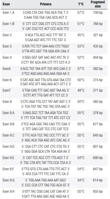
The DNA sequence from peripheral leukocytes showed a TtoA inversion at position 159 and an AtoT in version at position 160 in exon 2, leading to a valine toaspartic acid substitution at position 53 of the pro tein (p.V53D) (Figure 2A). This p.Val53Asp mutation was also found in the adenocarcinoma breast tissue and in the thyroid nodule, in a homozygous status (Fig ure 2B). We have not identiied this mutation in 200 control alleles. The patient’s father also presented this novel mutation in DNA extracted from peripheral leu
kocytes. Her son and mother showed normal sequenc es (Figure 2C).
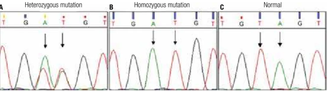
This Valine53 is a highly conserved amino acid among different species (Figure 3A). Predictive analysis of the protein secondary structure with the p.Val53Asp mutation showed minor structural changes in PTEN:
(1) strand elongation at position 124; (2) reduced coil at position 129; (3) coil stretching at positions 123 and 227 of the PTEN protein; (4) coil structure for the strand exchange at position 269; (5) reduced helix elongation at position 290 (Figure 3B).
Figure 2. Sequence analysis of exon 2 of the PTEN gene in: (A) index case-DNA from peripheral leukocytes: T-to-A inversion – at position 159 and A-to-T
inversion at position 160 (arrows), leading to a heterozygous missense mutation (GTA – GAT) at codon 53 (p.V53D). (B) breast tumor cells: homozygous
mutation (GTA – GAT) at codon 53, denoting loss of heterozygosity. (C) son (normal).
Heterozygous mutation
Amino acids sequence Reference
Normal protein
Mutated protein
Legend
= Helix Conf: = Conidence of prediction
Pred: predicted secondary structure AA: target sequence
= Strand = Coil
- +
Homozygous mutation Normal
Figure 3.(A) Alignment of sequences among different species to evaluate the conservation of the amino acid valine (V53). (B) Analysis of secondary structure of normal and mutated PTEN protein. Arrows indicate predicted changes in protein structure caused by p.Val53Asp mutation. H: helix, C: coil, E:
strand (β-sheet).
B A
Cop
yright
© ABE&M t
odos os dir
eit
os r
eser
vados
.
DISCUSSION
Germline mutations in PTEN have been described in a
variety of rare syndromes, collectively known as PTEN hamartoma tumor syndromes (PHTS). The deining clinical feature of PHTS is the presence of hamarto matous tumors, which are the disorganized growth of native cells in native tissues (11). The phenotypic spec trum of PHTS is seen in Cowden syndrome, Bannayan RileyRuvalcaba syndrome, and adult LhermitteDu clos disease. LhermitteDuclos disease is characterized by a dysplastic expansion of ganglion cells within the cerebellum, leading to replacement of the cerebellar internal granule cell layer and, consequently, various degrees of neurological signs, such as macrocephaly, mental retardation, seizures, and tremors (12). Bannay anRileyRuvalcaba syndrome presents macrocephaly, benign hamartomas, pigmented macules of the glans penis, lipomas, hemangiomas, and developmental delay or mental retardation (11). Cowden syndrome is char acterized by mucocutaneous features, including trichi lemmomas and papillomatous papules. PTEN hamar toma tumor syndromes are inherited in an autosomal dominant manner across all subtypes.
PTEN is a tumor suppressor gene with dual speci
icity phosphatase activity, and the Nterminal domain contains the phosphatase domain active site, where most PTEN mutations occur (13). PTEN negatively
regulates the phosphatidylinositol 3kinaseAKT and mammalian target of rapamycin (mTOR) signalling pathways, which are critical for cell proliferation, cell cycle progression, and apoptosis. Loss of function in this gene is a risk factor for oncogenesis and PTEN is
therefore considered a tumor suppressor gene (3). Loss of function in PTEN activates these pathways and leads
to increased cellular growth, migration, proliferation, and survival.
Gastrointestinal polyps, also typically found in CS, are usually asymptomatic and can occur anywhere in the tract, but mostly at the colon (13,14). Cowden syndrome patients are at increased risk of developing breast, thyroid, and endometrium cancer. Lifetime risk of breast cancer in women with CS is estimated to be as high as 50% as compared to 11% within the general population (15,16). As with other hereditary breast cancer syndromes, breast cancer is diagnosed at younger ages compared with the general population. The most frequently observed breast cancer histology is invasive ductal adenocarcinoma. As our patient had no familial history of breast cancer, but presented breast
cancer at a young age, the molecular study should cla rify the prognosis (16).
Benign thyroid lesions occur in up to 75% of pa tients with CS and may include adenomas, hamarto mas, multinodular goiter, and Hashimoto’s thyroiditis (17). If a molecular study is not available, total thy roidectomy is recommended, even after benign cyto logical diagnosis, given the increased risk of develop ing subsequent thyroid cancer (6). If molecular study on FNAB material is performed, total thyroidectomy should be indicated, as clinicians are aware of the in creased thyroid cancer risk when the PTEN mutation is
found. We studied the LOH in thyroid cells obtained from FNAB to clarify whether PTEN had a role in the
thyroid cell proliferation observed in our patient. The role of PTEN in her abnormal cellular proliferation was
consequently reinforced, despite being a benign lesion (adenomatous goiter).
It is noteworthy that the patient’s father also carried the p.V53D mutation and is apparently asymptomatic. To date, he has tested normal on prostate clinical evalu ation and serum PSA, as well as on kidney ultrasound and colonoscopy. These exams will be performed annu ally to exclude prostate, colorectal and kidney cancer, respectively. His thyroid ultrasound was also normal. As phenotype and genotype may be poorly related in CS, he is being followed up closely. As her son does not bear the PTEN mutation, he is not considered at
risk. As recommended by the International Cowden Consortium, our patient and her father must undergo cancer screening annually (endometrium, kidney, skin, prostate, colon, and thyroid) (6).
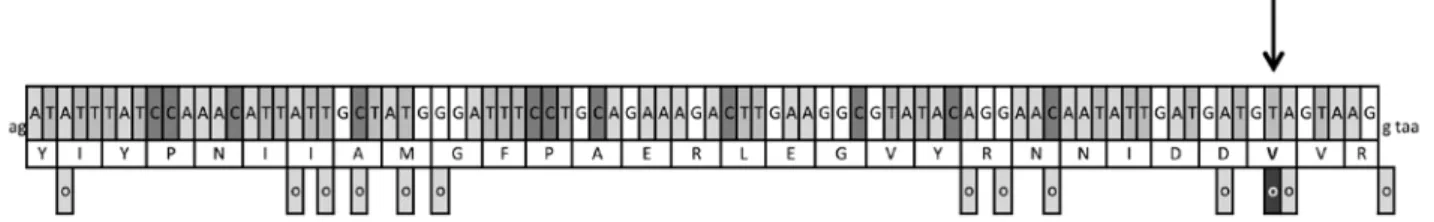
Most of the described mutations are frameshift or stop codon mutations throughout the whole gene, except exons 1 and 9 (17). A mutational hotspot was found in exon 5, which encodes the phosphatase cat alytic core motif (18). The p.Val53Asp mutation de scribed here is located at exon 2 of the PTEN gene, and
Cop
yright
© ABE&M t
odos os dir
eit
os r
eser
vados
.
In conclusions, we identiied a novel germline p.Val53Asp mutation in the PTEN gene in a patient
with Cowden syndrome, and demonstrated its involve ment in breast cancer and thyroid lesions. Clinical man agement of patients with CS should include early and frequent screening for associated malignancies.
Disclosure: no potential conlict of interest relevant to this article was reported.
REFERENCES
1. Lloyd KM, Dennis M. Cowden’s disease. A possible new symp-tom complex with multiple system involvement. Ann Intern Med. 1963(1);58:136-42.
2. Pilarski R, Eng C. Will the real Cowden syndrome please stand up (again)? Expanding mutational and clinical spectra of the PTEN hamartoma tumour syndrome. J Med Genet. 2004;41(5):323-6. 3. Pezzolesi MG, Zbuk KM, Waite KA, Eng C. Comparative genomic
and functional analyses reveal a novel cis-acting PTEN regulatory element as a highly conserved functional E-box motif deleted in Cowden syndrome. Hum Mol Genet. 2007;16(9):1058-71. 4. Rhei E, Kang L, Bogomolniy F, Federici MG, Borgen PI, Boyd J.
Muta-tion analysis of the putative tumor suppressor gene PTEN/MMAC1 in primary breast carcinomas. Cancer Res. 1997;57(17):3657-9. 5. Dahia PL, Marsh DJ, Zheng Z, Zedenius J, Komminoth P, Frisk T, et
al. Somatic deletions and mutations in the Cowden disease gene, PTEN, in sporadic thyroid tumors. Cancer Res. 1997;57(21):4710-3. 6. Blumenthal GM, Dennis PA. PTEN hamartoma tumor syndromes.
Eur J Hum Genet. 2008;16(11):1289-300.
7. Cibas ES, Ali SZ. The Bethesda System for Reporting Thyroid Cy-topathology. Thyroid. 2009;19(11):1159-65.
8. Abrao MG, Billerbeck AE, Nishi MY, Marui S, Mendonca BB. [Standardization of DNA extraction with NaCl from oral mucosa cells: application in PROP1 gene study]. Arq Bras Endocrinol Me-tabol. 2005;49(6):978-82.
9. Bielawski K, Zaczek A, Lisowska U, Dybikowska A, Kowalska A, Falkiewicz B. The suitability of DNA extracted from formalin-ixed, parafin-embedded tissues for double differential polymerase chain reaction analysis. Int J Mol Med. 2001;8(5):573-8.
10. Troncone G, Cozzolino I, Fedele M, Malapelle U, Palombini L. Prep-aration of thyroid FNA material for routine cytology and BRAF testing: a validation study. Diagn Cytopathol. 2010;38(3):172-6. 11. Marsh DJ, Kum JB, Lunetta KL, Bennett MJ, Gorlin RJ, Ahmed SF,
et al. PTEN mutation spectrum and genotype-phenotype correla-tions in Bannayan-Riley-Ruvalcaba syndrome suggest a single en-tity with Cowden syndrome. Hum Mol Genet. 1999;8(8):1461-72. 12. Zhou XP, Marsh DJ, Morrison CD, Chaudhury AR, Maxwell M,
Reifenberger G, et al. Germline inactivation of PTEN and dys-regulation of the phosphoinositol-3-kinase/Akt pathway cause human Lhermitte-Duclos disease in adults. Am J Hum Genet. 2003;73(5):1191-8.
13. Pilarski R, Stephens JA, Noss R, Fisher JL, Prior TW. Predicting PTEN mutations: an evaluation of Cowden syndrome and Ban-nayan-Riley-Ruvalcaba syndrome clinical features. J Med Genet. 2011;48(8):505-12.
14. Nelen MR, Padberg GW, Peeters EA, Lin AY, van den Helm B, Frants RR, et al. Localization of the gene for Cowden disease to chromosome 10q22-23. Nat Genet. 1996;13(1):114-6.
15. Agrawal S, Eng C. Differential expression of novel naturally oc-curring splice variants of PTEN and their functional consequen-ces in Cowden syndrome and sporadic breast cancer. Hum Mol Genet. 2006;15(5):777-87.
16. Liaw D, Marsh DJ, Li J, Dahia PL, Wang SI, Zheng Z, et al. Germ-line mutations of the PTEN gene in Cowden disease, an inherited breast and thyroid cancer syndrome. Nat Genet. 1997;16(1):64-7. 17. Harach HR, Soubeyran I, Brown A, Bonneau D, Longy M. Thyroid
pathologic indings in patients with Cowden disease. Ann Diagn Pathol. 1999;3(6):331-40.
18. Bonneau D, Longy M. Mutations of the human PTEN gene. Hum Mutat. 2000;16(2):109-22.
19. Tan MH, Mester J, Peterson C, Yang Y, Chen JL, Rybicki LA, et al. A clinical scoring system for selection of patients for PTEN muta-tion testing is proposed on the basis of a prospective study of 3042 probands. Am J Hum Genet. 2011;88(1):42-56.
Figure 4. Representation of exon 2 of the PTEN gene with described mutations (circles). The arrow shows the mutation at position 159 (T to A) leading