INVESTIGATION ON THE COMPLAINT
OF DYSPHAGIA IN APHASIC PATIENTS
Investigação da queixa de disfagia em pacientes afásicos
Karin Zazo Ortiz(1), Milena Ribeiro Marinelli(2)
(1) Speech Language Pathology Departament of Universidade
Federal de São Paulo, São Paulo, SP, Brazil.
(2) Universidade Federal de São Paulo, São Paulo, SP, Brazil.
Research developed at Núcleo de Investigação Fonoaudioló-gica em Neuropsicolinguistica of Universidade Federal de São Paulo, São Paulo, Brasil
Source: Fapesp
Conlict of interest: non-existent
The frequency of aphasics between patients that suffer of CVA is from 21% to 38% 3. There is an
association between the aphasic patients post-CVA and the high level of mortality4,5.
Some studies suggest that demographic factors
such as gender and age, inluence in the occur -rence, severity and frequency of aphasia6, while other studies do not conirm these indings7.
The aphasia can be deined as a neurological
language disorder, being several etiologies, among them the CVA8. In the same way, neurological
diseases are the common causes of dysphasia9 and
CVAs can represent more than 50% of this alteration cause8.
The dysphasia can be, in some cases, periodrary and the patient can receive a oral diet normally again. However, this disorder can also offer risks of
dehydration, nutritional deiciencies and pulmonary
complications. In neurogenic dysphasias, it is important to detect the aspiration risk in the patients
INTRODUCTION
Cerebral vascular accidents (CVAs), or strokes, comprise the vascular disorders in which a brain area is affected in transitory or permanent way1 .
The CVA is between the three main death causes in most of the developed and in development countries1,2. It can also cause global motor sequel, language modiications (aphasia), speech (apraxia
and dysarthria) and in the swallowing dynamic (dysphasia).
ABSTRACT
Purposes: to assess the presence and evolution of swallowing complaints in post-stroke aphasic patients and, based on reports by family members on symptom improvements, to determine the
inluence of aphasia on the prognosis of dysphagia. Methods: 30 post-stroke aphasic patients at the chronic phase were interviewed and, together with family members, answered a questionnaire on the presence and persistence of post-stroke swallowing complaints and related aspects, including improvements in dysphagia and aphasia. Kaplan-Meier curves of dysphagia complainers and
non-complainers were compared to verify the inluence of swallowing complaints on the prognosis of
aphasia. Results: 48% of patients reported swallowing problems after the stroke. Out of them, 93% showed positive changes (partial or total resolution of dysphagia symptoms). The average time for patient improvement (spontaneous or otherwise) was 76 days. 60% of the subjects reported total resolution of dysphagia symptoms, 47% of underwent speech therapy. Regarding aphasia, 87% of patients reported some improvement, although no patient reported full resolution of the symptoms. A total of 57% had undergone speech therapy to treat aphasia. The average time reported for positive
change in patients was 183 days. No signiicant difference in aphasia improvement was observed
between dysphagia complainers and non-complainers. Conclusions: out of the aphasic patients assessed in this study, 48% reported swallowing complaints. Based on reports by family members, aphasia had no impact on the prognosis of dysphagia.
prevent pulmonary complications and allow suitable therapeutic interventions10.
Many patients that experienced CVA do not
report dysphasia complaint, but they can show
dificulty to swallow. This dificulty can be masked
with adaptation and compensations performed by them11.
In neurological cases, sometimes, the dysphasia is associated to language disorders such as the aphasia. The cognitive and communicative capac-ities preservation of the patient comprises one of the indicators to classify the patient in the independence scale related to functional swallowing. The state of
consciousness luctuation or cognitive functions
can make the learning impossible disfavouring the swallowing12,13.
Thus, considering that possible alterations that can occur after cerebral injury, the present study aimed at verifying the presence and perception of the swallowing complaint evolution in aphasic
patients post- CVA. The following speciic objec -tives were linked:
To compare the average time of the dysphasia betterment perception and the aphasia perceived and reported by the family in aphasic patients post-CVA;
To analyze, from the relatives report regarding the manifestations betterment, if the aphasia occur-rence interferes in the dysphasia prognosis.
METHODS
The present study was performed at the Speech Therapy and Neurolinguistic Investigation Unit of the Department of Speech Therapy, Universidade Federal de São Paulo - Escola Paulista de Medicina (Unifesp-EPM).
To collect data, aphasic patients that presented as etiological cause a single CVA in the left hemisphere were selected, i.e., without other aphasia-caused etiologias, as brain injury or dementias, that looked spontaneously for assistance in the ambulatory of Acquired Neurogenic Disorders, totaling 30 post
CVA aphasic patients. There was no exclusion of
patients related to the CVA occurrence time criteria, but all patients must be in the chronic phase of the disease when interviewed. Patients must have language complaints to participate of the research.
All individuals selected to participate of the study, were invited to participate and signed the consent form free and cleared. These patients with their relatives answered a questionnaire about the
speech pathologist formally orally and individually. The questionnaires were applied as priority with the injured-brain patient. Only in the situations in which the patient showed compreenhension and/
or emission deiciencies that unabled the interview
with the same, the interview was performed with the relative. The presence of the relative at the interview moment aimed that all data collected would be
conirmed. In all interviews, even when the patient
answered to the questions, the relative was present to endure data reliability. The data related to the CVA history was removed from the patients evalu-ation protocols and noted down in a record sheet.
This record sheet was composed aiming to obtain the patient information, such as: personal data, CVA occurrence date, aphasia type, being this latter obtained by means of speech pathologist evalua-tions followed from the respective diagnostics.
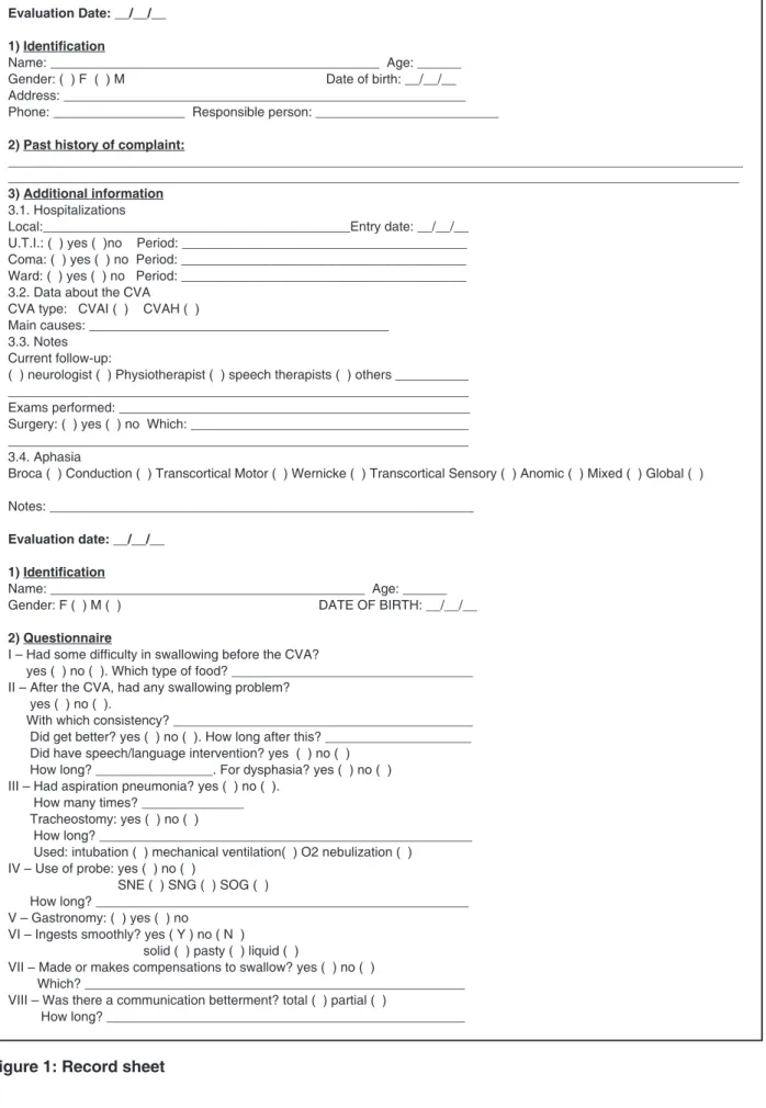
Regarding the questionnaire (Figure 1), it was elaborated from the manifestations already
described in patients that experienced post CVA
swallowing complaints.11
It contains questions that aimed to identify how the swallowing complaint associated to the aphasia occurred.
The questions contained in this questionnaire
covered the following aspects: swallowing dificulty
before and after CVA, with which consistency(Y)
has more dificulty if there was a need of phono -audiological intervention, if there was a communi-cation betterment and/or dysphasia and betterment
average time with or without speciic phonoau -diological intervention for such disorders. During the interview, the patient was asked if he had presented,
in any moment post-CVA, dificulty to swallow
or related complaints (coughing, choking, etc.) . When the answer was yes, further questions were followed, if these complaints (reported) improved, and if so, it was asked how was the betterment, in
order to conirm that the change reported by family/
patient would be a partial or total sign of betterment of the manifestation. In this study, we use the term “betterment” for all changes reported by patients/
relatives and identiied by the speech therapist
through the report as positive, even if they have been partial.
This study was approved by the Institution’s Ethics Committee of the Institution under CEP # 2041/08.
Evaluation Date: __/__/__
1) Identiication
Name: _____________________________________________ Age: ______ Gender: ( ) F ( ) M Date of birth: __/__/__ Address: _______________________________________________________ Phone: __________________ Responsible person: _________________________
2) Past history of complaint:
____________________________________________________________________________________________________ ____________________________________________________________________________________________________ 3) Additional information
3.1. Hospitalizations
Local:__________________________________________Entry date: __/__/__ U.T.I.: ( ) yes ( )no Period: _______________________________________ Coma: ( ) yes ( ) no Period: _______________________________________ Ward: ( ) yes ( ) no Period: _______________________________________ 3.2. Data about the CVA
CVA type: CVAI ( ) CVAH ( )
Main causes: _________________________________________ 3.3. Notes
Current follow-up:
( ) neurologist ( ) Physiotherapist ( ) speech therapists ( ) others __________ _______________________________________________________________
Exams performed: ________________________________________________
Surgery: ( ) yes ( ) no Which: ______________________________________ _______________________________________________________________ 3.4. Aphasia
Broca ( ) Conduction ( ) Transcortical Motor ( ) Wernicke ( ) Transcortical Sensory ( ) Anomic ( ) Mixed ( ) Global ( )
Notes: __________________________________________________________
Evaluation date: __/__/__
1) Identiication
Name: ___________________________________________ Age: ______ Gender: F ( ) M ( ) DATE OF BIRTH: __/__/__
2) Questionnaire
I – Had some dificulty in swallowing before the CVA?
yes ( ) no ( ). Which type of food? _________________________________ II – After the CVA, had any swallowing problem?
yes ( ) no ( ).
With which consistency? _________________________________________ Did get better? yes ( ) no ( ). How long after this? ____________________ Did have speech/language intervention? yes ( ) no ( )
How long? ________________. For dysphasia? yes ( ) no ( ) III – Had aspiration pneumonia? yes ( ) no ( ).
How many times? ______________
Tracheostomy: yes ( ) no ( )
How long? ___________________________________________________
Used: intubation ( ) mechanical ventilation( ) O2 nebulization ( ) IV – Use of probe: yes ( ) no ( )
SNE ( ) SNG ( ) SOG ( )
How long? ___________________________________________________
V – Gastronomy: ( ) yes ( ) no
VI – Ingests smoothly? yes ( Y ) no ( N )
solid ( ) pasty ( ) liquid ( )
VII – Made or makes compensations to swallow? yes ( ) no ( )
Which? ____________________________________________________ VIII – Was there a communication betterment? total ( ) partial ( )
How long? _________________________________________________
the log-rank test. Comparisons of survival curves were performed by the log-rank test and all tests
pre-conditions were veriied. The probability (p)
smaller than 0.05 was considered to indicate
statis-tical signiicance, all tests were two-tailed and all
analyze was calculated using the statistical package SPSS (Statistical Package for the Social Sciences) 13.0 for Windows.
General characteristics
The patients age ranged from 57,8 ± 13,7. The younger patient was 21 years and the older was 81 years.
A total of 30 were assessed, being that 50 of 3 % from the patients were man.
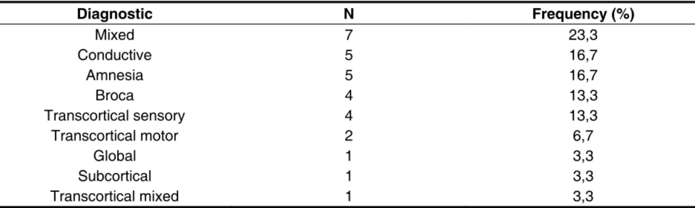
Aphasia types were represented in the Table 1.
Diagnostic N Frequency (%)
Mixed 7 23,3
Conductive 5 16,7
Amnesia 5 16,7
Broca 4 13,3
Transcortical sensory 4 13,3
Transcortical motor 2 6,7
Global 1 3,3
Subcortical 1 3,3
Transcortical mixed 1 3,3
Table 1 – Frequency of aphasia types
Betterment analysis reported of the aphasia regarding the time
Eighty-seven percent of the patients reported betterment of aphasia, although none of them reached the complete betterment. The minimum
Figure 2 - Kaplan-Meier curve for aphasia betterment data
time that the patient could present any betterment reported by the family was 15 days. Patients median that improved was equal to 183 days.
From the total of patients, 57% were submitted to speech therapy for aphasia.
Analyzing the Kaplan-Meier curves from the patients who were submitted to speech therapy
with those who were not, there was no signiicant
difference in the betterment between the two groups (94% vs. 78% of betterment, X2 (1) = 2.03, p = 0.155).
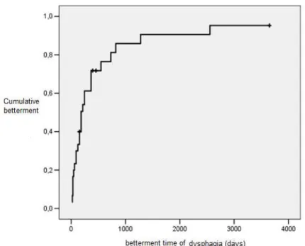
Betterment analysis of the dysphasia regarding the time
The dysphasia complaint was reported in 48% of the patients. From those, 93% reported betterment
Figure 3 - Kaplan-Meier curve for dysphasia betterment data
of dysphasia. The minimum time such that the patient report any kind of betterment was seven days, after the resumption of the consciousness level, post-CVA. The patients median who reported betterment was equal to 76 days. Among those with dysphasia, 43% reported they used the probe during hospitalization. The usage time of the probe ranged from 5 to 122 days (median = 22).
The Figure 3 presents the Kaplan-Meier curve for dysphasia betterment.
From the total with dysphasia, 60% reported overall betterment of dysphasia and 47% reported being treated for dysphasia. For the Kaplan-Meier curves of patients who were submitted to speech therapy with those who were not, there was no
signiicant difference in the betterment mentioned
by both groups (100% vs 87% of betterment, X2 (1) = 0.67, p = 0.411).
Analysis regarding the consistencies of food
Patients who complained of the dysphasia,
11 (73.33%) reported dificulties with the solid
consistency of foods, 11 (73.33%) with liquid and 7 (46.66%) with pasty, and from these, 4 (26.66%) complained with all kinds of consistency.
Five (5) patients complained of one of the
consis-tencies, 3 (three) of them reported dificulties with liquid and two (2) with solid, six (6) patients showed
dificulties with two consistencies, and three (3) had
problems with liquid and solid, two (2) with pasty and solid and one (1) with liquid and pasty.
None of the patients reported complaints of dysphasia prior to CVA.
Relations between entre aphasia and dysphasia To verify that the dysphasia seems to have inter-fered in the aphasia prognosis, we compared the Kaplan-Meier curves of the patients who complained of dysphasia with those who did not report. There
was no signiicant difference in the betterment of
aphasia between the two groups (93% vs. 81% of betterment, X2 (1) = 2.14, p = 0.143).
To verify that the betterment of aphasia affected the prognosis of the patients with dysphasia, compared the Kaplan-Meier curves of the patients reporting aphasia with those did not reported.
groups (93% vs. 100% betterment, X2 (1) = 0.04, p = 0.848).
DISCUSSION
Initially, regarding the characterization of the sample, it can be note that the average age of studied subjects was 57.8 years with a standard deviation of 13.7 years considered low compared to the age with the CVA highest prevalence in the population. According to the literature, more than half of the CVA cases occurs in people over 70 years14. It has been noted, however, that although
the CVAs occur predominantly in older individuals, it can often affect younger people5. In this study, the
average age of patients assessed corroborates with the fact that the CVA is affecting younger popula-tions in recent years.
Fifty-three percent of the patients assessed in this research were male gender. Indeed, there seems to be a predilection for male gender (1.3:1) 1.
Regarding aphasia betterment, 87% of the patients reported some type of aphasia evolution, being that none of them reported overall betterment of the condition. From 30 patients assessed, 57% were submitted to speech therapy and from these, 94% outlined a condition betterment and, from those that did not receive speech therapy, 78% showed some betterment. It is important to note that this study was performed in a public healthcare ambulatory.
The cases of aphasic patients exposed to speech
therapy relate to the ones who had a place in the outpatient care service. In turn, the cases that did not performed therapy, did not do it because there was no place available for the patient. Still, the assiduity and frequency of the treatment were non-controlled variables in this study. The betterment noted in
the cases exposed to the speech therapy can be justiied by the possible spontaneous recovery
occurrence, linked to the guidelines performed to the aphasic patient family at the end of the evaluation. The concept of spontaneous recovery, although much discussed and revised is accepted, because
sych recovery was veriied in many patients that
did not have any therapeutic stimulation and in the same way had a betterment. This type of recovery is assigned to the compensatory functioning of certain areas in the brain or other factors not determined yet16. In this way, the partial change of the language
condition reported by the patients or their relatives must have occurred due to spontaneous functional recovery in the acute phase of the disease. There was a percentage of patients reporting betterment of the aphasia condition against therapy and in
some patients who were not exposed to the speech
even with therapy did not outlined a betterment regarding the aphasia. However, as noted in Table 1, regarding the sample general characteristics, it appears that the majority of aphasic patients interviewed in this study, showed severe aphasias (N=19, 63.4%) which allows us to hypothesize that would have, related to the prognosis, a little chance of total spontaneous betterment of the condition, i.e. without speech therapy.
The median time that patients reported changes in the aphasic condition was 183 days (6 months). Comparing the patients who made speech therapy with those who did not, from the Kaplan- Meier curves
there was no signiicant difference in the betterment of these cases. Despite a signiicant difference
between the betterment of language disorder in
aphasic patients exposed, or not, to the speech
therapy, it is emphasized again that the sample in this study was very heterogeneous regarding to type of aphasia presented by patients. Mild cases usually have better chances of spontaneous betterment17
which makes the ideal comparison between the treated group and non-treated group impossible, since the aphasia severity was not controlled in this comparison. Likewise, it was not possible to control what each family considered a betterment or not of the patient during the interview, which gives
subjectivity to this analysis. And inally, this research
was not this study object of this research and it was performed only to compare the evolution of aphasia and dysphasia reported and observed by patients. In future researches, these variables, if of interest, can be objectively controlled.
At the beginning, regarding the sample, it can be seen that 48% complained of dysphasia. It is known that from the post-CVA patients, 25-50% complained of dysphasia18, and some authors even
suggest higher probabilities occurrence8. Speciic
studies about the epidemiology aspects and natural history of dysphasia associated with acute brain-vascular conditions suggest an incidence of around 50% for swallowing disorders19,20. It is
that 54.54% of the patients with dysphasia post-CVA were associated with aphasia, given that resembles to th noted in the present study.
From patients who complained of dysphasia, 93% reported some betterment and 60% from them reported an overall betterment. Many authors consider that dysphasia betterment spontaneously and the frequency of chronic cases is small21,22.
In relation to dysphasia, the median time that patients show a betterment was 76 days, after CVA. Remembering that, from the total with dysphasia, 47% were submitted to therapy and, when comparing patients who have speech therapy with those did not, it was noted that there was no signif-icant difference in the betterment between the two groups. There was 100% of betterment for patients who have speech therapy for dysphasia and 87% of betterment in patients who did not have the therapy. In a study that aimed to determine the incidence and prognosis of dysphasia in patients with CVA, it was noted that in 88% of the cases they were feeding orally without complications due to spontaneous betterment in the four months after CVA22.
In general, the literature indicates that patients may have one to four months to occur spontane-ously betterment of dysphasia23, 24. In the present
study, it was noted that patients took more than two months to present dysphasia betterment. Furthermore, it is emphasized again that this sample is different from the other analyzed in further studies, once patients were also aphasic and due to linguistic-cognitive changes associated to the condition, may have shown the motor rehabilitation process of dysphasia slightly delayed, because the
dificulty to compensate functionally the deicits and
understand the instructions and guidelines given by the therapists.
Among those with dysphasia, 43% needed to use probe during the hospitalization for an average time of 22 days. In a British research25 performed in
the periods between 1996 and 1999, we estimated a value around 1.7% of all patiensts who suffered from CVA and needed to receive food via probe. It was also found that these patients, after one year from CVA, 13% of them had already returned to oral feeding. In the present study, the recovery was faster, because patients returned to feed orally at an average time of 22 days. The amount higher of those who needed to use the probe as an alternative supply, maybe is due to the fact that patients were aphasic and needed greater preventive care by limiting communication. In turn, the lowest time for patients return to feed orally , may be due to greater assistance given or by the fact that, when presented betterment, even partial, of language disorder, and answered to dysphasia stimulation, swallowing
changes have been noted with fast recovery, which seems to agree with what the literature suggests25 .
Although the initial objective was to verify the possible interference of aphasia in the dysphasia recovery, it was also found that dysphasia inter-feres with aphasia rehabilitation. This last analysis was also made, since it is known that the higher the degree of functional dependency in patients, i.e., the greater the number of limitations, including the dysphasia, the greater the risk of depression26
that can interfere with the aphasia recovery. When patients who complain of dysphasia were compared with those who did not, in order to verify if the dysphasia interfered with the aphasia recovery, it was noted that there was no interference of this aspect in the aphasia rehabilitation. The same was found when comparing if the betterment of aphasia affected the prognosis of dysphasia. However, researches controlling aphasia severity, and inter-ference in the rehabilitation of dysphasia still need to be performed. Likewise, it would be required if studies could also control dysphasia severity.
Regarding the partial spontaneous betterment between aphasia and dysphasia, has a value of 78% of the betterment reported for the communi-cation condition and 87% for swallowing. Although
the difference was small, this was expected because
the recovery of the aphasia is slower, and also in relation to dysphasia, the incidence of spontaneous betterment is greater than 22. In the previous study,
it was noted that patients experiencing dysphasia,
whether presented aphasia or not, were those who presented the worst prognosis for hospital discharge.27
Study Limitations and Future Perspectives
This is a pilot study which analyzed the presence and evolution of the dysphasia complaint reported by aphasic relatives. A methodological limitation was the study being performed only with investigating complaints of dysphasia in chronic aphasic patients. There may have been false positives (patients who reported swallowing complaints of post-CVA) and false negatives (patients without swallowing complaints, but they might have some change), despite the risk of complaint has been overestimated or the problems have not been realized. Moreover, due to greater concern with dysphasia conditions,
many services have proposed screening exclu -sively using questionnaires28,29, different
profes-sionals training30, and special attention to dysphasia
patients with or without aphasia27,31. Future new
researches should be conducted in larger popula-tions to verify whether the co-occurrence of these disorders - evaluated in objective - interfere in the
From the aphasic patients assessed in this study, 48% showed dysphasia compliant.
The average time of the betterment reported for the dysphasia was 76 days and the average time of aphasia betterment reported by the family was 183 days.
RESUMO
Objetivos: veriicar a presença e evolução da queixa de deglutição em pacientes afásicos pós- AVE
e, a partir do relato dos familiares em relação à melhora das manifestações, analisar se a ocorrência da afasia interferiu no prognóstico da disfagia. Métodos: 30 pacientes afásicos pós-AVE na fase crô
-nica foram entrevistados e, juntamente com seus familiares, responderam a um questionário sobre a presença e a permanência de queixas de deglutição pós-AVE e aspectos relacionados, bem como melhoras ocorridas em relação aos quadros de disfagia e afasia. Para veriicar se a disfagia parece
ter interferido no prognóstico da afasia, comparou-se as curvas de Kaplan-Meier dos pacientes que
referiram queixas de disfagia com os que não referiram. Resultados: 48% dos pacientes tiveram
queixas de diiculdades de deglutição pós-AVE. Destes, 93% apresentaram mudanças positivas
(melhora parcial ou total do quadro). O tempo médio para que o paciente apresentasse qualquer tipo de mudança (espontânea ou não) foi de 76 dias. 60% referiu melhora total da disfagia, sendo que 47% foram submetidos à terapia fonoaudiológica. Em relação à afasia, 87% dos pacientes referiram
melhora, apesar de nenhum paciente ter referido melhora total. 57% haviam sido expostos à terapia
fonoaudiológica para a afasia. O tempo médio referido para que o paciente apresentasse qualquer
tipo de mudança positiva nas manifestações foi de 183 dias. Não houve diferença signiicante na melhora da afasia entre o grupo com e sem queixa de deglutição. Conclusões: dos pacientes afá
-sicos avaliados neste estudo, 48% apresentou queixa de disfagia. Veriicou-se, a partir do relato dos
familiares, que a ocorrência da afasia parece não ter interferido no prognóstico da disfagia.
DESCRITORES: Transtornos da Deglutição; Afasia; Reabilitação; Acidente Cerebral Vascular; Deglutição; Linguagem
relatives, positive correlations were not found, i.e., the presence of aphasia does not seem to have affected the dysphasia prognosis.
REFERENCES
1. Soler EP, Ruiz VC. Epidemiology and risk factors of cerebral ischemia and ischemic heart diseases: similarities and differences. Curr Cardiol Rev. 2010;6(3):138-49.
2. Fernadez JR. Acidente Cerebrovasculares (AVE)
en Cuidad de Concepicion y Áreas de Inluencia. Revista Medica de Tucumán. 2000;6(2):61-78.
3. Pedersen PM, Jorgensen HS, Nakayama H, Raaschou HO, Olsen TS. Aphasia in acute stroke: incidence, determinants, and recovery. Ann Neurol. 1995;38:659-66.
4. Paolucci S, Antonucci G, Pratesi L, Traballesi M, Lubich S, Grasso MG. Functional outcome in stroke inpatient rehabilitation: predicting no, low and high response patients. Cerebrovasc Dis. 1998;8:228-34.
5. Tilling K, Sterne JA, Rudd AG. A new method for predicting recovery after stroke. Stroke. 2001;32:2867-73.
6. Hier DB, Yoon WB, Mohr JP. Gender and aphasia in the Stroke Data Bank. Brain Lang. 1994;47:155-67.
7. Madalozzo D, Tognola WA. –Afasias: correlações
clínico-topográicas. Revista Brasileira de
Neurologia. 2006;42(2):5-13.
8. Ickenstein GW, Höhlig C, Prosiegel M, Koch H, Dziewas R, Bodechtel U, Muller R, Reichmann H, Riecker A. Prediction of Outcome in Neurogenic Oropharyngeal Dysphagia Within 72 Hours of Acute Stroke. J Stroke Cerebrovasc Dis. 2011 Jun 15. [Epub ahead of print].
processing in early subacute stroke.BMC Neurol. 2011;11(11):34.
10. Tippett DC.Clinical challenges in the evaluation and treatment of individuals with poststroke dysphagia.Top Stroke Rehabil. 2011 Mar-Apr;18(2):120-33.
11. Menezes FT, Gonçalvez MIR, Chiari B.M. – Adaptações alimentares em adultos pós AVEi sem
queixa de disfagia. Fono Atual. 2005;(34):14-21.
12. Luiz MR. – Atuação Fonoaudiológica em UTI. [levantamento parcial de dados para divulgação da atuação fonoaudiológica em UTI no Hospital das Clínicas da Faculdade de Medicina da USP, realizado em julho de 1999]. www.ufpe.br/utihc/ fono.htm, acesso em maio de 2008.
13. Crawford H, Leslie P, Drinnan MJ. Compliance with dysphagia recommendations by carers of adults with intellectual impairment. Dysphagia. 2007 Oct;22(4):326-34.
14. Tippett DC. Clinical challenges in the evaluation and treatment of individuals with poststroke Dysphagia. Top StrokeRehabil.2011;18(2):120-33. 15. Dulli D, Samaniego EA. Inpatient and community ischemic strokes in a university hospital. Neuroepidemiology. 2007;28(2):86-92.
16. Crinion JT, Leff AP. Recovery and treatment of aphasia after stroke: functional imaging studies. Curr Opin Neurol. 2007;20(6):667-73.
17. Berthier ML. Postsroke aphasia: epidemiology, pathophysiology and treatment. Drugs Aging. 2005;22(2):163-82.
18. Groher ME, Bukation R. The prevalence of swallowing disorders in two teaching hospitals. Dysphagia. 1986;1:3-6.
19. Langdon C, Blacker D. Dysphagia in stroke: a new solution. Stroke Res Treat. 2010;30: 570-3. 20. Barritt AW, Smithard DG. Role of
cerebral cortex plasticity in the recovery of
swallowing function following dysphagic stroke. Dysphagia.2009;24(1):83-90.
21. Fujishima I. Evaluation and management of dysphagia after stroke. Nippon Ronen Igakkai. 2003;40(2):130-4.
22. Meng NH, Wang TG, Lien IN. Dysphagia in patients with brainstem stroke: incidence and outcome. American Journal of Physical and Medicine Rehabilitation. Mar-April 2000;79(2):170-5.
23. Bravata DM, Daggett VS, Woodward-Hagg H, Damush T, Plue L, Russell S, Allen G, Williams LS, Harezlak J, Chumbler NR. Comparison of two approaches to screen for dysphagia among acute ischemic stroke patients: nursing admission screening tool versus National Institutes of Health stroke scale. Rehabil Res Dev. 2009;46(9):1127-34. 24. Martino R, Silver F, Teasell R, Bayley M, Nicholson G, Streiner DL, Diamant NE. The Toronto Bedside Swallowing Screening Test (TOR-BSST): development and validation of a dysphagia screening tool for patients with stroke. Stroke. 2009 Feb;40(2):555-61.
25. Elia M, StrattonRJ, Holden C, Meadows N, Micklewright A, Russell C et al. Home enteral tube feeding following cerebrovascular accident.
Committee of the British Artiicial Nutrition Survey
(BANS). 2001;20(1):27-30.
26. Han M, Ohnishi H, Nonaka M, Yamauchi R, Hozuki T, Hayashi T et al. Relationship between dysphagia and depressive states in patients with Parkinson’s disease. Parkinsonism Relat Disord. 2011 Jul;17(6):437-9.
27. Guyomard V, Fulcher RA, Redmayne O, Metcalf AK, Potter JF, Myint PK. Effect of dysphasia and dysphagia on inpatient mortality and hospital length of stay: a database study.J. Am Geriatr Soc. 2009 Nov;57(11):2101-6.
28. Schrock JW, Bernstein J, Glasenapp M, Drogell K, Hanna J. A novel emergency department dysphagia screen for patients presenting with acute stroke. Acad Emerg Med. 2011 Jun;18(6):584-9. 29. Smith-Tamaray M, Wilson L, McAllister L. Factors affecting dysphagia management and compliance with recommendations in non-metropolitan healthcare settings. Int J Speech Lang Pathol. 2011 Jun;13(3):268-79.
30. Byrne A, Pettigrew CM. Knowledge and attitudes of allied health professional students regarding the stroke rehabilitation team and the role of the Speech and Language Therapist. Int J Lang Commun Disord. 2010 Jul-Aug;45(4):510-21.
31. Falsetti P, Acciai C, Palilla R, Bosi M, Carpinteri F, Zingarelli A et al. Oropharyngeal dysphagia after stroke: incidence, diagnosis, and clinical predictors in patients admitted to a neurorehabilitation unit. J Stroke Cerebrovasc Dis. 2009 Sep-Oct;18(5):329-35. Received on: October 21, 2011
Accepted on: June 09, 2012 Mailing address:
Karin Zazo Ortiz
Rua Botucatu, 802 – Vila Clementino São Paulo – Brasil
CEP: 04023-900