Submitted 22 March 2015 Accepted 28 May 2015 Published30 June 2015
Corresponding author Ga¨elle Qu´er´e,
Academic editor M ´onica Medina
Additional Information and Declarations can be found on page 13
DOI10.7717/peerj.1034
Copyright 2015 Qu´er´e et al.
Distributed under
Creative Commons CC-BY 4.0
OPEN ACCESS
Histopathology of crustose coralline
algae a
ff
ected by white band and white
patch diseases
Ga¨elle Qu´er´e1,2, Anne-Leila Meistertzheim2, Robert S. Steneck3and Maggy M. Nugues2,4
1Leibniz Center for Tropical Marine Ecology (ZMT), Bremen, Germany
2Laboratoire d’Excellence ‘CORAIL’ and USR 3278 CRIOBE EPHE-CNRS-UPVD, Perpignan Cedex, France
3Darling Marine Center, School of Marine Sciences, University of Maine, Walpole, ME, USA 4Carmabi Foundation, Piscaderabaai z/n, Willemstad, Curac¸ao
ABSTRACT
Crustose coralline algae (CCA) are major benthic calcifiers that play crucial roles in marine ecosystems, particularly coral reefs. Over the past two decades, epizootics have been reported for several CCA species on coral reefs worldwide. However, their causes remain often unknown in part because few studies have investigated CCA pathologies at a microscopic scale. We studied the cellular changes associated with two syndromes: Coralline White Band Syndrome (CWBS) and Coralline White Patch Disease (CWPD) from samples collected in Curac¸ao, southern Caribbean. Healthy-looking tissue of diseased CCA did not differ from healthy tissue of healthy CCA. In diseased tissues of both pathologies, the three characteristic cell layers of CCA revealed cells completely depleted of protoplasmic content, but presenting an intact cell wall. In addition, CWBS showed a transition area between healthy and diseased tissues consisting of cells partially deprived of protoplasmic material, most likely corresponding to the white band characterizing the disease at the macroscopic level. This transition area was absent in CWPD. Regrowth at the lesion boundary were sometimes observed in both syndromes. Tissues of both healthy and diseased CCA were colonised by diverse boring organisms. Fungal infections associated with the diseased cells were not seen. However, other bioeroders were more abundant in diseased vs healthy CCA and in diseased vs healthy-looking tissues of diseased CCA. Although their role in the pathogenesis is unclear, this suggests that disease increases CCA susceptibility to bioerosion. Further investigations using an integrated approach are needed to carry out the complete diagnosis of these diseases.
Subjects Ecology, Marine Biology, Histology
Keywords Crustose coralline algae, Disease, Cell death, Boring fauna, Lesion, Histopathology, Regeneration
INTRODUCTION
Scientific awareness that marine diseases represent a major threat to coral reefs has led to the multiplication of disease investigations over the past three decades (Weil, 2001;
considerably increased our knowledge about macroscopic characteristics, abundance and distribution of coral reef diseases and the environmental factors influencing their dynamics (Gladfelter, 1982;Kuta & Richardson, 1996;Hayes & Goreau, 1998;Nugues, 2002;Willis, Page & Dinsdale, 2004;Aeby et al., 2008;Weil, Croquer & Urreiztieta, 2009;
Haapkyl¨a et al., 2010;Tribollet, Aeby & Work, 2011). However, little progress has been made in elucidating disease causation due to the lack of microscopic pathology (Work
& Meteyer, 2014). Coupled with microbial culture and molecular essays, histopathology
appears as a crucial tool to determine the association between a pathogen and a tissue lesion. It is therefore a vital step in any effective coral reef disease survey (Work & Meteyer, 2014). It provides insight into cell pathology and host response to help resolve the question of disease causation (Work et al., 2014). It can detect etiological microorganisms and propose or refute potential causative agents by their observationin situ. Furthermore, it provides a great amount of information on the cell and tissue damages associated with gross lesions (Peters, 1984;Ainsworth et al., 2007a;Burns & Takabayashi, 2011;Williams et al., 2011;Sudek et al., 2012). Sometimes, even in the absence of pathogens, changes in the host tissue histology hint at the type of infection and lead to a diagnostic (Gupta et al., 2009). It is therefore the only current diagnostic tool that allows the establishment of a link between the potential causative agent and the specific changes in cell and tissue (Work & Meteyer, 2014). For instance, histology has confirmed the association between a fungus and the blue-black band lesion in crustose coralline algae (CCA) affected by the Coralline Fungal disease (CFD) (Williams et al., 2014). However, an integrated approach (i.e., combining microbiological, microsensor, molecular and physiological techniques) is necessary in order to incriminate infectious agents as disease causation and thus complete the diagnostic picture (Richardson et al., 2001;Work & Meteyer, 2014).
Unfortunately, investigations at the cellular level are seriously lacking in diseases affecting CCA despite the importance of these calcifying algae in marine ecosystems, especially coral reefs. Along with scleractinian corals, CCA are important primary producers (Adey & Macintyre, 1973;Chisholm, 2003) and framework builders (Adey & Vassar, 1975) delivering significant functional services in coral reef ecosystems, including enhancing coral larval settlement (Morse et al., 1988;Heyward & Negri, 1999;Harrington et al., 2004;Ritson-Williams et al., 2010;Ritson-Williams et al., 2014). CCA are not spared by the increasing intensity and severity of marine diseases (Littler & Littler, 1995;Hayes
& Goreau, 1998) and field investigations on CCA diseases have multiplied in recent
years (Aeby et al., 2008;Vargas- ´Angel, 2010;Tribollet, Aeby & Work, 2011;Miller et al., 2013;Qu´er´e, Steneck & Nugues, 2015). At present, six disease categories have been reported (Vargas- ´Angel, 2010;Williams et al., 2014;Qu´er´e, Steneck & Nugues, 2015), but only CFD and coralline lethal orange disease (CLOD) have known causations. Virtually nothing is known about the other CCA disease categories and they remain histologically uncharacterized. Further knowledge on these diseases and the response of their host could be gained from studies at tissue and cellular levels.
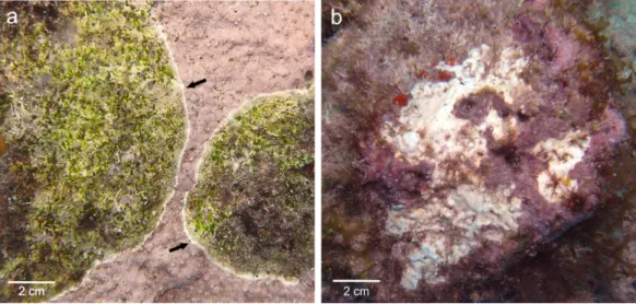
Figure 1 Gross lesions of CCA diseases. (A) CWBS inParagoniolithon solubile and (B) CWPD in
Hydrolithon boergeseniifrom Curac¸ao in 2012. Black arrow shows the white band in CWBS.
pathologies have the potential to reduce the survivorship and settlement of coral planulae and thus may have important implications for the maintenance and recovery of coral reefs (Qu´er´e & Nugues, 2015). They differ in gross symptoms, spatio-temporal variations and lesion spread, suggesting that they may have different causations (Qu´er´e, Steneck & Nugues, 2015). CWBS lesions are defined by a white-band that appears centrally or peripherally and advances slowly but steadily on the healthy tissue, while CWPD manifests by the presence of distinct white patches on the healthy crust, suggesting sudden losses of tissue (Figs. 1A
and1B). Both diseases result in tissue loss with subsequent colonization by endophytic algae often leading to the death of the diseased patch in the case of CWBS (Qu´er´e, Steneck
& Nugues, 2015). Visible symptoms may have a biotic or abiotic origin. On one hand,
thermal stress has been shown to cause bleaching in both corals and CCA in the laboratory
(Anthony et al., 2008) and algal necroses appear on CCA crust under elevated temperature
in aquaria (Martin & Gattuso, 2009). On the other hand, bacterial pathogens can also cause bleaching disease in the marine red algaeDelisea pulchra(Fernandes et al., 2011). Gross symptoms in the shape of rings are known to be caused by a bacterial infection in the case of CLOD (Littler & Littler, 1995) and by fungi in the case of CFD (Williams et al., 2014). The aim of this study was to describe CWBS and CWPD at the microscopic level in order to better understand these diseases and their effects on coralline algal tissues.
MATERIALS AND METHODS
Field collection
Crustose coralline algae were sampled in May 2012 at two sites along the leeward coast of Curac¸ao, Southern Caribbean (12◦N, 69◦W). Fragments (ca. 10–20 cm2) from four
Table 1 Number of healthy and diseased fragments collected from each species.
Healthy CWBS CWPD Total
Hydrolithon boergesenii 3 1 5 9
Neogoniolithon mamillare 2 4 3 9
Paragoniolithon solubile 1 3 0 4
Paragoniolithon accretum 1 0 0 1
Total 7 8 8 23
Playa Kalki (12◦22′30′′N, 69◦09′31′′W). Sampling was not targeted towards particular species, but we sought to have an approximately equal number of healthy and diseased samples. A total of 23 fragments, including 7 healthy fragments, 8 fragments affected by CWBS and 8 fragments affected by CWPD, were sampled (Table 1). For each diseased fragment collected, we made sure to incorporate healthy-looking tissue. Each replicate was selected from a distinct patch. Healthy and diseased fragments of each disease were placed in separated collecting bags to avoid contamination and transported in the dark to the laboratory.
Histology
Back in the laboratory, a sample (ca. 2–4 cm2) of each fragment was kept for taxonomic identification. The pieces used for taxonomic determination were rinsed with freshwater and dried for six hours in the oven at 60◦C before being checked under a dissecting
scope for reproductive and morphological features (Steneck, 1986). The rest was fixed in 4% Formalin-seawater solution and stored in the fridge until further use. Before decalcification, a small piece (ca. 1 cm2) was cut from each fragment so that only the crust of the CCA and a thin (ca. 5 mm) layer of limestone underneath remained. All superficial epibionts (i.e., mostly filamentous algae) present on the surface of the coralline algae were removed. In the case of diseased fragments, each piece was chipped so that it included the boundary between healthy and diseased tissues.
Each sample was then placed in an individual container with 5% L-ascorbic acid solution to gently decalcify over a period up to one week. The solution within each container was refreshed every two days. Once the skeleton and limestone were dissolved, the tissue samples were placed in individual embedding cassettes and dehydrated at room temperature in ascending grades of ethanol (70%, 80%, 95%, 100%, 100%) for 40 min each, followed by an immersion in limonene (three baths of 40 min each). Samples that could not be processed immediately were stored in 70% ethanol for a maximum of 5 days. This additional step did not affect the results (G Qu´er´e, pers. obs., 2014). CCA tissue was then placed in three successive baths of paraffin (Paraplast®PlusTM; Sigma-Aldrich, Seelze, Germany) each time 40 min before being embedded into paraffin blocks. Samples were orientated so that transverse sectioning was possible. The blocks were stored overnight at 4◦C in the fridge to ease withdrawal from the cassette the following day. The blocks
DB80 LX; Leica Biosystems GmbH, Wetzlar, Germany) mounted on a calibrated rotary microtome (LEICATMRM2245; Leica Microsystems GmbH, Wetzlar, Germany). Sections were floated onto water (20◦C), mounted onto clean slides and dehydrated on a slide
drying bench for minimum 40 min at 50◦C.
Sections were then rehydrated and stained following the Sharman staining series
(Sharman, 1943) modified fromRuzin (1999). This method stains the cell walls of plant
tissue in tannic acid and iron alum after the protoplasts have been stained in safranin and orange G (see Document S2 for detailed staining procedure). Several other staining methods were tried, but this method was the most effective to visualize the different cellular components of CCA. Sections were then dehydrated in successive baths of ethanol (45%, 90% and 100%) and cleared with limonene. Coverslips were finally mounted using adhesive resin. We examined and photographed 10 permanent histology sections of each CCA fragment using light microscopy (Leica DM750; Leica Microsystems GmbH,Wetzlar, Germany) with integrated camera (Leica ICC50 HD) using the Leica LAS EZ software.
Analyses
Host response was described at the microscopic level and interpreted by comparing normal healthy fragments paired with diseased ones. The presence of invading organisms, their type and localization within the tissue were recorded. In each fragment, organisms could be present in the CCA crust (i.e., epithallus, perithallus and/or hypothallus) or in the limestone underneath the crust. In addition, in diseased fragments, we noted whether they were located in the healthy-looking and/or diseased tissues of the fragments. The identification of the invading organisms was beyond the scope of this study and was restricted to two boring categories: macroborers (i.e., boring sponges, helminths and others) and microborers (i.e., cyanobacteria). Sample sizes were not sufficient to allow robust statistical analysis. All results are reported for pooled species of CCA as the number of replicates per species was too low to make comparisons between species, but species-specific data are listed inTable S1.
RESULTS
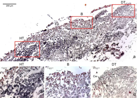
Figure 2 Transversal histological sections of the CCA,Paragoniolithon solubileaffected by CWBS stained in Sharman’s (1943) stain.(A) Overview with locations of the healthy, boundary and dead areas enlarged in (B), (C) and (D). Note the presence of a transition area with progressive loss of staining from healthy tissue (HT) to dead tissue (DT). B, Boundary; Ep, epithallial cells; Cw, cell wall (silver stain); P, protoplasm (orange to dark stain).
cells (Fig. 3C). In two cases of CWBS and one case of CWPD, we observed an overgrowth of the diseased/dead surface by the healthy crust, suggesting tissue recovery (Figs. 4Aand4B).
Figure 3 Transversal histological sections of the CCA,Hydrolithon boergeseniiaffected by CWPD stained in Sharman’s (1943) stain.(A) Overview with locations of the healthy (HT) and diseased areas (DT) enlarged in (B) and (D). (C) shows the boundary (B) between healthy and diseased areas. Note the absence of a transition area highlighted by sudden loss of staining. B, Boundary; Co, conceptacle; Ep, epithallial cells; Cw, cell wall (silver stain); P, protoplasm (orange to dark stain).
DISCUSSION
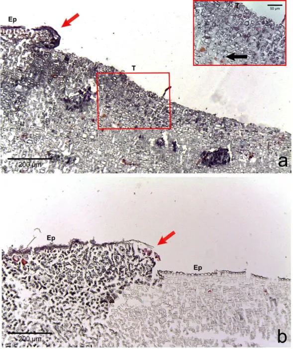
Figure 4 Regrowth of living crust.Regrowth of living crust in (A) CWBS and (B) CWPD. Remnant healthy crust (red arrows) regrew upward and laterally over dead/dying crust. Insert in (A) displays enlargement of transition area with cells showing a condensed nucleus or protoplasmic content (black arrows). T, Transition; Ep, epithallial cells.
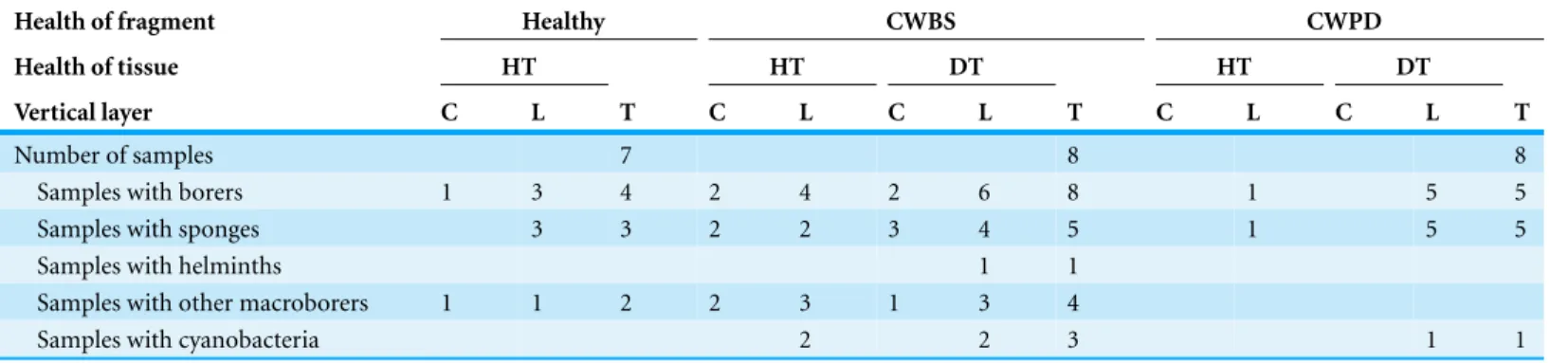
Table 2 Number of samples with boring organisms.Number of samples with boring organisms partitioned by health status of CCA fragments (i.e., healthy, CWBS vs. CWPD), health of tissue within fragment (i.e., healthy vs. diseased) and vertical layer within fragment (i.e., CCA crust vs. limestone).
Health of fragment Healthy CWBS CWPD
Health of tissue HT HT DT HT DT
Vertical layer C L T C L C L T C L C L T
Number of samples 7 8 8
Samples with borers 1 3 4 2 4 2 6 8 1 5 5
Samples with sponges 3 3 2 2 3 4 5 1 5 5
Samples with helminths 1 1
Samples with other macroborers 1 1 2 2 3 1 3 4
Samples with cyanobacteria 2 2 3 1 1
Notes.
HT, healthy tissue; DT, diseased tissue; C, crust; L, limestone; T, total fragment.
Note that the numbers can add up more than for the total fragments since the same fragment may have borers in different sections of the sample.
signs detected in this study differ from sloughing. During a sloughing event, epithallial cells are lost or appear loose (Keats, Knight & Pueschel, 1997;Garbary et al., 2013). Here, they remained present in all the diseased fragments as clearly visible inFig. 3. Additionally, all the different cell layers showed similar changes in the diseased part of the crust (difference in cell staining intensity) whereas in the case of a sloughing event, only the superficial epithallial cells would have shown deterioration.
In CWPD, healthy cells were in immediate vicinity of diseased empty cells whereas in CWBS, a transition area existed where cells had less protoplasmic content than healthy cells as highlighted by a weaker stain within the cells. This transition area could be the sign of a chronic, slowly progressing disease which is reflected in the slow but steady rates of CWBS progression on healthy tissue (i.e., 0.21±0.06 cm month−1inQu´er´e, Steneck & Nugues,
2015). In contrast, CWPD generally manifests by a sudden and extensive loss of tissue, often with a rapid turn-over (G Qu´er´e and M Nugues, pers. obs., 2012), characteristic of acute diseases (Work, Russell & Aeby, 2012;McCoy & Kamenos, 2015).
in Acroporid corals affected by white syndromes (Ainsworth et al., 2007b). However, several other distinct features are necessary to differentiate necrosis (e.g., vacuolization, cell rupture, tissue degradation) and PCD (e.g., cell shrinkage, formation of accumulation bodies) (Dunn et al., 2002;Franklin, Brussaard & Berges, 2006). Interestingly, plants can also present a hypersensitive response that consists of rapid death after infection by a pathogen (e.g., fungi, bacteria, viruses, nematodes) in order to prevent its spread (Garbary
et al., 2013). This phenomenon could constitute a plausible explanation for the CWPD
symptoms. However, further analyses are required to test this hypothesis.
We observed regrowth of healthy-looking tissue over diseased tissue in both diseases. In reef-building corals, an immune response and repair mechanism consisting of a locally accelerated growth has been shown in wounded colonies (D’Angelo et al., 2012). We could interpret this regrowth as a response of the remaining healthy tissue to counteract the progression of the lesion like a wound healing response in CCA. Similarly, CCA are capable of healing wounds caused by herbivores grazing on their crust by regeneration of perithallial cells within the thallus (Steneck, 1983). This healing response could explain the presence of CCA cells lining up the burrow around the invading organisms (Fig. 5B). CCA may have repaired cells around those damaged by the borer. Alternatively, the algal tissue could have grown around the invaders.
We found various metazoa (sponges, helminth, bivalve juveniles) and microrganisms (cyanobacteria) associated with both healthy and diseased CCA tissue. This is consistent with previous studies which have shown the presence of those organisms in healthy and diseased coral colonies (Work & Aeby, 2011;S´er´e et al., 2013) and in live and dead coralline thalli (Tribollet & Payri, 2001). These organisms were more abundant in diseased than healthy CCA fragments, and, within diseased fragments, they were more abundant in diseased vs healthy tissue, suggesting a potential link between CCA diseases and the presence of borers. However, it is unknown whether these borers are the cause of the disease or opportunistic secondary colonizers. Among the organisms observed here, several have been identified as pathogenic in other species. This is the case for helminths known to cause tissue loss inMontipora(Jokiel & Townsley, 1974) or cyanobacteria which appear to cause tissue lysis and necrosis in black band diseased corals (Ainsworth et al.,
2007a). Ciliates are also frequently associated with diseases and capable of invading animal
Microborers are also well-known agents of bioerosion in live and dead CCA thalli causing higher rates of erosion in dead versus live thalli (Tribollet & Payri, 2001). The same way dead coral skeletons are colonised at the surface and bored inwards, diseased crusts could become rapidly vulnerable to invaders (Tribollet & Payri, 2001). Previous studies looking at the association between host response and potential agents revealed that sponges, cyanobacteria and helminths are absent from acute lesions but often associated with chronic diseases, such as the slowly progressing phases of White Syndromes in Montipora capitata(Work, Russell & Aeby, 2012). Our observations confirm this pattern since sponges were often found spreading through the crust and the limestone in CWBS fragments. In contrast, in CWPD fragments, sponges were exclusively located in the limestone, suggesting that they did not have time to invade the crust. There is evidence that macroborers such as bivalves or sponges could take a couple of years to colonize dead skeleton, as they are long-lived, slow-growing organisms (Tribollet & Golubic, 2011).
The increase of borers within the coralline tissue could have a cascading effect by making carbonate substrata available to new borers, thus increasing their eroding action. Ocean acidification also accelerates reef bioerosion without necessarily affecting the health of bor-ing organisms (Wisshak et al., 2013). Furthermore, synergistic effects of ocean warming, ocean acidification and disease infection enhance the reduction in the calcification rates of CCA (Williams et al., 2014). In the face of climate change, disease outbreaks may thus, together with global stressors and boring organisms, aggravate reef degradation.
Histological observations of lesions from the two diseases did not reveal any evidence for the presence of fungi. A fungus belonging to the subphylumUstilaginomyceteshas been identified as the pathogenic agent responsible for CFD thanks to conventional histology
(Williams et al., 2014). We could deduce that fungi are not implicated in CWBS and
CWPD. Similarly, fungi were not observed in the white syndrome of Acroporid corals
(Ainsworth et al., 2007b). Our method did not allow for the visualization of bacteria
CONCLUSIONS
This study brings a descriptive distinction at the cellular level between CWBS and CWPD. Observations of the diseased tissues were consistent with the signs described in the field. CWBS known to progress slowly but steadily over the CCA in the field showed a transition zone in microscopy. In contrast, CWPD known to cause a sudden loss of tissue in CCA had no transition zone. Although boring organisms were observed at higher abundances within diseased tissues in comparison to healthy ones, we did not find evidence of a direct link between the presence of invaders and the disease lesion. However, the range of potential pathogens could be narrowed as no sign of fungal infection was observed. Standard techniques in histopathology alone cannot elucidate the question of disease causation. Additional methods are necessary to complete the diagnosis picture.
ACKNOWLEDGEMENTS
We wish to thank the Carmabi foundation and stafffor logistic support. Aline Tribollet, Elisabeth Faliex, and Thierry Work kindly assisted with identification of invading organisms and interpretation of the micrographs. We also thank Anna Le Ruz and Dr. Annette Peter for assistance in the laboratory.
ADDITIONAL INFORMATION AND DECLARATIONS
Funding
The research leading to these results has received funding from the European Union 7th Framework programme (P7/2007-2013) under grant agreement No. 244161. MMN also acknowledges support from the CNRS Chaire d’Excellence. The funders had no role in study design, data collection and analysis, decision to publish, or preparation of the manuscript.
Grant Disclosures
The following grant information was disclosed by the authors: European Union 7th Framework programme: (P7/2007-2013). CNRS Chaire d’Excellence.
Competing Interests
The authors declare there are no competing interests. Ga¨elle Qu´er´e is an employee of Leibniz Center for Tropical Marine Ecology (ZMT) and Maggy M. Nugues is an associate scientist of Carmabi Foundation.
Author Contributions
• Ga¨elle Qu´er´e conceived and designed the experiments, performed the experiments, analyzed the data, wrote the paper, prepared figures and/or tables, reviewed drafts of the paper.
• Anne-Leila Meistertzheim analyzed the data, contributed reagents/materials/analysis tools, reviewed drafts of the paper.
• Maggy M. Nugues conceived and designed the experiments, analyzed the data, contributed reagents/materials/analysis tools, wrote the paper, reviewed drafts of the paper.
Supplemental Information
Supplemental information for this article can be found online athttp://dx.doi.org/ 10.7717/peerj.1034#supplemental-information.
REFERENCES
Adey WH, Macintyre IG. 1973.Crustose coralline algae: a re-evaluation in the geological sciences.
Geological Society of America Bulletin84:883–904
DOI 10.1130/0016-7606(1973)84<883:CCAARI>2.0.CO;2.
Adey WH, Vassar JM. 1975.Colonization, succession and growth rates of tropical crustose
coralline algae (Rhodophyta, Cryptonemiales).Phycologia14:55–69
DOI 10.2216/i0031-8884-14-2-55.1.
Aeby GS, Work T, Fenner D, Didonato E. 2008.Coral and crustose coralline algae disease on the
reefs of American Samoa. In:Proceedings of the 11th international coral reef symposium, vol. 1. Available athttp://www.reefbase.org/resource center/publication/main.aspx?refid=27761.
Ainsworth TD, Kramasky-Winter E, Loya Y, Hoegh-Guldberg O, Fine M. 2007a.Coral disease
diagnostics: what’s between a plague and a band?Applied and Environmental Microbiology
73:981–992DOI 10.1128/AEM.02172-06.
Ainsworth TD, Kvennefors EC, Blackall LL, Fine M, Hoegh-Guldberg O. 2007b.Disease and
cell death in white syndrome of Acroporid corals on the Great Barrier Reef.Marine Biology
151:19–29DOI 10.1007/s00227-006-0449-3.
Anthony KRN, Kline DI, Diaz-Pulido G, Dove S, Hoegh-Guldberg O. 2008.Ocean acidification
causes bleaching and productivity loss in coral reef builders.Proceedings of the National Academy of Sciences of the United States of America105:17442–17446
DOI 10.1073/pnas.0804478105.
Burge CA, Mark Eakin C, Friedman CS, Froelich B, Hershberger PK, Hofmann EE, Petes LE,
Prager KC, Weil E, Willis BL, Ford SE, Harvell CD. 2014.Climate change influences on marine
infectious diseases: implications for management and society.Annual Review of Marine Science
6:249–277DOI 10.1146/annurev-marine-010213-135029.
Burns J, Takabayashi M. 2011.Histopathology of growth anomaly affecting the coral, montipora
capitata: implications on biological functions and population viability.PLoS ONE6:e28854
DOI 10.1371/journal.pone.0028854.
Bythell JC, Barer MR, Cooney RP, Guest JR, O’Donnell AG, Pantos O, Le Tissier MDA.
2002. Histopathological methods for the investigation of microbial communities
associated with disease lesions in reef corals.Letters in Applied Microbioloy 34:359–364
DOI 10.1046/j.1472-765X.2002.01097.x.
Chisholm JRM. 2003.Primary productivity of reef-building crustose coralline algae.Limnology
and Oceanography48:1376–1387DOI 10.4319/lo.2003.48.2.0787.
D’Angelo C, Smith EG, Oswald F, Burt J, Tchernov D, Wiedenmann J. 2012.Locally accelerated
growth is part of the innate immune response and repair mechanisms in reef-building corals as detected by green fluorescent protein (GFP)-like pigments.Coral Reefs31:1045–1056
Davy S, Burchett S, Dale A, Davies P, Davy J, Muncke C, Hoegh-Guldberg O, Wilson W.
2006. Viruses: agents of coral disease? Diseases of Aquatic Organisms69:101–110
DOI 10.3354/dao069101.
Dunn SR, Bythell JC, Le Tissier MD, Burnett WJ, Thomason JC. 2002.Programmed cell death
and cell necrosis activity during hyperthermic stress-induced bleaching of the symbiotic sea anemoneAiptasiasp.Journal of Experimental Marine Biology and Ecology272:29–53
DOI 10.1016/S0022-0981(02)00036-9.
Dunn SR, Pernice M, Green K, Hoegh-Guldberg O, Dove SG. 2012.Thermal stress promotes host
mitochondrial degradation in symbiotic cnidarians: are the batteries of the reef going to run out?PLoS ONE7:e39024DOI 10.1371/journal.pone.0039024.
Fernandes N, Case RJ, Longford SR, Seyedsayamdost MR, Steinberg PD, Kjelleberg S,
Thomas T. 2011. Genomes and virulence factors of novel bacterial pathogens
causing bleaching disease in the marine red algaDelisea pulchra.PLoS ONE6:e27387
DOI 10.1371/journal.pone.0027387.
Franklin DJ, Brussaard CPD, Berges JA. 2006.What is the role and nature of programmed
cell death in phytoplankton ecology? European Journal of Phycology 41:1–14
DOI 10.1080/09670260500505433.
Garbary DJ, Galway ME, Lord CE, Gunawardena AN. 2013.Advances in algal cell biology.
In: Heimann K, Katsaros C, eds.Programmed cell death in multicellular algae. Berlin: Walter de Gruyter GmbH, 1–20.
Gladfelter WB. 1982.White-band disease in acropora palmata: implications for the structure and
growth of shallow reefs.Bull Mar Sci32:639–643.
Greenberg JT. 1997.Programmed cell death in plant-pathogen interactions.Annual Review of
Plant Physiology and Plant Molecular Biology48:525–545
DOI 10.1146/annurev.arplant.48.1.525.
Gupta E, Balla P, Khurana N, Singh T. 2009.Histopathology for the diagnosis of infectious
diseases.Indian Journal of Medical Microbiology27:100–106DOI 10.4103/0255-0857.49423.
Haapkyl¨a J, Melbourne-Thomas J, Flavell M, Willis BL. 2010.Spatiotemporal patterns of coral
disease prevalence on Heron Island, Great Barrier Reef, Australia.Coral Reefs29:1035–1045
DOI 10.1007/s00338-010-0660-z.
Harrington L, Fabricius K, De’Ath G, Negri A. 2004.Recognition and selection of settlement
substrata determine post-settlement survival in corals.Ecology85:3428–3437
DOI 10.1890/04-0298.
Harvell D, Jord´an-Dahlgren E, Merkel S, Rosenberg E, Raymundo L, Smith G, Weil E, Willis B.
2007.Coral disease, environmental drivers, and the balance between coral and microbial
associates.Oceanography20:172–195DOI 10.5670/oceanog.2007.91.
Hayes RL, Goreau NI. 1998.The significance of emerging diseases in the tropical coral reef
ecosystem.Revista de Biologia Tropical46:173–185.
Heyward AJ, Negri AP. 1999.Natural inducers for coral larval metamorphosis.Coral Reefs
18:273–279DOI 10.1007/s003380050193.
Jokiel PL, Townsley SJ. 1974.Biology of the polyclad prosthiostomum (prosthiostomum) sp., a
new coral parasite from Hawaii. Pacific.Science28:361–373.
Keats DW, Knight MA, Pueschel CM. 1997.Antifouling effects of epithallial shedding in three
Kleppel GS, Dodge RE, Reese CJ. 1989.Changes in pigmentation associated with the bleaching of stony corals.Limnology and Oceanography34:1331–1335DOI 10.4319/lo.1989.34.7.1331.
Kuta KG, Richardson LL. 1996.Abundance and distribution of black band disease on coral reefs
in the northern Florida keys.Coral Reefs15:219–223DOI 10.1007/BF01787455.
Lazar B, Loya Y. 1991.Bioerosion of coral reefs-A chemical approach. Limnology and
Oceanography36:377–383DOI 10.4319/lo.1991.36.2.0377.
Littler MM, Littler DS. 1995.Impact of CLOD pathogen on Pacific coral reefs.Science
267:1356–1360DOI 10.1126/science.267.5202.1356.
Martin S, Gattuso J-P. 2009.Response of Mediterranean coralline algae to ocean acidification and
elevated temperature.Global Change Biology15:2089–2100
DOI 10.1111/j.1365-2486.2009.01874.x.
McCoy SJ, Kamenos NA. 2015.Coralline algae (Rhodophyta) in a changing world: integrating
ecological, physiological, and geochemical responses to global change.Journal of Phycology
51:6–24DOI 10.1111/jpy.12262.
Miller IR, Logan M, Johns KA, Jonker MJ, Osborne K, Sweatman HPA. 2013.Determining
background levels and defining outbreaks of crustose coralline algae disease on the Great Barrier Reef.Marine and Freshwater Research64:1022–1028DOI 10.1071/MF12330.
Morse DE, Hooker N, Morse ANC, Jensen RA. 1988.Control of larval metamorphosis and
recruitment in sympatric agariciid corals.Journal of Experimental Marine Biology and Ecology
116:193–217DOI 10.1016/0022-0981(88)90027-5.
Nugues MM. 2002.Impact of a coral disease outbreak on coral communities in St. Lucia: what and
how much has been lost?Marine Ecology Progress Series229:61–71DOI 10.3354/meps229061.
Pantos O, Cooney RP, Le Tissier MDA, Barer MR, O’Donnell AG, Bythell JC. 2003.The
bacterial ecology of a plague-like disease affecting the Caribbean coralMontastrea annularis. Environmental Microbiology5:370–382DOI 10.1046/j.1462-2920.2003.00427.x.
Peters EC. 1983.Possible agent of “White Band Disease” in Caribbean Acroporid corals.Journal of
Invertebrate Pathology41:394–396DOI 10.1016/0022-2011(83)90260-4.
Peters EC. 1984.A survey of cellular reactions to environmental stress and disease in Caribbean
scleractinian corals.Helgol¨ander Meeresunters37:113–137DOI 10.1007/BF01989298.
Pollock FJ, Morris PJ, Willis BL, Bourne DG. 2011.The urgent need for robust coral disease
diagnostics.PLoS Pathogens7:e1002183DOI 10.1371/journal.ppat.1002183.
Qu´er´e G, Nugues MM. 2015.Coralline algae disease reduces survival and settlement success
of coral planulae in laboratory experiments.Coral ReefsEpub ahead of print Mar 31 2015
DOI 10.1007/s00338-015-1292-0.
Qu´er´e G, Steneck RS, Nugues MM. 2015. Spatiotemporal and species-specific patterns
of diseases affecting crustose coralline algae in Curac¸ao. Coral Reefs34:259–273
DOI 10.1007/s00338-014-1225-3.
Richardson LL, Smith GW, Ritchie KB, Carlton RG. 2001. Integrating microbiological,
microsensor, molecular, and physiologic techniques in the study of coral disease pathogenesis. Hydrobiologia460:71–89DOI 10.1023/A:1013187723831.
Ritson-Williams R, Arnold SN, Paul VJ, Steneck RS. 2014.Larval settlement preferences of
Acropora palmataandMontastraea faveolatain response to diverse red algae.Coral Reefs
33:59–66DOI 10.1007/s00338-013-1113-2.
Ritson-Williams R, Paul V, Arnold S, Steneck R. 2010.Larval settlement preferences and
Rosenberg E, Kushmaro A, Kramarsky-Winter E, Banin E, Yossi L. 2009. The role of
microorganisms in coral bleaching.The ISME Journal3:139–146DOI 10.1038/ismej.2008.104.
Ruzin SE. 1999.Plant microtechnique and microscopy. Oxford: Oxford University Press.
S´er´e MG, Tortosa P, Chabanet P, Turquet J, Quod J-P, Schleyer MH. 2013.Bacterial
communities associated with porites white patch syndrome (PWPS) on Three Western Indian Ocean (WIO) Coral Reefs.PLoS ONE8:e83746DOI 10.1371/journal.pone.0083746.
Sharman BC. 1943.Tannic acid and iron alum with safranin and orange G in studies of the shoot
apex biotech histochem.Stain Technology18:105–111DOI 10.3109/10520294309105799.
Steneck RS. 1983.Escalating herbivory and resulting adaptive trends in calcareous algal crusts.
Paleobiology9:44–61.
Steneck RS. 1986.The ecology of coralline algal crusts: convergent patterns and adaptative
strategies.Annual Review of Ecology and Systematics17:273–303
DOI 10.1146/annurev.es.17.110186.001421.
Sudek M, Work TM, Aeby GS, Davy SK. 2012.Histological observations in the Hawaiian reef
coral,Porites compressa, affected by Porites bleaching with tissue loss.Journal of Invertebrate Pathology111:121–125DOI 10.1016/j.jip.2012.07.004.
Tribollet A, Aeby G, Work T. 2011.Survey and determination of coral and coralline algae
diseases/lesions in the lagoon of New Caledonia. Scientific Report. COMPONENT 3D—Project 3D3 Studies of coral diseases in New Caledonia. New Caledonia: CRISP.
Tribollet A, Golubic S. 2011.Reef bioerosion: agents and processes. In: Dubinsky Z, Stambler N,
eds.Coral reefs: an ecosystem in transition. Dordrecht: Springer, 435–449.
Tribollet A, Payri C. 2001. Bioerosion of the coralline alga Hydrolithon onkodes by
microborers in the coral reefs of Moorea, French Polynesia.Oceanologica Acta24:329–342
DOI 10.1016/S0399-1784(01)01150-1.
Vargas- ´Angel B. 2010.Crustose coralline algal diseases in the US-Affiliated Pacific Islands.Coral
Reefs29:943–956DOI 10.1007/s00338-010-0646-x.
Weil E. 2001.Caribbean Coral reef Diseases, Status, and Research Needs. Workshop priorities
for Caribbean Coral Reef Researchm Miami Priorities for Caribbean Coral Reef Research. A Workshop held October 3–5, 2001. Miami: RSMAS, University of Miami, Florida.Available at http://eprints.uberibz.org/27/1/Caribbean coral reef diseases.pdf.
Weil E, Croquer A, Urreiztieta I. 2009.Temporal variability and impact of coral diseases and
bleaching in La Parguera, Puerto Rico from 2003-2007.Caribbean Journal of Science45:221–246.
Williams GJ, Knapp IS, Maragos JE, Davy SK. 2011.Response to Gardner et al..Marine Pollution
Bulletin62:2878–2879DOI 10.1016/j.marpolbul.2011.10.002.
Williams GJ, Price NN, Ushijima B, Aeby GS, Callahan S, Davy SK, Gove JM, Johnson MD,
Knapp IS, Shore-Maggio A, Smith JE, Videau P, Work TM. 2014.Ocean warming and
acidifi-cation have complex interactive effects on the dynamics of a marine fungal disease.Proceedings of the Royal Society B: Biological Sciences281:20133069DOI 10.1098/rspb.2013.3069.
Willis BL, Page CA, Dinsdale EA. 2004.Coral disease on the Great Barrier Reef. In: Rosenberg E,
Loya Y, eds.Coral health and disease. Berlin: Springer, 69–104.
Wisshak M, Sch¨onberg C, Form A, Freiwald A. 2013.Effects of ocean acidification and global
warming on reef bioerosion—lessons from a clionaid sponge.Aquatic Biology19:111–127
DOI 10.3354/ab00527.
Work TM, Aeby GS. 2011. Pathology of tissue loss (white syndrome) in Acropora
sp. corals from the Central Pacific. Journal of Invertebrate Pathology 107:127–131
Work TM, Aeby GS, Lasne G, Tribollet A. 2014. Gross and microscopic pathology of hard and soft corals in New Caledonia.Journal of Invertebrate Pathology 120:50–58
DOI 10.1016/j.jip.2014.05.007.
Work T, Meteyer C. 2014.To understand coral disease, look at coral cells.EcoHealth11:610–618
DOI 10.1007/s10393-014-0931-1:1-9.
Work TM, Rameyer RA. 2005.Characterizing lesions in corals from American Samoa.Coral Reefs
24:384–390DOI 10.1007/s00338-005-0018-0.
Work TM, Russell R, Aeby GS. 2012.Tissue loss (white syndrome) in the coralMontipora capitata
is a dynamic disease with multiple host responses and potential causes.Proceedings of the Royal Society B: Biological Sciences279:4334–4341DOI 10.1098/rspb.2012.1827.
Zundelevich A, Lazar B, Ilan M. 2007.Chemical versus mechanical bioerosion of coral reefs by
boring sponges—lessons from Pione cf. vastifica.Journal of Experimental Biology210:91–96