Pluripotency and Differentiation
Alberto Miranda1,2*, Eva Pericuesta1, Miguel A´ ngel Ramı´rez1., Alfonso Gutierrez-Adan1.
1Departamento de Reproduccio´n Animal y Conservacio´n de Recursos Zoogene´ticos, Instituto Nacional de Investigacio´n y Tecnologı´a Agraria y Alimentaria, Madrid, Spain, 2Centro de Investigacio´n en Sanidad Animal, Instituto Nacional de Investigacio´n y Tecnologı´a Agraria y Alimentaria, Madrid, Spain
Abstract
Cellular prion protein (PRNP) is a glycoprotein involved in the pathogenesis of transmissible spongiform encephalopathies (TSEs). Although the physiological function of PRNP is largely unknown, its key role in prion infection has been extensively documented. This study examines the functionality of PRNP during the course of embryoid body (EB) differentiation in mousePrnp-null (KO) and WT embryonic stem cell (ESC) lines. The first feature observed was a new population of EBs that only appeared in the KO line after 5 days of differentiation. These EBs were characterized by their expression of several primordial germ cell (PGC) markers until Day 13. In a comparative mRNA expression analysis of genes playing an important developmental role during ESC differentiation to EBs, Prnp was found to participate in the transcription of a key pluripotency marker such asNanog. A clear switching off of this gene on Day 5 was observed in the KO line as opposed to the WT line, in which maximumPrnpandNanogmRNA levels appeared at this time. Using a specific antibody against PRNP to block PRNP pathways, reducedNanogexpression was confirmed in the WT line. In addition, antibody-mediated inhibition of ITGB5 (integrinavb5) in the KO line rescued the low expression ofNanogon Day 5, suggesting the regulation ofNanog transcription byPrnp via this Itgb5. mRNA expression analysis of the PRNP-related proteins PRND (Doppel) and SPRN (Shadoo), whose PRNP function is known to be redundant, revealed their incapacity to compensate for the absence of PRNP during early ESC differentiation. Our findings provide strong evidence for a relationship betweenPrnp and several key pluripotency genes and attributePrnpa crucial role in regulating self-renewal/differentiation status of ESC, confirming the participation of PRNP during early embryogenesis.
Citation: Miranda A, Pericuesta E, Ramı´rez MA´, Gutierrez-Adan A (2011) Prion Protein Expression Regulates Embryonic Stem Cell Pluripotency and Differentiation. PLoS ONE 6(4): e18422. doi:10.1371/journal.pone.0018422
Editor:Domingos Henrique, Instituto de Medicina Molecular, Portugal
ReceivedNovember 23, 2010;AcceptedMarch 6, 2011;PublishedApril 4, 2011
Copyright:ß2011 Miranda et al. This is an open-access article distributed under the terms of the Creative Commons Attribution License, which permits unrestricted use, distribution, and reproduction in any medium, provided the original author and source are credited.
Funding:This work was funded by Grant AGL2009-11358 from the Spanish Ministry of Science and Innovation. The funders had no role in study design, data collection and analysis, decision to publish, or preparation of the manuscript.
Competing Interests:The authors have declared that no competing interests exist.
* E-mail: [email protected]
.These authors contributed equally to this work.
Introduction
Cellular prion protein (PRNP) is a ubiquitous membrane glycoprotein whose abnormal self-replicating, misfolded form is widely believed to cause several central nervous system disorders, collectively known as Transmissible Spongiform Encephalopathies (TSE) [1]. PRNP is expressed in a broad range of vertebrate tissues such as spleen, lymph nodes, lung, heart, kidney, muscle and mammary glands [2,3]. It is also found in the gonads (spermatogenic cells and ovaries) [4] but most abundantly occurs in the central nervous system. In embryogenesis, mouse Prnp
mRNA is first highly expressed, mostly in the differentiating neuroepithelium, between E8.5 and E9, during the transition from anaerobic to oxidative metabolism [5] and thereafter expands to non-neuronal tissues at E13.5 [6]. Coupled to the fact that PRNP is highly conserved across species, these data point to an important role for this protein. However, actual evidences exist that make PRNP an absolute mystery. These evidences are that Prnp -knockout mice [5,7,8], cattle [9] and goats [10] show no drastically modified developmental phenotype and only display subtle changes such as synaptic transmission abnormalities [11,12], disturbed morphology [13], some alterations in circadian rhythm [14] and cognitive deficiency [15,16,17], suggesting mechanisms
able to withstand the absence of the protein. Similar observations were made when this gene was inhibited in adult neurons [18]. To explain these data, it has been hypothesized that another host-encoded protein is able to compensate for the lack of PRNP [19]. The main candidates for this compensating role are the PRNP-related proteins Doppel (PRND) and Shadoo (SPRN). The postembryonic expression of Doppel is restricted to the male testis, thoughPrndmRNA has been detected during embryogen-esis. Curiously, the absence of Doppel was observed not to provoke alterations in embryonic and postnatal development, but its deficiency causes male infertility in mice [20]. In contrast, Shadoo shows overlapping mRNA expression withPrnpin the brain and has revealed neuroprotective properties at this site. Shadoo has also been attributed a role in embryogenesis such that its downregulation in a Prnp-null embryo invariably gives rise to a lethal phenotype between E8 and E11 [19]. Hence, both these proteins play a crucial role in mammalian embryogenesis and could explain the lack of a severe phenotype in PRNP-knockout mammals as a step towards deciphering the biological role of this protein.
brain, yet is 1.5 times higher than its expression in somatic cells [21]. Moreover, during the immortalization of fibroblast MJ90 cells by Tert transfection, Prnp expression is almost three times greater [22]. In mouse ESC,Prnp is again elevated 1.4 and 6.2 times during the first two weeks of differentiation [23] while during their guided cardiogenic differentiation, the expression of the gene is more than 20-fold higher [24]. Curiously, direct reprogramming of somatic cells (MEFs/B-cells) to a pluripotent state (induced pluripotent stem cells, iPS) increases the expression ofPrnpup to 27-fold [25]. Recently, several studies have associated PRNP with pluripotency. For example, PRNP has been demonstrated to be a marker of haematopoietic stem cells, supporting their self-renewal [26]. Other data indicate that PRNP may be implicated in the biology of glioblastoma, breast cancer, prostate and gastric cancer [27,28] or, in other words, PRNP is involved in long term proliferation as are stem cells. In addition, it has been reported that the absence ofPrnpsignificantly slows the regeneration process of acutely damaged hind-limb tibialis anterior muscles of mice compared to wild-type muscles, affecting the myogenic precursor cells [29]. Collectively, these findings reveal a physiological function of PRNP in immortalized pluripotent ESC.
Despite advances made to date, the real function of PRNP is still unclear with ascribed roles in neuroprotection, the response to oxidative stress, cell proliferation and differentiation, synaptic function and signal transduction [30]. The study of the physiological functions of PRNP seems to be key to understanding both prion diseases and its possible implications in development and cancer biology. Given the widely described similarity of embryoid bodies (EBs) and early postimplantation embryos, we selected EBs as an ideal model [31]. Herein, by comparing stem cell differentiation to EBs inPrnp-KO and WT mouse cell lines, we identify the first non-redundant function of PRNP in ESC differentiation during early embryogenesis along with a relation-ship between the expression of Prnp and that of several key pluripotency markers.
Materials and Methods
Embryo Collection
Mice were kept on a 14-h light/10-h dark cycle. 10-week-old 129/OLA Prnp knock-out female mice were superovulated by intraperitoneal injections of 10 IU of pregnant mare serum gonadotropin (PMSG) (Folligon; Intervet, Boxmeer, Holland) followed 48 hours later by 10 IU of hCG (Chorulon; Intervet). On the day of hCG injection designated Day 0, the female mice were paired with males of the same strain to allow mating. After disinfecting with 70% alcohol and opening the abdominal wall, the oviducts were excised by clamping, dissecting the peritoneum and fat between the ovary and tube, and cutting the whole oviduct from the proximal end. After washing and flushing the oviduct from the proximal end by incising the whole oviduct with a 30-gauge needle, two-cell embryos were selected by 100x microscopy. All the animals were kept in an animal house and handled using procedures and protocols approved by the Animal Care and Ethical Committee (Informe CEEA 2009/009) of the Instituto Nacional de Investigacio´n y Tecnologı´a Agraria y Alimentaria (INIA, Madrid), guidelines of the European Union (Directive 86/609/ EEC) and current Spanish regulations (BOE 252/34367-91, 2005).
Production of ESC and EBs
For ESC production [32], two-cell embryos were collected 1.5 days after hCG injection and placed in a droplet of tempered KSOM-LIF medium under oil in a 5% CO2atmosphere at 37uC.
After 48 h in KSOM-LIF, the in vitrocultured blastocysts were plated individually in 96-well plates coated with 0.1% gelatine and containing mitomycin-C (Sigma-Aldrich corporation St. Louis, MO, USA) treated mouse embryonic fibroblasts (MEF). They were cultured in ESC medium composed of Dulbecco’s modified Eagle medium (DMEM plus 4500 mg/l glucose, glutaMAX, and pyruvate; Invitrogen, Carlsbad, CA, USA) supplemented with 20% FBS (PAA Laboratories Co¨lbe Germany), 2 mM glutamine, 1 mM MEM nonessential amino acids solution, 1 mM b -mercaptoethanol, 1000 U/ml LIF (leukemia inhibitory factor), and an antibiotic mixture containing 100 U/ml penicillin and 100mg/ml streptomycin. The blastocysts were allowed to attach to the MEF, until expansion without any further experimental interference for four days. After that time, cell clumps originating from the blastocysts were trypsinized and pipetted directly into a well of a 96-well plate containing MEFs and ESC medium. Approximately 4 days after trypsinization, compact cell colonies resembling ESC colony morphology could be detected and these cells were trypsinized in a 24-well plate containing MEFs and ESC medium. In this process, undifferentiated ESCs were separated from differentiated ones and from old MEF, and also expanded in number. Clones that were not confluent were replated on the same plate. When trypsinizing cells from a 35-mm dish for the first time for a particular clone, half the cells were frozen at280uC in foetal calf serum with 10% DMSO media. This was considered the first passage. For cell line expansion, cells were trypsinized at 80% of confluency.
For embryoid body (EB) differentiation [33], ESC colonies were digested with 0.05% trypsin-EDTA. The dissociated cells were collected in EB medium (DMEM plus (4500 mg/l glucose, glutaMAX, and pyruvate) supplemented with 20% FBS, 2 mM glutamine, 1 mM MEM nonessential amino acids solution, 1 mM b-mercaptoethanol, and an antibiotic mixture containing 100 U/ ml penicillin and 100mg/ml streptomycin and plated on a gelatine-coated dish for 45 min to allow the MEF cells to adhere. Non-adherent cells were collected and plated onto non adherent plastic Petri dishes in the same EB medium. The medium was replaced every 2 days.
RNA Extraction, Reverse Transcription, and mRNA Transcript Quantification
RNA was prepared from ESC or EBs of each experimental group (WT orPrnpKO) using theULTRASPEC total RNA Isolation Reagent Kit (BIOTECX laboratories, INC) according to the manufacturer’s instructions. Immediately after extraction, the RT reaction was carried out following the manufacturer’s instructions (Promega, Madrid, Spain) to produce cDNA. Tubes were heated to 70uC for 5 min to denature the secondary RNA structure, allowing Random Primer and Oligo dT attachment, and then the RT mix was completed with the addition of 5 units of Superscript RT enzyme. The tubes were then incubated at room temperature for 10 min and next at 42uC for 60 min to allow the reverse transcription of RNA, followed by 70uC for 10 min to denature the RT enzyme. To detect each transcript, we used 2ml
of the cDNA sample in the RT-PCR.
method was used to quantify expression levels (User Bulletin, Abi Prism 7700 Sequence Detection System, Applied Biosystems). Quanti-fication was normalized to the endogenous control H2az. Fluorescence was acquired in each cycle to determine the threshold cycle or the cycle during the log-linear phase of the reaction at which fluorescence increased above background for each sample. Within this region of the amplification curve, a difference of one cycle is equivalent to doubling the amplified PCR product. According to the comparative CT method, the CT value was determined by subtracting the H2az CT value for each sample from the CT value of each gene in the sample. CT was calculated using the highest sample CT value (i.e., the sample with the lowest target expression) as an arbitrary constant to be subtracted from all other CT sample values. Fold changes in the relative gene expression of the target were determined using the formula 2–CT.
Statistical Analysis
Data were analyzed using theSigmaStat(Jandel Scientific, San Rafael, CA) package by one-way repeated-measures ANOVA with arcsine data transformation.
Semiquantitative PCR
cDNA was obtained as described above and PCR was performed with a PCR mix (Go taq Flexi DNA polymerase Kit, Promega) following the manufacturer’s instructions. The PCR products were analyzed on a 2% agarose gel by ethidium bromide staining. The gel was visualized under ultraviolet light to identify the specific band for each gene analyzed.
Antibody Inhibition Experiments
EBs of both ESC lines were re-suspended on Day 5 in EB medium alone or supplemented with SHA31 anti-mouse PRNP antibody (CISA, INIA), with anti-rabbit ITGB5 antibody (ABCAM) or control antibody (mouse IgG) at final concentrations in the medium of 0.2mg/ml. The EBs were collected 6 or 24 hours later and frozen for mRNA extraction.
Terminal Deoxynucleotidyl Transferase-Mediated dUTP Nick End-Labelling (TUNEL) Assay
For the TUNEL method of detecting apoptosis, EBs cultured for 5 days were fixed in 4% paraformaldehyde in PBS, pH = 7.4, for 1 h, and then washed in PBS/1%BSA three times. The fixed EBs were permeabilized with 0.1% Triton X-100 in PBS for 15 min at 37uC and then washed again in PBS/1%BSA three times. TUNEL assays were performed using theIn Situ Cell Death Detection Kit, TMR Red (Roche) according to the manufacturer’s protocol. As control, it was checked that a lack of TdT in the TUNEL mix completely abolished labelling.
Results
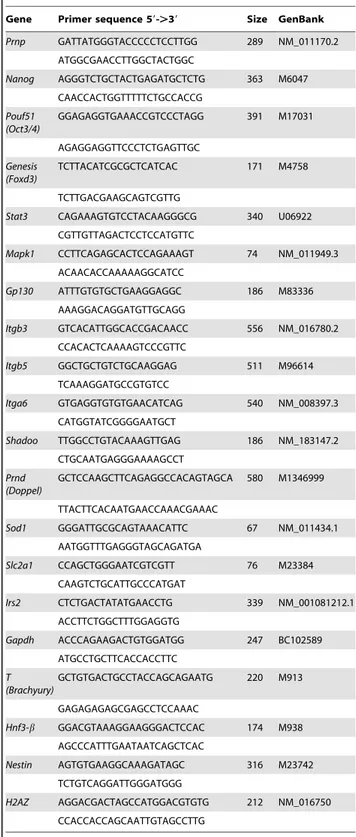
Two distinct types ofPrnp-KO EBs exist
Two lines of Prnp-KO ESC were produced in a 129Ola background (that of the WT control line R1). Since both these lines develop in the same way, the results for each line were collectively considered. Confluent WT and KO ESC lines were trypsinized and grown in a LIF-free medium without the feeder layer to form EBs. From Days 1 to 3, large numbers of unclustered cells could be seen among the KO EBs. This could be the reason of a significantly reduced EB numbers on Day 5 (Fig. 1A). TUNEL apoptosis analysis on Day 5 of ESC differentiation revealed a low number of isolated cells positive for immunostaining in the WT line (Fig. 1B) (2–5% of the WT EB surface). Extensive apoptotic
red zones were detected in the KO EBs on Day 5 of differentiation (10–15% of the KO EB surface), which could explain the high number of floating cells observed in the early EB culture supernatants. Interestingly, two types of EBs were observed on Day 5 in the KO group. About 40% of the EBs were white showing no apoptosis signal, and 60% were morphologically
Table 1.Primers used for RT-PCR.
Gene Primer sequence 59-.39 Size GenBank
Prnp GATTATGGGTACCCCCTCCTTGG 289 NM_011170.2
ATGGCGAACCTTGGCTACTGGC
Nanog AGGGTCTGCTACTGAGATGCTCTG 363 M6047 CAACCACTGGTTTTTCTGCCACCG
Pouf51 (Oct3/4)
GGAGAGGTGAAACCGTCCCTAGG 391 M17031
AGAGGAGGTTCCCTCTGAGTTGC
Genesis (Foxd3)
TCTTACATCGCGCTCATCAC 171 M4758
TCTTGACGAAGCAGTCGTTG
Stat3 CAGAAAGTGTCCTACAAGGGCG 340 U06922 CGTTGTTAGACTCCTCCATGTTC
Mapk1 CCTTCAGAGCACTCCAGAAAGT 74 NM_011949.3
ACAACACCAAAAAGGCATCC
Gp130 ATTTGTGTGCTGAAGGAGGC 186 M83336 AAAGGACAGGATGTTGCAGG
Itgb3 GTCACATTGGCACCGACAACC 556 NM_016780.2
CCACACTCAAAAGTCCCGTTC
Itgb5 GGCTGCTGTCTGCAAGGAG 511 M96614 TCAAAGGATGCCGTGTCC
Itga6 GTGAGGTGTGTGAACATCAG 540 NM_008397.3 CATGGTATCGGGGAATGCT
Shadoo TTGGCCTGTACAAAGTTGAG 186 NM_183147.2
CTGCAATGAGGGAAAAGCCT
Prnd (Doppel)
GCTCCAAGCTTCAGAGGCCACAGTAGCA 580 M1346999
TTACTTCACAATGAACCAAACGAAAC
Sod1 GGGATTGCGCAGTAAACATTC 67 NM_011434.1 AATGGTTTGAGGGTAGCAGATGA
Slc2a1 CCAGCTGGGAATCGTCGTT 76 M23384 CAAGTCTGCATTGCCCATGAT
Irs2 CTCTGACTATATGAACCTG 339 NM_001081212.1
ACCTTCTGGCTTTGGAGGTG
Gapdh ACCCAGAAGACTGTGGATGG 247 BC102589 ATGCCTGCTTCACCACCTTC
T (Brachyury)
GCTGTGACTGCCTACCAGCAGAATG 220 M913
GAGAGAGAGCGAGCCTCCAAAC
Hnf3-b GGACGTAAAGGAAGGGACTCCAC 174 M938
AGCCCATTTGAATAATCAGCTCAC
Nestin AGTGTGAAGGCAAAGATAGC 316 M23742 TCTGTCAGGATTGGGATGGG
H2AZ AGGACGACTAGCCATGGACGTGTG 212 NM_016750 CCACCACCAGCAATTGTAGCCTTG
similar to the WT EBs. The white EBs persisted after a long period of differentiation (more than 80 days) while in the WT line, only a small number of EBs looked white after 5 days of differentiation (1–2%) and these disappeared over the following 2 days (Fig 1C). In addition, after 30 days of differentiation, mature EBs in both KO populations were plated on a feeder layer in complete stem cell medium (including LIF and bFGF), and after 3 passages, we could only recover cells positive for alkaline phosphatase of the white KO population. mRNA was extracted from the different types of EB and it was noted that Gp130 and Stat3 (proteins involved in maintaining pluripotency) were upregulated only in the white KO population (data not shown).
Prnp–KO ESC and EBs express different pluripotent markers to the WT line during differentiation
The mRNA expression of pluripotency markers was examined in both the WT andPrnp–KO lines. We selected the genesOct3/4
(Pou5f1),Nanog, andFoxD3(Genesis) because the feedback regulating cycle of these genes is among the most important for maintaining stem cell pluripotency [35]. Stem cells from the WT line showed significantly higherNanogandOct3/4and lowerFoxD3transcript levels than KO ESC (Fig. 2A and 2B), indicating a different background of pluripotency in the KO and WT lines.
In order to better understand the role of PRNP in differentiation,
PrnpmRNA was quantified from the ESC stage (Day 0) to Day 13 EBs. Stem cell lines were cultured in ESC medium without LIF and samples were taken on Days 0, 1, 2, 3, 5, 7 and 13. Next, mRNA was extracted and transcripts were quantified by qRT-PCR. During
WT EBs differentiation,Prnpshowed increasing levels of expression starting on Day 2 until Day 7, showing peak expression on Day 5 (Fig. 2C). In the WT line, no significant differences could be detected in mRNA levels ofFoxD3until Day 13 when an expression increase was observed (Fig. 2A). However, Prnp expression was negatively correlated with that of the pluripotency markerOct-4. Thus, higherOct-4expression levels were detected on Days 0 and 1 followed by reduced expression until Day 13, following an opposite expression pattern toPrnpand confirming that ES cells had indeed differentiated.Nanogwas also downregulated on Day 13, accord-ingly with the differentiation status. Interestaccord-ingly,Nanogexpression showed a similar pattern toPrnp,with increasing expression levels observed from Day 2 to Day 7 peaking on Day 5 (Fig. 2A). In contrast, in the KO line,Oct-4andFoxD3expression levels showed some variation during early differentiation but high mRNA levels persisted on Days 7 and 13 (Fig. 2B). Further,Nanog expression levels were highest on Day 13, indicating the pluripotent state of these cells even after 13 days of differentiation (Fig. 2B).
To understand this initial developmental status of the different pluripotency markers in the KO ESC, we examined the expression of bone morphogenetic protein (BMP) genes, which are related to the germ cell program. These genes were chosen because KO EBs show characteristics comparable to those of germ cells, as the only cells in the organism that repress their somatic differentiation program in favour of a germline-specific network of RNA regulation. Indeed, BMPs regulate the differentiation of PGCs and are also able to potentiate pluripotency in the presence of LIF. Our semiquantitative PCR revealed increased expression levels of all the PGC markers in the KO stem cells, regardless of the stem cell line (Fig. 3A). The expression of two later PGC markers,Mvh4
(Ddx4) andDazl (Fig. 3B), was lacking in the WT line but was detected in WT cells grown in a GS medium and in the KO lines. When we assessed the same markers during the time course of EB differentiation, it was found that early (Bmp4) and intermediate [Fragillis (Ifitm3) and Stella (Dppa3)] PGC markers (Fig. 3B) were upregulated on Day 5 in WT EBs and that these were gradually replaced with the later expressedMvh4andDazlfrom Day 7 to 13 (Fig. 3C). The same occurred in normal-appearance KO EBs, but in the white EB population all these markers persisted at all the time points. For this reason, we called the white EBs derived from the KO ES cellsPGC-liketo differentiate them from the KO EBs of similar morphological appearance to WT EBs (designatedWT-likeKO EBs). Characterizing the WT andPrnp-KO lines during
differentiation to EBs
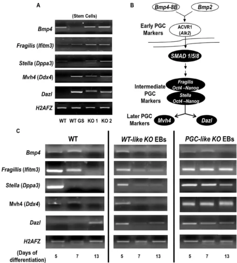
The appearance of two EB populations in the KO line could be the consequence of two different ways of compensation and expression ofShadoo and Doppel, two genes whose functions are redundant to those of Prnp in some tissues and developmental stages. Accordingly, we then examined mRNA levels of these genes during the time course of EB differentiation. In addition, to determine if the differences between the three types of EBs were due to differences in tissue differentiation, we analyzed the mRNA expression of early markers of ectoderm (Nestin), mesoderm (Brachury -T-) and endoderm (Hnf3) (Fig. 4). We considered it might be also interesting to determine the metabolic state of these EB populations since PRNP is thought to be involved in protection from oxidative stress [30]. Thus, we examined mRNA levels of glucose capturing genes (Slc2a and Irs2) and glycolysis genes (Gapdh) (Fig. 5) giving information about oxidative metabolism, and mRNA levels of the SOD1 gene (Sod1), which codes for a protein whose antioxidant activity is regulated by PRNP [36,37]. WT ESC expressed all the transcripts except Nestin in higher amounts than the KO ESC (Fig. 4, Day 0). Interestingly, during Figure 1. Apoptotic and macroscopic effects produced by the
delection ofPrnp.A) Comparing the numbers of EBs in the WT and KO lines (* P,0.05. Error bars, s.e.m.; ‘‘Y’’ axis represents ‘‘number of EBs per plate’’) B) TUNEL analysis of wild type (WT) and Prnp-null (KO) embryoid bodies (EBs). C) Morphology of EBs on Day 7 and Day 80 of differentiation. The KO ESC line produced 2 types of EBs: one similar to WT (*) and the other called PGC-likeKO EBs (arrowheads).
differentiation, mRNA levels of all markers were significantly raised and maintained from Day 5 in thePGC-likeKO EBs (Fig. 4) compared to WT and WT-like KO EBs, which showed similar expression among each other. However, in theWT-likeKO EBs the absence of PRNP resulted in non-expression ofNestinon Day 13 (Fig 4) suggesting a role for PRNP in neural cell differentiation during early embryogenesis. This phenomenon was not observed in the PGC-like KO EBs since these cells maintained similar transcription levels during the differentiation time examined.
On the other hand, the mRNAs ofIrs2, Slc2a, GapdhandSod1
were augmented in thePGC-likeKO EBs on Days 5 and 7 (Fig. 5).
GapdhmRNA was also elevated on Day 5 inWT-likeKO EBs but reached baseline levels on Day 7, suggesting a transition feature. This metabolic situation was maintained until Day 13 in both kinds of KO EB (data not shown) and indicates enhanced glycolytic and oxidative metabolism in thePGC-likeKO EBs. Stat3mRNA is upregulated in the later stages of differentiation
Our experimental protocol includes the use of LIF-starved ES medium, so the receptor (Gp130) and main effector genes (Erk1/2
andStat3) of the LIF pathway were analysed. ERK 1/2 is known to be involved in the loss of pluripotency and is also a downstream component of the PRNP biochemical matrix, while STAT3 contributes to the maintenance of self-renewal. WT EBs showed lower expression of Erk1/2 mRNA than PGC-like KO EBs, disappearing on Day 13 (Fig. 6). The same lower levels were observed for Stat3 in the WT EBs, but disappearing on day 7. However, in thePGC-likeKO EBs, increased transcription of this gene was observed on Day 13, indicating and supporting the previously described pluripotent state of these EBs. In contrast, Gp130 expression failed to vary or only showed subtle variations during the time course of differentiation, indicating that the upregulation ofStat3transcription is not related to activation of the LIF pathway.
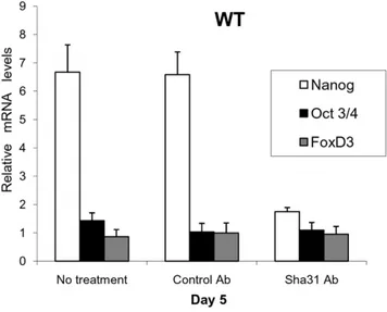
PrnpregulatesNanogmRNA expression
On Day 5 of EB differentiation, the highest levels ofPrnpand
Nanog were detected in the WT EBs. Similar waves in their mRNA expression pattern were also observed suggestingPrnp -dependent Nanog transcription (Fig. 2A and Fig. 2C). To confirm this relationship, we conducted Prnp inhibition Figure 2. Pluripotency in the WT andPrnp-null cell lines.WT EB mRNA (A) and KO EB mRNA (B) expression patterns of three pluripotency genes during differentiation. Note the lack in the KO line (B) of an increase inNanogexpression on Day 5. C) WT EB mRNA expression pattern ofPrnp during differentiation (Days 0, 1, 2, 3, 5, 7 and 13).Prnpis expressed at its highest levels on Day 5 (Error bars, s.e.m.).
experiments. Several studies have shown that the addition of specific PRNP antibody to the culture medium inactivates this protein’s signal transduction pathways [38,39] so we proceeded to add the monoclonal antibody Sha31 to WT EBs on Day 5. Interestingly, a significant reduction inNanogmRNA levels was detected, even over the shorter time interval (6 h) (Fig. 7), and this effect persisted for 24 hours without refreshing the antibody (data not shown). Further, mRNA expression patterns of important PRNP related markers on Day 5 (e.g. integrins avb5 and a6 and the metabolic gene Sod1), which were differentially expressed between thePGC-likeKO EBs and WT EBs, became similar (data not shown). This inhibition ofNanog
expression confirms the involvement of Prnp during EB differentiation.
Nanogexpression on Day 5 is blocked by integrin beta5 inPrnp-KO EBs
Integrins are essential for PGC functionality [40,41] and it has been reported that the absence ofPrnpmay be compensated by these proteins [42,43]. Since integrin signalling is crucial in promoting mouse ESC self-renewal [44], we speculated thatPrnp
expression might also be related to this integrin expression during ESC differentiation. So we examined by real time PCR the
transcription levels of three genes coding for integrinsavb3 (Itgb3), avb5 (Itgb5), and a6 (Itga6) in stem cells and during early EB differentiation (Days 5, 7, and 13). We found thatItgb3was only detected inPGC-like KOEBs on Day 5. Similar transcription levels of Itga6 were found in the KO stem cells and in the WT line. During differentiation, significantly elevated expression was observed on Day 5 in thePrnp-KO background population; this transcription gradually fell from Day 7 to 13. In the WT, expression was increased only on Day 13 (Fig. 8A). Interestingly, mRNA forItgb5, described to be a gonadal tissue marker [41], appeared in higher amounts in the WT ESC than in the KO ESC; however, expression was higher in thePGC-like KOEBs at all the differentiation times (Fig. 8A).
Because the expression ofItgb5on Day 5 differed significantly between WT and KO, we designed an Itgb5 antibody mediated inhibition experiment to analyse its effect on the transcription patterns observed on Day 5 of EB differentiation. Surprisingly, when ITGB5 was blocked at the membrane level in thePGC-like KOEBs,NanogmRNA transcription was significantly augmented, while in the WT line, this treatment had no significant effect (Fig. 8B). These results indicate that in the absence ofPrnp, the expression ofNanogon Day 5 of EB differentiation may be partially mediated by the inhibition of this integrin.
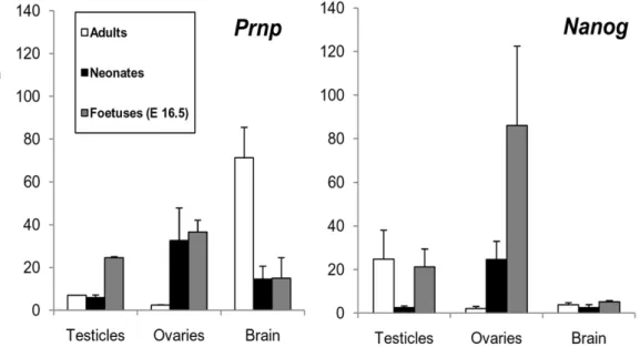
CorrelatedPrnpandNanogexpression in WT mice points to a role for both genes in the development of the foetal gonads
We quantified the in vivo transcription ofPrnpandNanogin the brains, testicles and ovaries of E16.5 WT mouse foetuses, neonates and adults. These tissues were selected becausePrnp-KO ESC and their derived EBs showed an ectodermal gene expression pattern. In brain, as expected, Prnpwas highly expressed, with noNanog
expression detected in any case (Fig. 9). In the testicles, a clear increase inPrnpmRNA levels was seen during the foetal period;
and in the ovaries, this was observed in the foetal and neonatal period.Nanogexpression in these two tissues was upregulated at the same time points as Prnp except in adult testicles, where Nanog
showed clear upregulation contrary to the behaviour of Prnp. However,Doppel was upregulated at this point (data not shown) and could explain the absence of Prnp expression in the adult testicle. These results indicate that correlativeNanog-PrnpmRNA expression, possibly due toNanog Prnp-dependant transcription as was seen in the ‘‘in vitro’’ experiments, occurs in the gonads but not in neuronal tissues.
Figure 4. Differentiation transcription behaviour of the WT andPrnp-null cell lines.mRNA expression patterns of PRNP related proteins (Doppel and Shadoo) and early markers of endoderm (Hnf3), mesoderm (Brachyury -T-) and ectoderm (Nestin) in WT and KO stem cells and in WT, WT-likeKO andPGC-likeKO EBs during differentiation (Days 5, 7 and 13). (* indicates statistical differences for the transcription of each gene between WT and KO ESC at P,0.05) (Error bars, s.e.m).
Discussion
The key role of prion protein in infection has been expansively reported, yet its physiological function remains elusive. To address this issue, we designed a comparative analysis during differentiation of WT and Prnp-KO ESC. In the current study, we employed ESC to assess the potential role of PRNP in early embryo development, examining how this locus becomes transcriptionally activated when potential transient compensatory mechanisms are still not established. We here present the first evidence that Prnp is involved in regulating the self-renewal/differentiation status of stem cells. Its expression was found to affect that of several pluripotent genes, including Nanog, a protein that drives constitutive stem
cell self-renewal and safeguards pluripotency [45,46] through its involvement in the Sox2-Oct3/4 transcription factor network [47,48,49,50,51].
The main pathway through which ESC-derived cells become differentiated cells involves the formation of embryoid bodies, thus denoted because of their similarity to the early postimplan-tation embryo. Embryoid bodies are formed ‘‘in vitro’’ after several days of growth of ESC in the absence of LIF (leukemia inhibitory factor). These bodies are the final ESC population that has the potential to give rise to all the cell types of adult tissues and have been described as the developmental equivalent of the egg cylinder-stage mouse embryo, with an outer endodermal layer and a core of differentiating cells, often comprised of epithelial-lined cavities [52]. Hence the data obtained in this Figure 5. Metabolic transcription behaviour of the WT andPrnp-null cell lines.WT,WT-likeKO EB andPGC-likeKO EB mRNA expression patterns forIrs2(Insulin Receptor Substrate 2),Slc2a(Glucose Transporter 1 -Glut1-),Gapdh(Glyceraldehyde 3-phosphate Dehydrogenase) andSod1 (Superoxide Dismutase 1) on Days 5 (A) and 7 (B). (Error bars, s.e.m.).
work may represent what happens ‘‘in vivo’’ during embryo development.
Our results indicate that despite the widespread expression of key genes of pluripotency in WT and Prnp-KO ESC lines (e.g., Oct4) and their characteristic undifferentiated cell morphology, functional ESC may represent only a small fraction of the total number of cells grown under self-renewal conditions. Effectively, in our KO ESC, a large PGC-like stem cell population was identified. Further, mRNA levels of early endoderm and mesoderm markers were significantly lower, while early neuronal ectoderm marker mRNA levels were fairly similar to those in WT ESC. Taken together with the fact that PGC markers were also expressed, anectodermalstem cell type was defined in the KO ESC, according to the simple absence ofPrnp. Indeed, a heterogeneity in the stem cell population leading to different transcription behavior has been previously described based onNanogmRNA levels [53]. This could explain the appearance of two different kinds of EB when LIF was removed from the culture medium in the KO line, based on the detected absence ofNanogtranscription in this EB population. Thus, phenotypically ‘‘undifferentiated’’ cells may
consist of a heterogeneous population of functionally distinct cell types, and small changes in gene expression could lead to the predominance of one of these types over the others.
Interestingly, in Prnp-KO ESC, Doppel and/or Shadoo were clearly downregulated. However, on Day 5, PGC-like KO EBs overexpressed Doppel and Shadoo, showing the altered expression of early embryo layer markers and also enhanced glucose and oxidative metabolism. This last feature has been related to a high rate of proliferation in cancer or stem cells (Warburg effect) [54,55] and to increased protection against oxidative stress, according to the high pluripotency level of these PGC-like KO
EBs and to compensation for the high sensitivity to oxidants of the KO lines, respectively. Until now, Doppel and Shadoo, ascribed to the PRNP family, were known to compensate for the absence of PRNP, having redundant (in the case of Shadoo in brain [19]) or exclusive (in the case of Doppel in male testicles [20]) functions. However, we noted that WT-like KO EBs exhibited similar expression patterns of these genes with subtle differences in comparison to WT EBs. Thus, we could speculate that an ESC, lacking Prnp yet showing increased Doppel and Shadoo gene expression during differentiation, will give rise to a living system closely resembling PGCs, morphologically different to WT cells, more metabolically active in terms of differentiation [55] and with a high degree of pluripotency. Conversely, if Doppel and Shadoo were not overexpressed, KO EBs would be practically identical to their WT counterparts. Our results therefore suggest that the absence of PRNP is not counteracted by the presence of Doppel and Shadoo, as previously thought. The two different populations of KO EBs with a completely different expression pattern for these genes, further supports the idea of an irreplaceable role of PRNP in this process, most likely in coordinating the expression of indispensable proteins for the appropriate behaviour of the stem cells.
Recently, it has been indicated that during certain embryogen-esis processes some redundancy for PRNP interactions resides within the integrin pathway [43], and that integrin signalling is crucial for promoting mouse ESC self-renewal [44]. Here, we analysed the expression of three integrins during PRNP-null stem cell differentiation to investigate if these integrins play some sort of Figure 6. LIF pathway regulation in the WT andPrnp-null cell
lines. WT EB mRNA (A) andPGC-like KO EB mRNA (B) expression patterns of some LIF pathway genes during differentiation (Days 0 [(Stem Cell], 5, 7 and 13). (*indicates significant differences for a particular gene between WT andPGC-likeKO EBs (P,0.05)) (Error bars, s.e.m.).
doi:10.1371/journal.pone.0018422.g006
Figure 7. Blocking of PRNP by the monoclonal antibody Sha31. On Day 5 and after 6 hours of antibody treatment, it is revealed that NanogmRNA transcription is PRNP-dependent during differentiation and independent of feedback from the pluripotency cluster genesOct3/ 4andFoxD3. (Error bars, s.e.m.).
role in maintaining the stemness of these ESC. Integrin avb3 (ITGB3) and integrinavb5 (ITGB5) are expressed in mouse foetal developing gonads (from Days 10.5 to 13.5); ITGB3 is specific to primordial germ cells [41] and ITGB5 plays important roles in maintaining stemness in undifferentiated mouse ESC [56]. The observation of Itgb3transcription only on Day 5 in the KO EB population indicated primordial germ cell transcription activity at this stage. Moreover, the higher expression ofItgb5on Days 5, 7 and 13 was in agreement with the presence of the primordial germ cell markers identified in thePGC-like KOEBs. It has been reported that integrin-a6 (ITGA6) function is required for the early stages of lens cell differentiation [57]. The higher expression ofItga6we identified on Day 5 in KO EBs, and on Day 13 in the WT EBs, could point to the cell differentiation activity of PGCs in the KO EBs, and of the three embryonic layers in the WT line.
Our results also indicate a relationship between PRNP and integrins in ES cells and during differentiation. It has been shown that PRNP binds some extra cellular matrix (ECM) proteins [43,58,59], and other important ECM proteins, such as integrins,
could also interact or cooperate with PRNP in this process. Integrins have been the focus of several studies over the past few years, not only because of their capacity to bind ECM, but also because of their ability to activate a number of cell signaling pathways, including those of differentiation and pluripotency [44,56]. It is also known that Prnpdownregulates Itgb3 mRNA expression in Mesenchymal Embryonic Cells (MEC) [42], highlighting the importance of integrins in the PRNP biochemical matrix. Other authors have determined that PGCs contain subpopulations showing low or null integrin-a6 (ITGA6) expres-sion with a greater ability to develop into pluripotent stem cells [40]. The fact that several authors have also related the upregulation of certain integrin transcription with a loss of pluripotency and self-renewal [44], suggest a pivotal link between PRNP or integrins and these processes. Our results revealed that
Itga6,Itgb3andItgb5were deregulated in the KO line. Specifically,
times (Days 7 and 13). Since, as mentioned earlier, these integrins are confirmed PGC and gonadal tissue markers, respectively, this suggests the idea of arrested EBs in that ectoderm cell line, also consistent with the loss of self-renewal in a situation of high integrin expression [44].
In addition, the expression ofErk1/2, previously incriminated in differentiation, was significantly downregulated on Day 13 in the
PGC-like KO EBs, at the same time an increase in Stat3
transcription and downregulation of Itga6 were observed. These results suggest thatPGC-like KOEBs seemed to acquire pluripotent and PGC self-renewal characteristics on Day 13. Further, in our antibody-mediated inhibition experiments, when ITGB5 was blocked on Day 5 at the membrane level in the PGC-like KO
EBs, the important pluripotency mRNA transcript Nanog was significantly elevated, while this treatment had no effect in the WT line, though a non-significant upward trend was observed. Similar results were reported when mouse ESC cultured on laminin or fibronectin (activators of integrin pathways) in ESF7 medium showed low immunostaining for alkaline phosphatase, weak immunostaining in immunocytochemical experiments detecting NANOG and a low population of NANOG positive cells in flow cytometric analysis [44]. However, when the mouse ESC was treated with an anti-b1 integrin antibody (blocking the interaction ECM-integrins) these findings were reversed [44].
We also observed here that the disruption of PRNP by knocking out through transgenesis or knocking down through antibody inhibition led to diminishedNanogmRNA levels (especially on Day 5, when peak Prnp and Nanog were observed in the WT line), accompanied by high levels of Itgb5 mRNA. These data thus indicate the possiblePrnpregulation ofNanogtranscription viaItgb5
during early differentiation to EBs. Moreover, the results of our ‘‘in vivo’’ experiments suggest that this relationship during early differentiation between Prnp and Nanog specifically occurs in
gonadal but not in brain tissue. In summary, our data point to the first known relationship between PRNP and NANOG, one of the most important proteins needed to maintain pluripotency, with a major role in the self-renewal of ESC and their differentiation into gonadal cells.
A loss of PRNP functionality was largely proposed as one of the main causes of a prion disease physiopathology. There is a lack of apremortemway of detecting them and bearing in mind thatNanog
levels remain low or disappear during the first stages of differentiation ofPrnp-KO ESC, the use of this gene as a marker of prion disease and, more importantly, as an early detection method could be investigated. Otherwise, PRNP is not essential for ESC survival since other pathways could support its absence, although a significant percentage of cells die in the first stages of differentiation to EBs. In contrast,Prnpknockout or downregula-tion modifies the normal macroscopic and transcripdownregula-tional behaviour of the stem cell during differentiation, confirming the participation of PRNP in early embryogenesis [19]. The association betweenPrnpand pluripotency marker expression also provides evidence of this contribution of Prnp to stem cell differentiation (e.g. the role of PRNP in neural cell differentiation [60]). This feature is mediated byNanogand not compensated by the PRNP-related proteins Doppel and Shadoo, suggesting for the first time a non-redundant function of PRNP in maintaining pluripotency and differentiation during early embryogenesis.
Author Contributions
Conceived and designed the experiments: AM MAR AG-A. Performed the experiments: AM MAR. Analyzed the data: AM AG-A. Contributed reagents/materials/analysis tools: AM EP MAR AG-A. Wrote the paper: AM EP MAR AG-A.
References
1. Castilla J, Gutierrez Adan A, Brun A, Pintado B, Ramirez MA, et al. (2003) Early detection of PrPres in BSE-infected bovine PrP transgenic mice. Arch Virol 148: 677–691.
2. Horiuchi M, Yamazaki N, Ikeda T, Ishiguro N, Shinagawa M (1995) A cellular form of prion protein (PrPC) exists in many non-neuronal tissues of sheep. J Gen Virol 76(Pt 10): 2583–2587.
Figure 9.PrnpandNanogexpression in testicles, ovaries and brain in adult, neonates and foetal mice at embryonic day 16.5 (E16.5).
3. Castilla J, Gutierrez-Adan A, Brun A, Pintado B, Parra B, et al. (2004) Different behavior toward bovine spongiform encephalopathy infection of bovine prion protein transgenic mice with one extra repeat octapeptide insert mutation. J Neurosci 24: 2156–2164.
4. Fujisawa M, Kanai Y, Nam SY, Maeda S, Nakamuta N, et al. (2004) Expression of Prnp mRNA (prion protein gene) in mouse spermatogenic cells. J Reprod Dev 50: 565–570.
5. Bueler H, Fischer M, Lang Y, Bluethmann H, Lipp HP, et al. (1992) Normal development and behaviour of mice lacking the neuronal cell-surface PrP protein. Nature 356: 577–582.
6. Hajj GN, Santos TG, Cook ZS, Martins VR (2009) Developmental expression of prion protein and its ligands stress-inducible protein 1 and vitronectin. J Comp Neurol 517: 371–384.
7. Manson JC, Clarke AR, Hooper ML, Aitchison L, McConnell I, et al. (1994) 129/Ola mice carrying a null mutation in PrP that abolishes mRNA production are developmentally normal. Mol Neurobiol 8: 121–127.
8. Sakaguchi S, Katamine S, Nishida N, Moriuchi R, Shigematsu K, et al. (1996) Loss of cerebellar Purkinje cells in aged mice homozygous for a disrupted PrP gene. Nature 380: 528–531.
9. Richt JA, Kasinathan P, Hamir AN, Castilla J, Sathiyaseelan T, et al. (2007) Production of cattle lacking prion protein. Nat Biotechnol 25: 132–138. 10. Yu G, Chen J, Xu Y, Zhu C, Yu H, et al. (2009) Generation of goats lacking
prion protein. Mol Reprod Dev 76: 3.
11. Collinge J, Whittington MA, Sidle KC, Smith CJ, Palmer MS, et al. (1994) Prion protein is necessary for normal synaptic function. Nature 370: 295–297. 12. Mallucci GR, Ratte S, Asante EA, Linehan J, Gowland I, et al. (2002) Post-natal
knockout of prion protein alters hippocampal CA1 properties, but does not result in neurodegeneration. Embo J 21: 202–210.
13. Colling SB, Khana M, Collinge J, Jefferys JG (1997) Mossy fibre reorganization in the hippocampus of prion protein null mice. Brain Res 755: 28–35. 14. Tobler I, Gaus SE, Deboer T, Achermann P, Fischer M, et al. (1996) Altered
circadian activity rhythms and sleep in mice devoid of prion protein. Nature 380: 639–642.
15. Walz R, Amaral OB, Rockenbach IC, Roesler R, Izquierdo I, et al. (1999) Increased sensitivity to seizures in mice lacking cellular prion protein. Epilepsia 40: 1679–1682.
16. Criado JR, Sanchez-Alavez M, Conti B, Giacchino JL, Wills DN, et al. (2005) Mice devoid of prion protein have cognitive deficits that are rescued by reconstitution of PrP in neurons. Neurobiol Dis 19: 255–265.
17. Coitinho AS, Roesler R, Martins VR, Brentani RR, Izquierdo I (2003) Cellular prion protein ablation impairs behavior as a function of age. Neuroreport 14: 1375–1379.
18. Fuhrmann M, Bittner T, Mitteregger G, Haider N, Moosmang S, et al. (2006) Loss of the cellular prion protein affects the Ca2+homeostasis in hippocampal CA1 neurons. J Neurochem 98: 1876–1885.
19. Young R, Passet B, Vilotte M, Cribiu EP, Beringue V, et al. (2009) The prion or the related Shadoo protein is required for early mouse embryogenesis. FEBS Lett 583: 3296–3300.
20. Behrens A, Genoud N, Naumann H, Rulicke T, Janett F, et al. (2002) Absence of the prion protein homologue Doppel causes male sterility. Embo J 21: 3652–3658.
21. Grskovic M, Chaivorapol C, Gaspar-Maia A, Li H, Ramalho-Santos M (2007) Systematic identification of cis-regulatory sequences active in mouse and human embryonic stem cells. PLoS Genet 3: e145.
22. Hiyama K, Otani K, Ohtaki M, Satoh K, Kumazaki T, et al. (2005) Differentially expressed genes throughout the cellular immortalization processes are quite different between normal human fibroblasts and endothelial cells. Int J Oncol 27: 87–95.
23. Hailesellasse Sene K, Porter CJ, Palidwor G, Perez-Iratxeta C, Muro EM, et al. (2007) Gene function in early mouse embryonic stem cell differentiation. BMC Genomics 8: 85.
24. Faustino RS, Behfar A, Perez-Terzic C, Terzic A (2008) Genomic chart guiding embryonic stem cell cardiopoiesis. Genome Biol 9: R6.
25. Mikkelsen TS, Hanna J, Zhang X, Ku M, Wernig M, et al. (2008) Dissecting direct reprogramming through integrative genomic analysis. Nature 454: 49–55. 26. Zhang CC, Steele AD, Lindquist S, Lodish HF (2006) Prion protein is expressed on long-term repopulating hematopoietic stem cells and is important for their self-renewal. Proc Natl Acad Sci U S A 103: 2184–2189.
27. Pan Y, Zhao L, Liang J, Liu J, Shi Y, et al. (2006) Cellular prion protein promotes invasion and metastasis of gastric cancer. Faseb J 20: 1886–1888. 28. Mehrpour M, Codogno P (2009) Prion protein: From physiology to cancer
biology. Cancer Lett.
29. Stella R, Massimino ML, Sandri M, Sorgato MC, Bertoli A (2010) Cellular prion protein promotes regeneration of adult muscle tissue. Mol Cell Biol 30: 4864–4876.
30. Westergard L, Christensen HM, Harris DA (2007) The cellular prion protein (PrP(C)): its physiological function and role in disease. Biochim Biophys Acta 1772: 629–644.
31. Qin J, Guo X, Cui GH, Zhou YC, Zhou DR, et al. (2009) Cluster characterization of mouse embryonic stem cell-derived pluripotent embryoid bodies in four distinct developmental stages. Biologicals 37: 235–244.
32. Ramirez MA, Fernandez-Gonzalez R, Perez-Crespo M, Pericuesta E, Gutier-rez-Adan A (2009) Effect of stem cell activation, culture media of manipulated embryos, and site of embryo transfer in the production of F0 embryonic stem cell mice. Biol Reprod 80: 1216–1222.
33. Ramirez MA, Pericuesta E, Fernandez-Gonzalez R, Pintado B, Gutierrez-Adan A (2007) Inadvertent presence of pluripotent cells in monolayers derived from differentiated embryoid bodies. Int J Dev Biol 51: 397–407.
34. Bermejo-Alvarez P, Rizos D, Rath D, Lonergan P, Gutierrez-Adan A (2008) Epigenetic differences between male and female bovine blastocysts produced in vitro. Physiol Genomics 32: 264–272.
35. Hatano SY, Tada M, Kimura H, Yamaguchi S, Kono T, et al. (2005) Pluripotential competence of cells associated with Nanog activity. Mech Dev 122: 67–79.
36. Brown DR, Besinger A (1998) Prion protein expression and superoxide dismutase activity. Biochem J 334(Pt 2): 423–429.
37. Klamt F, Dal-Pizzol F, Conte da Frota MJ, Walz R, Andrades ME, et al. (2001) Imbalance of antioxidant defense in mice lacking cellular prion protein. Free Radic Biol Med 30: 1137–1144.
38. Monnet C, Gavard J, Mege RM, Sobel A (2004) Clustering of cellular prion protein induces ERK1/2 and stathmin phosphorylation in GT1-7 neuronal cells. FEBS Lett 576: 114–118.
39. Mouillet-Richard S, Ermonval M, Chebassier C, Laplanche JL, Lehmann S, et al. (2000) Signal transduction through prion protein. Science 289: 1925–1928. 40. Morita-Fujimura Y, Tokitake Y, Matsui Y (2009) Heterogeneity of mouse primordial germ cells reflecting the distinct status of their differentiation, proliferation and apoptosis can be classified by the expression of cell surface proteins integrin alpha6 and c-Kit. Dev Growth Differ.
41. Ishii M, Tay TW, Matsui T, Kidokoro T, Mizukami T, et al. (2006) Expression pattern of alphavbeta3 and alphavbeta5 integrin mRNA in mouse fetal gonads. J Reprod Dev 52: 461–468.
42. Muras AG, Hajj GN, Ribeiro KB, Nomizo R, Nonogaki S, et al. (2009) Prion protein ablation increases cellular aggregation and embolization contributing to mechanisms of metastasis. Int J Cancer 125: 1523–1531.
43. Hajj GN, Lopes MH, Mercadante AF, Veiga SS, da Silveira RB, et al. (2007) Cellular prion protein interaction with vitronectin supports axonal growth and is compensated by integrins. J Cell Sci 120: 1915–1926.
44. Hayashi Y, Furue MK, Okamoto T, Ohnuma K, Myoishi Y, et al. (2007) Integrins regulate mouse embryonic stem cell self-renewal. Stem Cells 25: 3005–3015.
45. Chambers I, Colby D, Robertson M, Nichols J, Lee S, et al. (2003) Functional expression cloning of Nanog, a pluripotency sustaining factor in embryonic stem cells. Cell 113: 643–655.
46. Chambers I, Silva J, Colby D, Nichols J, Nijmeijer B, et al. (2007) Nanog safeguards pluripotency and mediates germline development. Nature 450: 1230–1234.
47. Wang J, Rao S, Chu J, Shen X, Levasseur DN, et al. (2006) A protein interaction network for pluripotency of embryonic stem cells. Nature 444: 364–368. 48. Rodda DJ, Chew JL, Lim LH, Loh YH, Wang B, et al. (2005) Transcriptional
regulation of nanog by OCT4 and SOX2. J Biol Chem 280: 24731–24737. 49. Pan G, Thomson JA (2007) Nanog and transcriptional networks in embryonic
stem cell pluripotency. Cell Res 17: 42–49.
50. Masui S, Nakatake Y, Toyooka Y, Shimosato D, Yagi R, et al. (2007) Pluripotency governed by Sox2 via regulation of Oct3/4 expression in mouse embryonic stem cells. Nat Cell Biol 9: 625–635.
51. Boiani M, Scholer HR (2005) Regulatory networks in embryo-derived pluripotent stem cells. Nat Rev Mol Cell Biol 6: 872–884.
52. O’Shea KS (2004) Self-renewal vs. differentiation of mouse embryonic stem cells. Biol Reprod 71: 1755–1765.
53. Tanaka TS (2009) Transcriptional heterogeneity in mouse embryonic stem cells. Reprod Fertil Dev 21: 67–75.
54. Kondoh H, Lleonart ME, Nakashima Y, Yokode M, Tanaka M, et al. (2007) A high glycolytic flux supports the proliferative potential of murine embryonic stem cells. Antioxid Redox Signal 9: 293–299.
55. Kondoh H (2008) Cellular life span and the Warburg effect. Exp Cell Res 314: 1923–1928.
56. Lee ST, Yun JI, Jo YS, Mochizuki M, van der Vlies AJ, et al. (2010) Engineering integrin signaling for promoting embryonic stem cell self-renewal in a precisely defined niche. Biomaterials 31: 1219–1226.
57. Walker JL, Zhang L, Zhou J, Woolkalis MJ, Menko AS (2002) Role for alpha 6 integrin during lens development: Evidence for signaling through IGF-1R and ERK. Dev Dyn 223: 273–284.
58. Graner E, Mercadante AF, Zanata SM, Forlenza OV, Cabral AL, et al. (2000) Cellular prion protein binds laminin and mediates neuritogenesis. Brain Res Mol Brain Res 76: 85–92.
59. Schmitt-Ulms G, Legname G, Baldwin MA, Ball HL, Bradon N, et al. (2001) Binding of neural cell adhesion molecules (N-CAMs) to the cellular prion protein. J Mol Biol 314: 1209–1225.